jarlife journal
Sample text

AND option

OR option

A CROSS-SECTIONAL ANALYSIS OF APOE GENE POLYMORPHISM AND THE RISK OF COGNITIVE IMPAIRMENTS IN THE ALZHEIMER’S DISEASE NEUROIMAGING INITIATIVE STUDY


G. Wang1, D.E. Vance2, W. Li3, for the Alzheimer’s Disease Neuroimaging Initiative*


1. Tongji Hospital, Tongji Medical College, Huazhong University of Science and Technology, Wuhan, China; 2. Office of Research and Scholarship, School of Nursing, University of Alabama at Birmingham, Birmingham, Alabama; 3. Physician Assistant Studies, School of Health Professions, University of Alabama at Birmingham, Birmingham, Alabama

Corresponding Author: Dr. Wei Li, SHPB 485, 1720 2nd Avenue South, Birmingham, AL 35294, USA, Phone: 205-996-2656, Fax: 205-975-7302, Email: wli@uab.edu

J Aging Res & Lifestyle 2021;10:26-31
Published online April 26, 2021, http://dx.doi.org/10.14283/jarlife.2021.5


Abstract

Background: It is inconclusive on how apolipoprotein epsilon (APOE) gene polymorphism is associated with the risk of having mild cognitive impairment (MCI) or Alzheimer’s disease (AD). Objectives: To investigate how APOE genotype is associated with the risk of MCI or AD using the data collected from the Alzheimer’s Disease Neuroimaging Initiative (ADNI) participants. Methods: A cross-sectional design was used to analyze the baseline data collected from the 1,720 ADNI participants. APOE gene polymorphism was analyzed on how they are related to the risk of cognitive impairments of either MCI or AD using a percent yield (PY) method. Then cognitive functions were compared among six different APOE genotypes using a two-way ANCOVA by controlling possible confounding factors. Results: The prevalence of six APOE genotypes in 1,720 participants is as following: e2/e2 (0.3%), e2/e3 (7.4%), e3/e3 (45.4%), e2/e4 (2%), e3/e4 (35%) and e4/e4 (9.9%). The e2/e2 and e4/e4 genotypes were associated with the lowest and the highest risk respectively for cognitive impairments of either MCI or AD. Further, a worse cognitive diagnosis was associated with an increasing number of APOE e4 allele in a dose dependent manner. Participants with genotype e3/e3 had a better memory measure than those with the genotype of e3/e4. Conclusions: APOE gene polymorphism is associated with different level of risks for cognitive impairments. The heterozygous genotype e3/e4 is associated with a worse memory function compared to the genotype of e3/e3. Further investigations are needed to intervene the cognitive deteriorations in those with at risk APOE genotypes.

Key words: Alzheimer’s disease (AD), apolipoprotein epsilon (APOE), mild cognitive impairment (MCI), aging, lifestyle.


In 1993, the apolipoprotein epsilon (APOE) gene polymorphism was reported to be associated with the risk of late-onset Alzheimer’s disease (AD) (1). APOE, the major susceptibility gene for late-onset, sporadic AD, is located on chromosome 19q13.2 (2). There are three common APOE alleles: e2, e3, and e4, which are defined by two single nucleotide polymorphisms in APOE (rs429358/e4, rs7412/e2) (3). As a result, there are six different APOE genotypes: e2/e2, e2/e3, e2/e4, e3/e3, e3/e4, and e4/e4. Three of them are homozygous (e2/e2, e3/e3, and e4/e4) and the remaining three are heterozygous (e2/e3, e2/e4, and e3/e4) genotypes. In 1994, allele e2 was demonstrated to have protective effects against AD (4). By contrast, allele e4 could increase the risk of sporadic AD in a dose dependent manner (4). In 2003, one study confirmed the association between allele e4 and the risk of AD (5). In 2018, AD risk was shown to be increased with APOE genotype varying from e2/e3 to e2/e4 to e3/e3 to e3/e4 to e4/e4 in a population-based cohort study (6). However, it is still not clear how APOE genotype is associated with the risk of mild cognitive impairment (MCI) and memory function. Using the data collected from the Alzheimer’s Disease Neuroimaging Initiative (ADNI), the purpose of this secondary data analysis study was to categorize how APOE genotype is associated with: 1) the risk of MCI or AD; and 2) cognitive performance. Interestingly, the APOE gene polymorphism was shown to correlate with the outcome in 58 individuals with mild-to-moderate AD after a 10-weeks-long multidimensional stimulation therapy (7). The ADNI participants represent an elderly group who are or were living a relatively healthy lifestyle. The potential findings of our study would be meaningful to direct the care for those with cognitive impairments based on the information of APOE gene polymorphism and cognitive status (MCI or AD). For example, besides living a healthy lifestyle, the clinical management of individuals with cognitive impairments can be optimized by considering the current cognitive diagnosis and the risk of cognitive deterioration associated with a certain APOE genotype.


Methods

ADNI

Data were downloaded from the ADNI database (adni.loni.usc.edu) on October 6, 2019. As an ongoing project, ADNI was launched in 2003 and have been sponsored by the following agencies: National Institute on Aging (NIA), National Institute of Biomedical Imaging and Bioengineering (NIBIB), Food and Drug Administration (FDA), private pharmaceutical companies, and non-profit organizations. The primary goal of the ADNI has been to test whether serial magnetic resonance imaging (MRI), positron emission tomography (PET), biomarkers, and clinical and neuropsychological assessment can be combined to measure the progression of MCI and early AD (8). In the first three phases (1, GO, and 2), the ADNI recruited over 1,700 adult participants from over 50 sites across the United States and Canada. The participants were people (55 to 90 years old), and they consisted of people with different cognitive diagnosis at the baseline visit. Further information about this parent study can be found at http://www.adni-info.org/ and in previous reports (8-13).

APOE Genotyping

APOE genotyping was done using DNA from blood samples collected from ADNI participants. For ADNI-1 participants, APOE genotyping was done through polymerase chain reaction (PCR) amplification, Hhal restriction enzyme digestion, and subsequent standard gel resolution processes (14, 15). For ADNI-GO and ADNI-2 participants, genotyping was carried out by Prevention Genetics and LGC Genomics. Prevention Genetics employed array processing using allele-specific PCR with universal molecular beacons (16, 17). At LGC, assays were performed using competitive allele-specific PCR, enabling bi-allelic scoring of single nucleotide polymorphisms. Genotypes were called and returned to the ADNI Genetics Core after manual quality control.

Baseline Cognitive Diagnosis

For ADNI phase 1, participants were recruited with three cognitive diagnoses at baseline: healthy control (HC), MCI, and AD. The recruitment criteria for HC participants included MMSE scores between 24-30 (inclusive), a clinical dementia rating (CDR) of 0, non-depressed, no diagnosis of either MCI or dementia. The recruitment criteria for participants with MCI included Mini Mental Status Examination (MMSE) scores between 24-30 (inclusive), a memory complaint, have objective memory loss measured by education adjusted scores on Wechsler Memory Scale Logical Memory II, a CDR of 0.5, absence of significant levels of impairment in other cognitive domains, essentially preserved activities of daily living, and an absence of dementia. The recruitment criteria for participants with AD included MMSE scores between 20-26 (inclusive), CDR of 0.5 or 1.0, and meeting NINCDS/ADRDA criteria for probable AD.
For phases GO and 2, the diagnosis of MCI was separated into early MCI (EMCI) and late MCI (LMCI). The criteria for EMCI were: MMSE scores between 24-30 (inclusive), a memory complaint (reported by subject or informant), must have objective memory loss measured by education adjusted scores on delayed recall of one paragraph from Wechsler Memory Scale Logical Memory II (between approximately 0.5 and 1.5 SD below the mean of Cognitively Normal), a CDR of 0.5, absence of significant levels of impairment in other cognitive domains, essentially preserved activities of daily living, and an absence of dementia.
For phase 2, significant memory concern (SMC) was added as one separate category of baseline cognitive diagnosis. Participants with SMC had self-reported memory concern, quantified by using the Cognitive Change Index and the CDR of Zero. However, they scored normally for cognitive tests, and the informant did not equate the expressed concern with progressive memory impairment.
The detailed information on baseline cognitive diagnosis and APOE genotype was provided in Table 1 for the 1,720 ADNI participants. Since cognitive diagnoses of SMC, EMCI, and LMCI were added after the ADNI phase 1, participants with cognitive diagnoses of SMC and HC were combined into one group: Cognitively normal (CN) for our data analysis purpose. As such, EMCI and LMCI were also combined into the MCI group (Table 1).
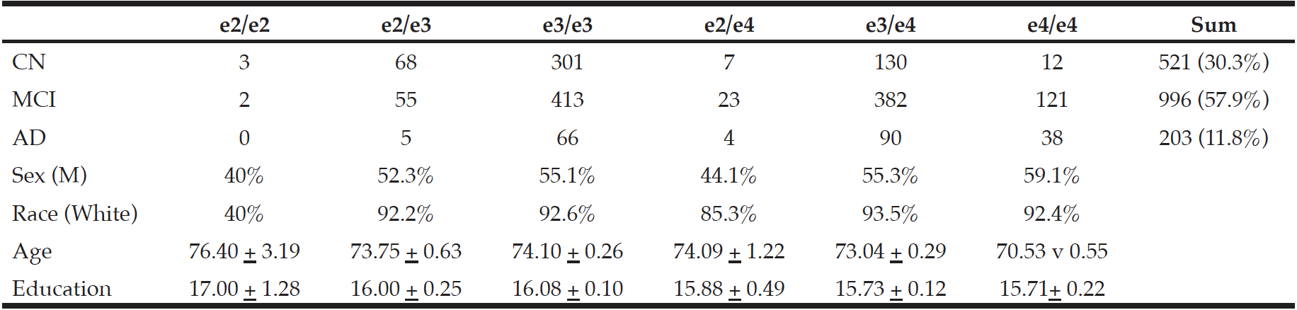
Table 1
Demographic information and baseline cognitive diagnosis were compared among groups of participants with
different APOE genotypes

AD: Alzheimer’s disease; APOE = Apolipoprotein Epsilon; CN: Cognitively normal; M = male; MCI: mild cognitive impairment


Cognitive Measures

The cognitive assessment raw data from the baseline visit were processed and converted into composite scores using validated methods (18-20). The ADNI participants had a comprehensive neuopsychological assessment at the baseline. Individual tests were chosen for analyzing cognitive functions in domains of executive function, language, memory, and visuospatial function. A bi-factor model was used to calculate a composite score for each of the cognitive functions defining the mean at 0 and standard deviation of 1. A lower composite score corresponds with a worse cognitive performance in each of the cognitive domains.

Data Analysis

For calculating the relative risk of each APOE genotype associated with cognitive impairment of MCI or AD, we used a method called percent yield (PY) (21). For the ADNI, all participants were recruited into the study based on their clinical diagnosis of CN, MCI, and AD. The enrollment ratios of these three baseline diagnoses of CN, MCI, and AD were 30.3%, 57.9%, and 11.8% respectively (Table 1).
For each APOE genotype, the PY was calculated with dividing the actual count of participants with each cognitive diagnosis (CN, MCI, or AD) by the theoretical allocation based on the enrollment ratio and the total count of participants for each APOE genotype. For example, there were 5 participants with the genotype e2/e2. Based on the enrollment ratio, there should be 30.3% * 5 = 1.51 persons being allocated to the CN group. Actually, there were 3 participants with the genotype e2/e2 and who were also cognitively normal. Therefore, the PY is 3/1.51=1.98 for participants with the genotype e2/e2, who were cognitive normal (Table 2).

Table 2
The risk of cognitive impairments shown as percent yield was associated with APOE genotype

AD = Alzheimer’s disease; CN = cognitively normal; MCI = mild cognitive impairment


SPSS (version 26.0) was used to conduct all statistical analyses. A one-way analysis of variance (ANOVA)was used to compare age at baseline or education among the six APOE genotype groups (Table 3). Chi-square tests were used to examine the relationship of the APOE genotype with either sex or race (Table 3). Then a two-way analysis of covariance (ANCOVA) model was utilized to evaluate how APOE genotype interacts with baseline diagnostic group (CN, MCI, AD) to affect the cognitive functions with controlling age at baseline, gender, education, and race. Bonferroni post-hoc correction was used for comparing the cognitive functions across the six APOE genotype groups. Data were shown in the form of mean ± standard deviation for both age and education, and p < 0.05 was considered as significant for all statistical analyses.

Table 3
Cognitive functions were compared among participant groups with different APOE genotypes


Data Availability Statement

Data and analytical methods are carefully documented for the performed study. Any data-sharing request can only be submitted to the ADNI for approval purpose.


Results

For the 1,720 ADNI study participants with their APOE genotypes determined, e3/e3 (45.4%) and e3/e4 (35%) were most commonly seen (Table 1). By contrast, the homozygous e2/e2 genotype was the least common (0.3%). The second least common seen genotype was e2/e4 (2%). The percentages for genotypes of e2/e3 and e4/e4 were 7.4% and 9.9%, respectively (Table 1).
At baseline, participants from different APOE genotype groups were significantly different pertaining to either age or race but not education or sex (Table 1). For the baseline age in years, participants in the e4/e4 group had an average age of 70.53 ± 0.55, which was significantly younger than that for the e2/e3 group of 73.75 ± 0.63 (p=0.002) or e3/e3 of 74.10 ± 0.26 (p<0.001). For race, most APOE genotype groups were composed of mainly Whites except the e2/e2 group, which had two Whites out of a total of five participants.
For AD, the relative risk (RR) was in an increasing trajectory in the order of e2/e2, e2/e3, e3/e3, e2/e4, e3/e4, and e4/e4. Genotype e3/e4 had a RR of 1.27, which was lower than the same measure for the e4/e4 group of 1.88 (Table 2). Interestingly, genotype e2/e4 did not increase the AD risk with the RR of 1. For MCI, the RRs for e2/e4, e3/e4, and e4/e4 were 1.17, 1.10, and 1.22, respectively.
At last, cognitive functions were compared among the six APOE genotype groups (Table 3). For the executive function, the APOE gene polymorphism had significant effects (F=2.97, p=0.011) with the e2/e2 group had lower a low composite score than the same measure for the group of e2/e3, e3/e3 or e3/e4. Similarly, there was a significant effect of APOE gene polymorphism on the memory function (F=2.75, p=0.018). The e3/e3 group had a mean memory composite score of 0.37 ± 0.03 (N=780, 95% confidence interval (CI): 0.32-0.42), which was significantly higher than the same measure for the e3/e4 group of 0.23 ± 0.03 (95% CI: 0.17-0.29, N=602, p=0.007) (Table 3). By contrast, neither language (F=0.75, p=0.59) nor visuospatial function (F=1.38, p=0.23) were significantly different among the six different APOE genotype groups (Table 3).


Discussion

Out of the three APOE alleles: e2, e3, and e4, the e3 allele is the most common one followed by e4 then e2 (2), which is consistent with our observations from the ADNI data as e3/e3 and e3/e4 are the most commonly seen genotypes (Table 1). The most common e3 allele was considered to be neutral regarding to AD risk. By contrast, the e2 allele was considered to be protective and associated with a lower risk of AD (4). In addition, each additional copy of APOE e4 was associated with a higher risk and younger age at onset for late onset-AD (4). As expected, the e2/e2 genotype had the most protective effects against MCI or AD. However, the genotype e2/e2 was reported for having a slightly higher 10-year absolute risk for AD than that for the genotype of e2/e3 (6). For either MCI or AD, both e2/e3 and e3/e3 genotypes had protective roles as shown in Table 2. Interestingly, the e2/e4 genotype did not increase the risk for AD but did so for MCI with a PY of 1.17 (Table 2). As expected, both e3/e4 and e4/e4 genotypes were associated with an increased risk for either MCI or AD. A dose dependent effect was observed for the e4 allele for being a risk factor for AD as reported previously (22). It was also reported that e4 allele was associated with an increased risk and e2 allele had a protective role for AD development (23). In another study, individuals with subjective cognitive decline were also shown to have a higher e4 allele frequency than the controls (24).
The e2/e2 had worse executive functions than the genotype groups of e2/e3, e3/e3, or e3/e4, which was unexpected and the findings might be skewed due to the small sample size of the e2/e2 group (n=5). The e3/e3 group had a better performance than the e3/e4 group on memory, which indicated allele e4 had deteriorative effects on this cognitive function (Table 3). It was reported that the protective effects of allele e2 on memory was only observed in females and the effects of allele e4 were unobserved (25).
The e4/e4 group had a significantly younger baseline age than all other groups. Plus, the e4/e4 group had the worst memory performance among all APOE genotype groups (Table 26). In a previous report, even for participants with normal cognitive functions, the e4 allele carriers had a faster declining rate of cognitive performance than the non-carriers (26).
The relation between APOE gene polymorphism and cognitive function had been studied before. For example, individuals carrying the e4 allele showed contextual cueing deficits compared to those who did not carry the e4 allele (27). In addition, an APOE genotype containing e4 allele was shown as an independent risk factor for cognitive decline from a longitudinal study of 14 years (28). In participants with MCI, APOE e4 carrier genotype was associated with a poorer frontal executive function than the e4 non-carrier genotype (29). Further, performance on the MMSE was significantly poorer for e4/e4 homozygotes than e4 heterozygotes or e4 non-carriers (30). Structurally, an e4 allele dose effect was observed for accelerating hippocampal atrophy in participants with different baseline cognitive diagnosis (31). In the current report, we observed the deteriorative effect from the e4 allele but not the protective effect from the e2 allele for the memory function.
Our study had some limitations. The number of participants were small for some genotype groups. For example, there were only 5 participants in the genotype e2/e2 group. In addition, the e2/e4 group had 34 participants. The ADNI participants were mainly composed of Whites and recruited based on the baseline cognitive diagnosis for a prospective cohort study. The participants are generally well educated, have a decent socioeconomic status, and living a healthy lifestyle. Therefore, it is worthy to note that our study was not based on a randomly selected, population-based sample, which could limit the generality of our findings.
In conclusion, our findings supported the e2 allele had protective role for reducing the risk for MCI or AD. At the same time, the data provided evidence on the e4 allele’s deteriorative role for cognitive impairments in memory. However, our findings have the following implications for the elderly population especially for those with at risk APOE genotypes. First, it might be crucial to practice precision medicine using cognitive stimulation training to help those who already developed or are at risk for developing cognitive impairments based on their genetic information. For example, the e4 allele carriers should be monitored closely on cognitive impairment appearance and given corresponding stimulation training/treatment. Second, the APOE gene polymorphism is more closely associated with cognitive performance in some domains (for example memory) than others. Thus, it is important to consider the gene polymorphism factor for doing cognitive stimulation therapy targeting on a specific domain as other researchers begun to consider (32). At last, adopting or maintaining a healthy lifestyle is important to reduce the risk of cognitive impairments despite the genetic predisposition.


*Data used in preparation of this article were obtained from the Alzheimer’s Disease Neuroimaging Initiative (ADNI) database (adni.loni.usc.edu). As such, the investigators within the ADNI contributed to the design and implementation of ADNI and/or provided data but did not participate in analysis or writing of this report. A complete listing of ADNI investigators can be found at: http://adni.loni.usc.edu/wp- content/uploads/how_to_apply/ ADNI_Acknowledgement_ List.pdf

Author contributions: All authors contributed to data analysis and interpretation, draft and critical revision of the manuscript for important intellectual content.

Conflict of interest: All authors have nothing to disclose.

Ethical standard: Written informed consent was obtained from all participants (or guardians of participants) participating in the study (consent for research). The IRB approval was also obtained from each participating clinical/research site.

Acknowledgments: The NACC database is funded by NIA/NIH Grant U01 AG016976. NACC data are contributed by the NIA-funded ADCs: P30 AG019610 (PI Eric Reiman, MD), P30 AG013846 (PI Neil Kowall, MD), P50 AG008702 (PI Scott Small, MD), P50 AG025688 (PI Allan Levey, MD, PhD), P50 AG047266 (PI Todd Golde, MD, PhD), P30 AG010133 (PI Andrew Saykin, PsyD), P50 AG005146 (PI Marilyn Albert, PhD), P50 AG005134 (PI Bradley Hyman, MD, PhD), P50 AG016574 (PI Ronald Petersen, MD, PhD), P50 AG005138 (PI Mary Sano, PhD), P30 AG008051 (PI Thomas Wisniewski, MD), P30 AG013854 (PI M. Marsel Mesulam, MD), P30 AG008017 (PI Jeffrey Kaye, MD), P30 AG010161 (PI David Bennett, MD), P50 AG047366 (PI Victor Henderson, MD, MS), P30 AG010129 (PI Charles DeCarli, MD), P50 AG016573 (PI Frank LaFerla, PhD), P50 AG005131 (PI James Brewer, MD, PhD), P50 AG023501 (PI Bruce Miller, MD), P30 AG035982 (PI Russell Swerdlow, MD), P30 AG028383 (PI Linda Van Eldik, PhD), P30 AG053760 (PI Henry Paulson, MD, PhD), P30 AG010124 (PI John Trojanowski, MD, PhD), P50 AG005133 (PI Oscar Lopez, MD), P50 AG005142 (PI Helena Chui, MD), P30 AG012300 (PI Roger Rosenberg, MD), P30 AG049638 (PI Suzanne Craft, PhD), P50 AG005136 (PI Thomas Grabowski, MD), P50 AG033514 (PI Sanjay Asthana, MD, FRCP), P50 AG005681 (PI John Morris, MD), P50 AG047270 (PI Stephen Strittmatter, MD, PhD).


References

1. Corder EH, Saunders AM, Strittmatter WJ, et al. Gene dose of apolipoprotein E type 4 allele and the risk of Alzheimer’s disease in late onset families. Science 1993; 261:921-923.
2. Giau VV, Bagyinszky E, An SS, Kim SY. Role of apolipoprotein E in neurodegenerative diseases. Neuropsychiatr Dis Treat 2015; 11:1723-1737.
3. Sidaraite A, Vilkeviciute A, Glebauskiene B, Kriauciuniene L, Zaliuniene D, Liutkeviciene R. Association of ApoE haplotype with clinical evidence of pituitary adenoma. Gene 2019; 706:154-161.
4. Corder EH, Saunders AM, Risch NJ, et al. Protective effect of apolipoprotein E type 2 allele for late onset Alzheimer disease. Nat Genet 1994; 7:180-184.
5. Engelborghs S, Dermaut B, Goeman J, et al. Prospective Belgian study of neurodegenerative and vascular dementia: APOE genotype effects. J Neurol Neurosurg Psychiatry 2003; 74:1148-1151.
6. Rasmussen KL, Tybjærg-Hansen A, Nordestgaard BG, Frikke-Schmidt R (2018) Absolute 10-year risk of dementia by age, sex and APOE genotype: a population-based cohort study. CMAJ 2018; 190:E1033-E1041.
7. Guerini FR, Farina E, Costa AS, et al. ApoE and SNAP-25 polymorphisms predict the outcome of multidementional stimulation therapy rehabilitation in Alzheimer’s Disease. Neurorehabil Neural Repair 2016; 30: 883-893.
8. Weiner MW, Aisen PS, Jack CR Jr, et al. The Alzheimer’s disease neuroimaging initiative: progress report and future plans. Alzheimers Dement 2010; 6:202-211.e7.
9. Jagust WJ, Bandy D, Chen K, et al. The Alzheimer’s Disease Neuroimaging Initiative positron emission tomography core. Alzheimers Dement 2010; 6:221-229.
10. Petersen RC, Aisen PS, Beckett LA, et al. Alzheimer’s Disease Neuroimaging Initiative (ADNI): clinical characterization. Neurology 2010; 74:201-209.
11. Saykin AJ , Shen L, Foroud TM, et al. Alzheimer’s Disease Neuroimaging Initiative biomarkers as quantitative phenotypes: genetics core aims, progress, and plans. Alzheimers Dement 2010; 6:265-273.
12. Trojanowski JQ, Vandeerstichele H, Korecka M, et al. Update on the biomarker core of the Alzheimer’s Disease Neuroimaging Initiative subjects. Alzheimers Dement 2010; 6:230-238.
13. Jack Jr CR, Bernstein MA, Borowski BJ, et al. Update on the magnetic resonance imaging core of the Alzheimer’s disease neuroimaging initiative. Alzheimers Dement 2010; 6:212-220.
14. Reymer PW, Groenemeyer BE, van de Burg R, Kastelein JJ. Apolipoprotein E genotyping on agarose gels. Clinical chemistry 1995;41:1046-1047.
15. Hixson JE, Vernier DT. Restriction isotyping of human apolipoprotein E by gene amplification and cleavage with HhaI. Journal of lipid research 1990;31:545-548.
16. Hawkins JR, Khripin Y, Valdes AM, Weaver TA. Miniaturized sealed-tube allele-specific PCR. Human mutation 2002;19:543-553.
17. Myakishev MV, Khripin Y, Hu S, Hamer DH. High-throughput SNP genotyping by allele-specific PCR with universal energy-transfer-labeled primers. Genome research 2001;11:163-169.
18. Gibbons LE, Carle AC, Mackin RS, et al. A composite score for executive functioning, validated in Alzheimer’s Disease Neuroimaging Initiative (ADNI) participants with baseline mild cognitive impairment. Brain Imaging Behav 2012; 6:517-527.
19. Crane PK, Carle A, Gibbons LE, et al. Development and assessment of a composite score for memory in the Alzheimer’s Disease Neuroimaging Initiative (ADNI). Brain Imaging Behav 2012; 6:502-516.
20. Choi SE, Mukherjee S, Gibbons LE, et al. Development and validation of language and visuospatial composite scores in ADNI. Alzheimers Dement (N Y) 2020; 6: e12072.
21. Diabetes Screening Among Immigrants: A population-based urban cohort study Creatore MI, Booth GL, Manuel DG, Moineddin R, Glazier RH. Diabetes Care 2012; 35: 754-761.
22. Borroni B, Grassi M, Costanzi C, Archetti S, Caimi L, Padovani A. APOE genotype and cholesterol levels in lewy body dementia and Alzheimer disease: investigating genotype-phenotype effect on disease risk. Am J Geriatr Psychiatry 2006; 14:1022-1031.
23. Wang JC, Kwon JM, Shah P, Morris JC, Goate A. Effect of APOE genotype and promoter polymorphism on risk of Alzheimer’s disease. Neurology 2000; 55:1644-1649.
24. Moreno-Grau S, Rodríguez-Gómez O, Sanabria Á, et al. Exploring APOE genotype effects on Alzheimer’s disease risk and amyloid β burden in individuals with subjective cognitive decline: The FundacioACE Healthy Brain Initiative (FACEHBI) study baseline results. Alzheimers Dement 2018; 14:634-643.
25 McFall GP, Bäckman L, Dixon RA. Nuances in Alzheimer’s Genetic Risk Reveal Differential Predictions of Non-demented Memory Aging Trajectories: Selective Patterns by APOE Genotype and Sex. Curr Alzheimer Res 2019; 16:302-315.
26. Albert M, Anja Soldan, Gottesman R, McKhann G, et al. Cognitive changes preceding clinical symptom onset of mild cognitive impairment and relationship to ApoE genotype. Curr Alzheimer Res 2014; 11:773-784.
27. Negash S, Petersen LE, Geda YE, et al. Effects of ApoE genotype and mild cognitive impairment on implicit learning. Neurobiol Aging 2007; 28:885-893.
28. Knopman DS, Mosley TH, Catellier DJ, Coker LH. Fourteen-year longitudinal study of vascular risk factors, APOE genotype, and cognition: the ARIC MRI Study. Alzheimers Dement 2009; 5:207-214.
29. Seo EH, Kim SH, Park SH, Kang S, Choo IH. Independent and Interactive Influences of the APOE Genotype and Beta-Amyloid Burden on Cognitive Function in Mild Cognitive Impairment. J Korean Med Sci 2016; 31:286-295.
30. Christensen H, Batterham PJ, Mackinnon AJ, et al. The association of APOE genotype and cognitive decline in interaction with risk factors in a 65-69 year old community sample. BMC Geriatr 2008; 8:14.
31. Li B, Shi J, Gutman BA, et al. Influence of APOE Genotype on Hippocampal Atrophy over Time – An N=1925 Surface-Based ADNI Study. PLoS One 2016; 11:e0152901.
32. Roheger M, Kessler J, Kalbe E. Structured Cognitive Training Yields Best Results in Healthy Older Adults, and Their ApoE4 State and Baseline Cognitive Level Predict Training Benefits. Cogn Behav Neurol 2019;32:76-86.

TECHNOLOGY AND SPECIALIST CARE FOR OLDER PATIENTS IN THE POST COVID-19 ERA


K. Dervan1, G. Mulkerrin2, T. McDonnell1, E.C. Mulkerrin1


1. Department of Geriatric Medicine, University Hospital Galway, Republic of Ireland; 2. Department of Neurology, National Hospital for Neurology and Neurosurgery, Queen’s Square, London, United Kingdom

Corresponding Author: Dr. K Dervan, Department of Geriatric Medicine, University Hospital Galway, Republic of Ireland- killiandervan@gmail.com

J Aging Res & Lifestyle 2020;9:35-39
Published online October 22, 2020, http://dx.doi.org/10.14283/jarlife.2020.8


Abstract

The COVID-19 pandemic poses a major challenge to delivering multi-disciplinary complex care for older patients. Modern technology can assist in providing such care. This article highlights efforts to bridge the “digital divide” between generations and addresses the beneficial impact telemedicine has on older people’s lifestyles. Novel triage models for accessing emergency care which were successful for patients of all age groups including those over 65 years are described. Moreover, innovative successful approaches to the outpatient assessment of older patients with complex chronic health conditions using telemedicine are highlighted. Furthermore, innovative solutions piloted in remote areas of Australia offer promise for telemedicine-lead remote assessment of older patients, and the results are encouraging compared to in-person consultations. The experience with a General Practitioner (GP)/specialist online real-time interaction model for remote management of HF in mainly older people has also been encouraging. The use of telemedicine in falls prevention has produced impressive results in high-risk older patients albeit with some ambivalence from older participants. Virtual reality rehabilitation programmes have produced better physical outcomes than traditional rehabilitation programmes. Furthermore, telerehabilitation for chronic obstructive pulmonary disease (COPD) and HF have shown to be both feasible and effective. To maximise their benefits in the difficult post-COVID-19 period, technologies must be embraced by both physicians and older patients. Online community care platforms discussed have demonstrated a positive, tangible impact on the lifestyle of the older generation. Furthermore, educational guidelines can assist in implementing the fundamentals of telemedicine, but for the widespread successful and safe integration of these services, adoption of regulatory frameworks with a focus on ethical issues of telehealth is imperative.

Key words: COVID-19, aging, technology, virtual assessment, telemedecine.


Introduction

Since January 2020, a seismic shift has occurred in the way in which healthcare providers, patients, and the wider community interact with one another and the world around them. The outbreak of novel coronavirus-associated acute respiratory disease, known as COVID-19 is a “once-in-a-century pandemic” (1). Older patients, especially those with significant co-morbidities are at increased risk of severe disease and the cascade of associated complications, including disability and death (2). Strict social distancing measures have been implemented, while hospitals minimise in-person office visits in a bid to decrease the potential risk of cross-contamination. Dr. Marjory Warren the mother of British geriatric medicine formulated the early principles of Geriatric Medicine and, almost 70 years on, her teachings remain valid today (3). The importance of multidisciplinary teams, attention to diverse issues (medical, social, and functional), and active involvement of the older person in their clinical progress is paramount. The recent measures implemented in response to COVID-19 have added significant strain to these services (4), whilst also having a negative knock-on effect on older people’s livelihood. Below strategies enabling technology to assist in the provision of specialist care to older patients in the post-COVID-19 pandemic era are discussed.


Bridging the “Digital-Divide”

Despite barriers to technology for older people, the current pandemic may act as a catalyst for bridging the digital divide, as older people have become more digitally connected, with many partaking in “Zoom” calls to see family and friends. Interestingly, the proportion of people using the internet aged 65 years or older has been rapidly increasing in recent years, with 80% of people aged 65-74 having used the internet in the previous 3 months (5). While a level of ambivalence amongst the older population regarding new technologies is understandable, numerous studies have reported high satisfaction rates with telemedicine (6). Older patients have reported a positive impact on their livelihoods, with an increased sense of well-being and safety, along with improvements in social cohesion thanks to online community care platforms (7). Furthermore, a study focussing on barriers to interacting with technology noted apprehension about the lack of clarity in the support and instructions provided. Despite this, most participants were eager to adopt new technology and willing to learn (8). To bridge the digital divide, EU initiatives like the “Grandparents & Grandchildren Project”, aim to encourage the younger generation to help and provide reassurance, in an attempt to improve digital literacy amongst older people (9). Whilst further research is necessary to successfully negotiate the “bridge”, the above findings suggest significant progress and a positive attitude towards new technologies with a beneficial impact on lifestyle in the older population.


Role of Telemedicine

The unprecedented emergence of COVID-19 has transformed current clinical practice and now more than ever it is imperative to explore alternative options for providing specialist care in the future. Of particular importance in the case of older patients is ensuring that the person consents to their involvement in the virtual assessment process. Indeed a strong regulatory framework for digital health applications, processes, and software such as that of the US Food and Drug Administration should be adopted internationally. (10)

Acute Care

A “Forward Triage Model” – whereby patients are stratified before arrival in the Emergency Department (ED) with the use of smart devices or webcam-enabled computers, can act as a central strategy for surge control amidst a global pandemic. It focuses on efficient screening, is patient-centred, and conducive to self-quarantine measures, thus protecting patients, clinicians, and the community from exposure (11). An example of a similar successful system is the “ETHAN Project” in Houston, Texas (12). This initiative incorporates telecommunications technology to triage patients with non-life-threatening illnesses, who were being attended by emergency medical services (EMS) via telemedicine with an ED Physician. Whilst this study primarily focussed on EMS utilization and outcomes, patients in the study reported an 88% overall satisfaction rate with telemedicine-enabled EMS response. Furthermore, there was a significant reduction in ED ambulance transfers from 74% to only 18% and there was a 44-minute reduction for the EMS unit back in-service time, resulting in two-fold greater productivity (12). Although further research is needed, it is worthwhile noting that patients >65 years old were included in the above study, an important factor for consideration when determining how clinicians can help provide specialist care with the use of technology to older patients during and post-COVID-19.

Outpatient Assessments

Providing outpatient services to an older cohort of patients is often difficult due to the complexity of their healthcare needs and adverse outcomes are common after discharge from ED (13). Some benefits of telemedicine for older patients are already documented (14). Early studies found that video conference-enabled telemedicine was as accurate as in-patient clinical examination for establishing a diagnosis of dementia (15). Additionally, patients reported a high degree of satisfaction and a willingness to participate in telemedicine-enabled clinical care in the future (15). Furthermore, at-home telemonitoring of chronically ill patients has been shown to have a positive impact on healthcare expenditure, the number of hospital admissions, reduction in length of stay(LOS) in hospital, and most importantly a reduction in mortality (16). Similarly, a 2015 Cochrane Systematic Review demonstrated similar health outcomes for patients with chronic health conditions including HF and DM when comparing telehealth-enabled remote monitoring and videoconferencing to in-person or telephone visits (17). Remaining independent and living in one’s own home is often a key desire of older patients; outpatient virtual assessments facilitate this and could positively influence an older person’s manner of living.
A recent pilot study in Western Australia compared the effectiveness of two alternative geriatrician models of care; the tele-geriatric service (TGS) and visiting geriatrician (VG) (18). In this study, GPs were encouraged to refer patients early during the course of symptom evolution, which subsequently translated into improved care coordination and slower disease progression. Furthermore, whilst both TGS and VG had similarly reduced rates of health service use, a higher volume of patients across a broader geographical area and improved waiting list management occurred using TGS. Consequently, TGS reduced avoidable hospitalisations and subsequent health deterioration (18).
Another key area where technology is applicable to healthcare is in the role of virtual consultations (VC), multidisciplinary meetings, and rehabilitation, with VC having been rapidly deployed in response to COVID-19 (19). The success of VC has also been demonstrated with the implementation of a GP-specialist on-line, real-time interaction for optimal management of outpatients with HF. Analysis of outcomes of this innovative approach shows a very positive impact on the provision of HF care in the community with high acceptability of its (mainly older) users, with only 17% requiring subsequent review in OPD (20).

Table 1
Potential technological modalities for older patient assessments


Rehabilitation

Falls are a common presenting complaint of older people both from the community and NH, occurring in up to 30% of that population. Recent studies have shown that a telerehabilitation home-based programme, with integrated tele-surveillance, is feasible and effective in preventing falls in older patients with a high risk of falling (21), yet there remains a level of ambivalence among older participants around the utility of technology and their capacity to adapt it to address falls prevention (22).
After hospitalisation in an acute geriatric ward, over 11% of older patients are referred to rehabilitation facilities (23). A recent meta-analysis and systematic literature review of virtual reality rehabilitation (VRR) programmes, demonstrated that VRR programmes are more effective than traditional rehabilitation programmes for physical outcomes (24). Furthermore, other studies have shown that home-based telerehabilitation in older patients with COPD and HF was both feasible and effective (25).


Potential Barriers

The COVID-19 pandemic has catalysed the rapid adoption of telehealth, and whether healthcare providers are ready or not, the new reality is that virtual care has arrived (26). The advantages of telemedicine can be summarised as the 5 C’s: accessible care, increased convenience, enhanced comfort, greater confidentiality, and now reduced risk of contagion (27). Despite this telemedicine has its limitations, including the inability to perform a physical examination and inequitable access to the internet and related technologies for older people which again highlights the need to bridge the “digital divide”. Furthermore, effective and efficient integration of telemedicine programmes require extensive staff and patient education, and accessory platforms to facilitate video and audio communications. Comprehensive guides and toolkits can help rapidly integrate telemedicine into practice (28), but for our older patients embracing new technologies it can be a steep learning curve and one could argue are we attempting to “sow a seed and reap a harvest simultaneously?”
For successful implementation of telemedicine globally, it needs to be included in the essential levels of care granted to all citizens and countries must adopt the necessary framework of regulations for supporting the wide integration of telemedicine into current health care systems (29). In addition, in the growing age of information technologies, it is imperative patient’s information is kept confidential and secure, and that informed consent is obtained. Abiding by guidelines and attention to ethical issues in telemedicine helps to ensure safer use of these services.(30).
If such a remote, tele-healthcare management approach is successful, it should impact positively on health, promote active involvement in decision making, help older patients adopt a healthy lifestyle and outlook, reduce unplanned hospitalisations, and may also herald other novel approaches to managing this vulnerable population.


Summary

In summary, COVID-19 has resulted in a dramatic change in the way in which we as clinicians provide specialist care which will endure. The aftermath of this pandemic will present an unprecedented challenge in adapting to new modalities for delivering healthcare. Dr. Marjory Warren’s identification of the need for interdisciplinary care to older frail people remains relevant today. Addressing the optimal role of technology in the provision of specialist care for older patients can have a positive impact on the future delivery of care to older patients and one’s livelihood.


Learning Points

• The COVID-19 pandemic should act as a catalyst to advance the use of digital technologies for older people.
• Successful remote telemedicine-enhanced initiatives for emergency management of patients, including older ones, have emerged during the pandemic.
• Online community care platforms have been shown to impact positively on the livelihood of older patients, providing an increased sense of well-being and safety whilst improving social cohesion.
• Initiatives such as “tele-geriatric services” should be evaluated as mechanisms to enhance community access to specialist geriatric assessment.
• Existing and experimental models of remote technologically enhanced care in rehabilitation, outpatient (many chronic disease-specific) and residential settings should be trialed for generic older patients.
• Adopting necessary regulatory frameworks for implementing telemedicine services, with attention to ethical issues is imperative for safe use of these services.


Ethical standards: Adhered to high ethical standards.

Conflicts of Interest: No conflict of interest.


References

1. Gates B. Responding to Covid-19 — A Once-in-a-Century Pandemic? New England Journal of Medicine. 2020;382(18):1677-9.
2. Garnier-Crussard A, Forestier E, Gilbert T, Krolak-Salmon P. Novel Coronavirus (COVID-19) Epidemic: What Are the Risks for Older Patients? J Am Geriatr Soc. 2020;68(5):939-40.
3. St John PD, Hogan DB. The relevance of Marjory Warren’s writings today. Gerontologist. 2014;54(1):21-9.
4. Ó Flatharta T, Mulkerrin EC. Back to basics: Giant challenges to addressing Isaac’s “Geriatric Giants” post SARS-CoV-2 crisis. The Journal of Nutrition, Health and Aging. 2020;”In-Press”.
5. Davidson S. Digital Inclusion Evidence Review 2018: ageuk.org.uk; 2018 [Available from: https://www.ageuk.org.uk/globalassets/age-uk/documents/reports-and-publications/age_uk_digital_inclusion_evidence_review_2018.pdf.
6. Vincent C, Reinharz D, Deaudelin I, Garceau M, Talbot LR. Public telesurveillance service for frail elderly living at home, outcomes and cost evolution: a quasi experimental design with two follow-ups. Health Qual Life Outcomes. 2006;4:41.
7. Willard S, Cremers G, Man YP, van Rossum E, Spreeuwenberg M, de Witte L. Development and testing of an online community care platform for frail older adults in the Netherlands: a user-centred design. BMC Geriatr. 2018;18(1):87.
8. Vaportzis E, Clausen MG, Gow AJ. Older Adults Perceptions of Technology and Barriers to Interacting with Tablet Computers: A Focus Group Study. Front Psychol. 2017;8:1687-.
9. Grandchildren Ga. Digital Literacy Training for Adults: Initiative, Actors, Strategies. Guidelines Concerning Adult Literacy Teaching Strategies for People Aged Over 55.; 2013.
10. (FDA). FaDA. Developing Software Precertification Program: A Working Model.; 2018.
11. Hollander JE, Carr BG. Virtually Perfect? Telemedicine for Covid-19. New England Journal of Medicine. 2020;382(18):1679-81.
12. Langabeer JR, 2nd, Gonzalez M, Alqusairi D, Champagne-Langabeer T, Jackson A, Mikhail J, et al. Telehealth-Enabled Emergency Medical Services Program Reduces Ambulance Transport to Urban Emergency Departments. West J Emerg Med. 2016;17(6):713-20.
13. Salvi F, Morichi V, Grilli A, Giorgi R, De Tommaso G, Dessì-Fulgheri P. The elderly in the emergency department: a critical review of problems and solutions. Intern Emerg Med. 2007;2(4):292-301.
14. Batsis JA, DiMilia PR, Seo LM, Fortuna KL, Kennedy MA, Blunt HB, et al. Effectiveness of Ambulatory Telemedicine Care in Older Adults: A Systematic Review. J Am Geriatr Soc. 2019;67(8):1737-49.
15. Shores MM, Ryan-Dykes P, Williams RM, Mamerto B, Sadak T, Pascualy M, et al. Identifying undiagnosed dementia in residential care veterans: comparing telemedicine to in-person clinical examination. Int J Geriatr Psychiatry. 2004;19(2):101-8.
16. Celler B, Varnfield M, Nepal S, Sparks R, Li J, Jayasena R. Impact of At-Home Telemonitoring on Health Services Expenditure and Hospital Admissions in Patients With Chronic Conditions: Before and After Control Intervention Analysis. JMIR Med Inform. 2017;5(3):e29.
17. Flodgren G, Rachas A, Farmer AJ, Inzitari M, Shepperd S. Interactive telemedicine: effects on professional practice and health care outcomes. Cochrane Database Syst Rev. 2015;2015(9):Cd002098.
18. Lillicrap L, Hunter C, Goldswain P. Improving geriatric care and reducing hospitalisations in regional and remote areas: The benefits of telehealth. J Telemed Telecare. 2019:1357633×19881588.
19. Gilbert AW, Billany JCT, Adam R, Martin L, Tobin R, Bagdai S, et al. Rapid implementation of virtual clinics due to COVID-19: report and early evaluation of a quality improvement initiative. BMJ Open Quality. 2020;9(2):e000985.
20. Gallagher J, James S, Keane C, Fitzgerald A, Travers B, Quigley E, et al. Heart Failure Virtual Consultation: bridging the gap of heart failure care in the community – A mixed-methods evaluation. ESC Heart Fail. 2017;4(3):252-8.
21. Bernocchi P, Giordano A, Pintavalle G, Galli T, Ballini Spoglia E, Baratti D, et al. Feasibility and Clinical Efficacy of a Multidisciplinary Home-Telehealth Program to Prevent Falls in Older Adults: A Randomized Controlled Trial. J Am Med Dir Assoc. 2019;20(3):340-6.
22. Mackenzie L, Clifford A. Perceptions of older people in Ireland and Australia about the use of technology to address falls prevention. Ageing and Society. 2020;40(2):369-88.
23. Marengoni A, Agüero-Torres H, Timpini A, Cossi S, Fratiglioni L. Rehabilitation and Nursing Home Admission after Hospitalization in Acute Geriatric Patients. Journal of the American Medical Directors Association. 2008;9(4):265-70.
24. Howard MC. A meta-analysis and systematic literature review of virtual reality rehabilitation programs. Computers in Human Behavior. 2017;70:317-27.
25. Bernocchi P, Vitacca M, La Rovere MT, Volterrani M, Galli T, Baratti D, et al. Home-based telerehabilitation in older patients with chronic obstructive pulmonary disease and heart failure: a randomised controlled trial. Age Ageing. 2018;47(1):82-8.
26. Wosik J, Fudim M, Cameron B, Gellad ZF, Cho A, Phinney D, et al. Telehealth transformation: COVID-19 and the rise of virtual care. J Am Med Inform Assoc. 2020;27(6):957-62.
27. Dorsey ER, Okun MS, Bloem BR. Care, Convenience, Comfort, Confidentiality, and Contagion: The 5 C’s that Will Shape the Future of Telemedicine. J Parkinsons Dis. 2020;10(3):893-7.
28. Smith WR, Atala AJ, Terlecki RP, Kelly EE, Matthews CA. Implementation Guide for Rapid Integration of an Outpatient Telemedicine Program During the COVID-19 Pandemic. J Am Coll Surg. 2020;231(2):216-22.e2.
29. Ohannessian R, Duong TA, Odone A. Global Telemedicine Implementation and Integration Within Health Systems to Fight the COVID-19 Pandemic: A Call to Action. JMIR Public Health Surveill. 2020;6(2):e18810.
30. Langarizadeh M, Moghbeli F, Aliabadi A. Application of Ethics for Providing Telemedicine Services and Information Technology. Med Arch. 2017;71(5):351-5.

ASSOCIATIONS BETWEEN MULTIDOMAIN LIFESTYLE INTERVENTIONS AND INTRINSIC CAPACITY DOMAINS DURING AGING: A NARRATIVE REVIEW


M. Fourteau1, K. Virecoulon Giudici1, Y. Rolland1,2, B. Vellas1,2, P. de Souto Barreto1,2


1. Gerontopole of Toulouse, Institute of Ageing, Toulouse University Hospital (CHU Toulouse), Toulouse, France; 2. UPS/Inserm UMR1027, University of Toulouse III, Toulouse, France.

Corresponding Author: Marie Fourteau, Gérontopôle de Toulouse, Institut du Vieillissement, 37 Allées Jules Guesde, F-31000 Toulouse, France, Phone: (+33) 561 145 664, Fax: (+33) 561 145 640, e-mail: mariefourteau@hotmail.com

J Aging Res & Lifestyle 2020;9:16-25
Published online September 3, 2020, http://dx.doi.org/10.14283/jarlife.2020.6


Abstract

Background: Recently, the World Health Organization defined five domains of intrinsic capacity (IC), composed of physical and mental capacities linked to body functions, and that contribute to healthy aging: locomotion, cognition, psychological, vitality and sensorial. In the past decade, studies investigating the effects of concomitant lifestyle interventions (also called multidomain interventions) on one or several IC domains have been developed. The aim of this study is to synthetize the scientific literature about the associations between multidomain lifestyle interventions and IC domains. Methods: We conducted a narrative review of randomized controlled trials examining the effects of multidomain lifestyle interventions on at least one IC domain among older people. Multidomain intervention was defined as the presence of at least two of the following lifestyle interventions: physical activity/exercise, nutrition, cognitive stimulation, and management of cardiovascular risk factors (eg, smoking, alcohol consumption). Results: Multidomain interventions were associated with improvements on locomotion (as measured by performance-based tests of lower-limb function) and vitality (as measured by handgrip strength); benefits on cognitive function were also found, in particular among populations at increased risk of dementia and when operationalizing strong multidomain interventions (eg, using regular exercise training instead of physical activity advices). No study investigated the effects of multidomain lifestyle interventions on the sensorial domain (hearing and/or vision). The modalities composing the multidomain interventions and intervention length, as well as study population, substantially varied across studies; the most common combination of interventions was physical activity- and nutritional-related interventions. Conclusion: Available evidence is still limited, but literature suggests a positive effect of multidomain lifestyle interventions on IC domains, in particular locomotion. Further studies are still needed on this topic, in particular, studies exploring the effects of multidomain lifestyle interventions on the sensorial domain, as well as on a composite measurement of all IC domains.

Key words: Intrinsic capacity, aging, multidomain intervention, locomotion, cognition, psychological, vitality, vision, hearing.

Abbreviations: ADL: Activity of Daily Living; CAIDE: Cardiovascular Risk Factors, Aging, and Incidence of Dementia; FINGER: Finnish Geriatric Intervention Study to Prevent Cognitive Impairment and Disability; FIT: Frailty Intervention Trial; IADL: Instrumental Activities of Daily Living; IC: Intrinsic Capacity; MAPT: Multidomain Alzheimer Preventive Trial; MDI: Multidomain Intervention pre; DIVA: Preventive of Dementia by Intensive Vascular care; RCT: Randomized Controlled Trial; SPPB: Short Physical Performance Battery; WHO: World Health Organization.


Introduction

Functional decline often occurs during aging. Recently, the World Health Organization (WHO) (1) supported the idea that healthy aging should not be defined as the absence of diseases, but as a process to develop and maintain functional abilities during aging. In this context, experts from WHO, in collaboration with academic researchers around the world, developed the theoretical framework of intrinsic capacity (IC) (2), ie, the combination of all physical and mental capacities of an individual. These experts proposed to divide IC through five domains that strongly contribute to healthy aging (1, 2): psychological, cognitive, locomotion, vitality and sensory. Therefore, developing strategies that benefit multiple domains of IC would probably lead to the promotion of healthy aging.
Several lifestyle interventions have been found to benefit specific domains of IC. Physical exercise improves locomotion (3) and may improve cognitive function (4, 5) as well as psychological outcomes, whereas cognitive training improves cognitive function6; nutritional aspects are also associated with different IC domains (7, 8). From the observation that different lifestyle interventions may lead to improvements in different clinical outcomes, the benefits of the combination of different lifestyle interventions, the so-called “multidomain intervention”, have been recently explored. Multidomain lifestyle interventions would potentially have synergistic and positive effects on IC domains. However, as far as we know, no study gathered the available scientific evidence of the effects of multidomain lifestyle interventions on the five domains of IC. Moreover, the best combination of lifestyle interventions to improve older adults’ function remains to be elucidated.
This narrative review aimed to synthetize the scientific findings regarding the effects of randomized controlled trials (RCT) of multidomain lifestyle interventions on the domains of IC among older adults.


Methods

Eligibility criteria

Articles were eligible for this review if they were written in English or French; reported the results of a RCT regarding the effects of multidomain lifestyle interventions on one or several IC domains; included older adults (participants’ minimum age or the mean age of the study population ≥ 60 years). Multidomain lifestyle intervention was defined as the concomitant presence of at least two of the following lifestyle interventions: physical activity/exercise, nutrition, cognitive stimulation, and management of cardiovascular risk factors (eg, smoking, alcohol consumption).
The domains of IC were defined as follows:
1. Locomotion. Measured using the Short Physical Performance Battery9 (SPPB) or gait speed.
2. Cognition. Measured using a validated neuropsychological test or a battery of tests for older adults.
3. Psychological. Measured using a validated scale of depressive symptoms for the elderly (such as the Geriatric Depression Scale – GDS).
4. Vitality. A consensual way of measuring the concept of vitality is not yet established10. We opted to operationalize this domain by using the handgrip strength10, which is a vital sign during aging11 and is associated to nutritional status12.
5. Sensory. Measured using validated tests for vision (eg, near/distance visual acuity) and hearing (eg, audiometry, Whisper test) capacities in older people.

The exclusion criteria comprised studies using a single lifestyle intervention or comparing different types of lifestyle interventions without assessing the effects of the combination of at least two interventions; mean age of the study population< 60 years; and samples of participants specifically presenting MCI or dementia.

Search strategy

Potentially eligible studies were searched on PubMed database from November 2019 to July 2020, and in the reference lists of previous literature reviews and other publications (13, 14), as well as authors’ personal files. For the search, key-words related to the population (eg, elderly), the intervention (eg, multidomain), and the study design (eg, random) were used. All the search terms are summarized in Supplementary Table 1.

Table 1
Characteristic of the included studies

a. Singapore; b. Japan; c. Taïwan; d. Sweden; e. Austria; f. Australia; g. Finland; h. France; i. Holland; j. Korea;


Data extraction

One author extracted the information of selected publications on: study population characteristics, intervention, IC domains investigated, and findings of the effects of multidomain lifestyle intervention on the IC domain. This review included 23 articles. Characteristics of key articles/studies and interventions are, respectively, presented in Table 1 and Table 2 (15–38).

Table 2
Summary of Interventions and Results of the included studies

k. Instrumental Activity of Daily Living; l. Activity of Daily Living; m. Mini Mental State Examination; n. Primary Care Evaluation of Mental Disorders; o. Body Mass Index; p. Fat Free Mass; q. Bone Mineral Density; r. x2 factorial design; s. The modified organic Brain Syndrome Scale; t. Geriatric depression Scale 15 items; u. Swedish version of the clinical outcome variables; v. MA-I : Mental activity Intervention; w. MA-C: Mental Activity Control; x. EX-I : Exercise intervention; y. EX-C : Exercise Control; z. Physiological Profile Assessment; aa. Basic activities of daily living; bb. Further stratified into free recall; cc. Digital Substitution Symbol Test; dd. Trail Making test; ee. Categorical Naming Test; ff. Controlled Oral World Association Test;; gg. Trunk Fat Mass; hh. Diagnotic and Statistical manuel of Mental Disorders IV; ii. Visual Association test


Results

The articles/studies were performed in Japan (18, 20), Australia (15, 16), Korea (37), Singapore (17), Taiwan (19), Austria (22), Finland (21, 27, 28, 35), France (24–26, 32, 33, 38), the Netherlands (29, 30), the United States (36) and Sweden (21, 23). The sample size varied from 80 to 1680 participants across studies, with a mean age higher than 65 years old for all studies. Intervention length varied from 2 months to 6 years. The most used multidomain lifestyle intervention was a combination of physical activity, nutritional and cognitive interventions (17, 24–28, 35, 38); a combination of physical activity and nutritional intervention (18–21) or individually-tailored multifactorial interventions (including exercise, nutritional and psychological interventions depending on individual’s needs) (15, 16, 34). A combination of physical activity and cognitive intervention (36) or physical activity, nutritional intervention and health education (22, 23); as well as assessments of cardiovascular risk factor and adapted advice were also observed (29, 30). Thus, the advices and encouragement in physical, cognitive and social activities, in combination with rewards for good health behaviours (37) were included.
About the IC domains investigated, locomotion (15–18, 20, 21, 24, 27, 34, 35) and cognition (23–30, 36, 37) capacities were the most frequent IC domains investigated, followed by the vitality domain (as measured by handgrip strength) (16, 18–20, 22, 32), and then the psychological domain (15, 16, 19, 23, 27, 33). One study has investigated the effects of a multidomain lifestyle intervention on a composite score of IC (38.) This analysis, using data of the MAPT study, has included the following four IC domains: locomotion, cognition, psychological and vitality.
No study has assessed the effects of multidomain lifestyle interventions on a valid measurement of vision or hearing capacities (sensorial domain).
Effects of multidomain lifestyle intervention on IC domains in elderly persons

IC Z-score

Recently, one study from MAPT38 has investigated the effects of a multidomain lifestyle intervention (composed by physical activity advices, nutritional counselling, and cognitive training), with or without omega-3 supplementation, on an IC Z-score including four domains (ie. locomotion, psychology, cognition and vitality). After three years, the IC Z-score decreased among all groups, but no significant difference was found between groups.
This article was the first to study the effects of a multidomain lifestyle intervention on a composite score of intrinsic capacity. Further studies are needed in order to be able to conclude on this topic.

Locomotion

Some publications (15–18, 20, 21, 24, 27, 34, 35) evaluated the effects of multidomain lifestyle interventions on locomotion. Regarding gait speed, results of at least three articles (15, 18, 34) showed an improvement of gait speed in the multidomain intervention group. This significant increase was observed in particular among participants with slow gait speed and participants with higher levels of frailty. Besides, the combined physical, nutritional and psychological intervention (15) showed also an improvement of gait speed in frailty older adults. However, other studies (17, 20, 21, 24) did not find any effects of multidomain intervention on walking speed.
At least five articles (15, 16, 24, 34, 35) used the SPPB score in order to measure the mobility of participants. The multidomain approach composed of individually-tailored interventions (including exercise, nutritional, and psychosocial support depending on individual’s needs) described in three publications (15, 16, 34) had positive effects on the SPPB score after 12 months, but not at 3 months of follow-up. Additionally, the combination of physical activity, nutritional counselling, cognitive training, social activities and management of metabolic and vascular risk in the Finnish Geriatric Intervention Study to Prevent Cognitive Impairment and Disability (FINGER) (35) showed a protective effect of physical decline for chair rise test, but no effects were observed for SPPB score (27). However, another study with an intervention composed by physical activity, nutritional counselling and cognitive training, with or without omega-3 polyunsaturated fatty acid supplementation, had no effects on the SPPB score (24).
Overall, despite still limited findings, part of the currently scientific evidence about SPPB supports a positive effect of multidomain lifestyle intervention. In contrast, concerning gait speed, most of the included studies found no effects of multidomain lifestyle intervention.

Cognition

Several articles (23–30, 36, 37) assessed cognitive capacity after a multidomain intervention. The assessment of cognitive function varied across studies. Executive function was an outcome in two studies (26, 27), and was improved in one of them (27). In the FINGER Study (27), both intervention and control groups (usual health advice) had an increase on cognitive function (executive function and processing speed), but this improvement was significantly higher in the intervention group after 24 months. Despite using magnetic resonance imaging (MRI) (28), authors were unable to explain the effects of this multidomain intervention on global cognitive function by some structural brain mechanisms. One study (37)proposing bimonthly counseling-visits with rewards for good behavior showed superior cognitive function (ie. improvement of MMSE score) compared to the control group (ie. standards and usual care), identifying participation at cognitive activities as a protector factor against cognitive decline. The Mental Activity and eXercise (MAX) Trial (36) enrolled inactive older adults with cognitive complaints in home-based mental activity and class-based physical activity for 12 weeks, and found an improvement of global cognitive scores over time but with no differences between intervention and active control groups, what may suggest that, for this population, the amount of activity would be more important than its type.
Moreover, analysis of the effects of interventions in the Multidomain Alzheimer Preventive Trial (MAPT study) (25) restricted to the subgroup of older adults with higher risk of dementia due to cardiovascular risk factors (CAIDE dementia risk score) showed an improvement of the cognitive function in multidomain groups; similar results were found in the subgroup with high β-amyloid load in the brain (24). However, it is important to highlight that in the full sample of the MAPT study (24) as well as in the Preventive of Dementia by Intensive Vascular care (preDIVA) (29, 30) no effects were observed on cognitive function, including among those with an increased dementia risk score. Besides, the addition of rewards for good behaviours at the combined nutritional and cognitive activities (23) did not show beneficial effect on cognitive function.
Therefore, the effects of multidomain lifestyle intervention on cognitive function are not a consensus. On the one hand, two studies presented positive effects for this type of intervention. These results corroborate with those of another study comparing the effects of physical activity, cognitive training and the combination of both with a control group (39). On the other hand, two cohorts did not find any results on cognitive function (24, 25, 29, 30). Thus, the effectiveness of multidomain lifestyle interventions on cognitive function still demands further exploring.

Psychological

Only few studies (15, 16, 19, 23, 27, 33) tested the effects of multidomain interventions on psychological function among elderly subjects. The results of these studies (15, 16, 19, 23, 27, 33) revealed no effects of multidomain interventions on depressive symptoms (measured by the GDS or the Center for Epidemiologic Studies Depression Scale – CES-D) in elderly people with frailty, memory complains or at risk of cognitive decline, after follow-ups varying from 3 months to 3 years.
So far, the available scientific evidence did not support any effects of multidomain intervention on the psychological domain of IC among older adults.

Vitality

Some studies of multidomain RCT (16, 18–20, 22, 32) evaluated the effects of the intervention on handgrip strength. At least three studies showed significant positive effects of interventions – including individually-tailored interventions (physical activity, nutritional intervention, psycho-social support according to participants’ needs) (16); exercise and cognitive intervention (19); and exercise associated with nutritional intervention (22) – on handgrip strength. Effects were significant in follow-ups of 12 months or over.
We found two studies that showed no improvement from the multidomain intervention (including exercise training and nutritional supplementation of milk fat globule membrane (18); or physical activity, nutritional counselling, and cognitive training (32)) on handgrip strength. However, one study (20) comprising nutritional and physical intervention showed a decline in handgrip during post-intervention follow-up, after maintenance during the combined intervention.
In summary, effects of multidomain lifestyle intervention on handgrip strength in older adults are still mixed. Twos studies found positive effects, other two studies found no effects on vitality domain and one study did not find significant benefits on handgrip during intervention but showed a significant decline post-intervention. Thus, it is not possible to conclude on the effectiveness of multidomain lifestyle interventions on handgrip measure.

Final considerations

This is the first review focused on current evidence of the effects of multidomain lifestyle interventions on IC among elderly people. One of the findings revealed herein is the complete absence of studies considering the sensory domain (hearing and vision) as an outcome. Although positive effects of multidomain interventions on locomotion, cognition and vitality (handgrip strength) were observed, findings were globally mixed. Another important finding of this review is that, as far as we know, only one study investigated the effects of the multidomain intervention on a global IC score, operationalized by considering four of the five domains of IC, except the sensory domain (38). The limited evidence about this topic does not allow us to conclude whether or not multidomain interventions would globally affect intrinsic capacity. Further studies operationalizing IC are therefore necessary.
The absence of multidomain RCT examining intervention effects on the sensorial IC domain is understandable given the nature of this domain: improving vision and hearing would not be expected with nutritional counselling or stimulating physical activity, for example. However, it is possible that multidomain interventions could contribute to preventing sensorial impairments, in particular for vision, through the prevention and control of cardiovascular and metabolic conditions, such as hypertension and diabetes. The well-known associations of physical activity/exercise and the prevention/management of cardiovascular and metabolic diseases are well-established (40–42). In addition, studies including antioxidant supplementations (43, 44), or multidomain lifestyle intervention (including Mediterranean diet, physical activity, avoided smoking and sedentary behaviours) (45) suggested positive effects on the progress and prevalence of aging related macular degeneration. It can also be noted that in MAPT Study, preventive consultations (which were part of the multidomain intervention) included evaluation of vision and hearing deficits, with recommendations for management where necessary (46).
Although still limited, available evidence supports an effect of multidomain interventions on locomotion when exercise training is present. SPPB was improved in multidomain RCTs that operationalized exercise sessions (not only advices on physical activity), even though other trials with exercise sessions found no effects on locomotion outcomes. It is possible that multidomain interventions have an effect on global locomotion (SPPB) only when a comprehensive exercise training, with strength but also balance exercises, is comprised in the intervention; indeed, a multidomain trial (34), including exercise training with strength and balance exercises, had positive effects on SPPB, in particular among frail participants and those who were more compliant with the intervention.
Multidomain lifestyle interventions improved cognition among older adults when operationalizing strong interventions (27) or for subpopulation of individuals at increased risk of cognitive decline (25, 47). Indeed, the FINGER trial (27) had supervised strength and aerobic training, compared to other RCTs that restricted the physical part of the intervention to counselling (24). Moreover, FINGER (27) and MAPT (24), this latter showing positive effects of the multidomain intervention on cognition among people at high risk for dementia (ie, high both CAIDE dementia score and amyloid load in the brain), had strong cognitive training and were among the largest (well-powered) and longest multidomain RCTs. Therefore, it is possible that lifestyle multidomain interventions should have strong components for both cognitive and exercise training to increase cognitive function. Multidomain interventions may still benefit cognition with a less strong physical component in subpopulations at increased risk for clinically meaningful cognitive decline.
For the psychological IC domain, we did not find improvements of multidomain interventions on depressive symptoms (15, 19, 23, 33). It is possible that in order to improve the psychological domain of IC, an intervention with more important psychological content (eg, group-based activities focusing on social support) would be needed. It is also plausible to think, as it seems to occur for the cognitive IC domain, that multidomain lifestyle interventions would be more effective in a subpopulation at increased risk for depression.
The handgrip strength, composing the vitality domain, was improved in three studies (16, 19, 22), but not in two others trials (18, 32); therefore, literature on this topic is mixed. One major point regards the non-consensual definition of vitality: although we opted to operationalise this IC domain using handgrip strength, as in previous publications (10), another operational definition using nutritional status is attracting further attention, including the World Health Organization (48). Although handgrip is associated with nutritional status (12), they are distinct measurements. It is thus possible that nutritional status would respond differently to a multidomain lifestyle intervention; this is still truer since a nutrition-related component is found in nearly all multidomain interventions operationalized to date.
Taken together, findings suggest that, in the long term, multidomain interventions can bring beneficial effects to health. The magnitude of these benefits will vary according to the modalities of intervention composing the multidomain approach. Globally, long-term (one year or over) and strong (eg, composed by exercise training instead of physical activity counselling) interventions lead to benefits on specific IC domains. It should be noted, however, that this review found an important variability across studies regarding the modalities of intervention composing the multidomain intervention. This variability makes it difficult to compare their findings and to generalize their results. Moreover, the small quantity of studies with multidomain interventions in elderly people do not allow us to precisely define the more effective protocol to each IC domain. Such methodological differences may probably have contributed to the mixed findings gathered in the present work.


Conclusion

IC tend to decline during aging, reducing individuals’ resilience and increasing their vulnerability to adverse health outcomes. Multidomain interventions are an interesting approach to optimize the effects of combined lifestyle interventions on the different IC domains. This review gathered heterogeneous findings on the effects of multidomain interventions on the different IC domains. Although still limited, the evidence suggest multidomain interventions may benefit locomotion and, to a lesser extent, cognition and vitality (handgrip strength). Developing strategies for preserving IC is crucial and of high clinical interest in the scenery of integrated care for older adults. Therefore, further investigating the links of multidomain interventions with each IC domain, but also with a global measurement of IC (combining all domains) would importantly contribute to the topic.


Acknowledgments: The present work was performed in the context of the Inspire Program, a research platform supported by grants from the Region Occitanie/Pyrénées-Méditerranée (Reference number: 1901175) and the European Regional Development Fund (ERDF) (Project number: MP0022856).

Conflict of interest: The author(s) declare(s) that there is no conflict of interest regarding the publication of this article.

Ethical standard: All procedures followed were in accordance with the ethical standards.


SUPPLEMENTARY MATERIAL


References

1. World Health Organization. World report on ageing and health. WHO. Published 2015. Accessed November 25, 2019. http://www.who.int/ageing/events/world-report-2015-launch/en/
2. Cesari M, Araujo de Carvalho I, Amuthavalli Thiyagarajan J, et al. Evidence for the Domains Supporting the Construct of Intrinsic Capacity. J Gerontol Ser A. 2018;73(12):1653-1660. doi:10.1093/gerona/gly011
3. Pahor M, Guralnik JM, Ambrosius WT, et al. Effect of Structured Physical Activity on Prevention of Major Mobility Disability in Older Adults: The LIFE Study Randomized Clinical Trial. JAMA. 2014;311(23):2387. doi:10.1001/jama.2014.5616
4. Young J, Angevaren M, Rusted J, Tabet N. Aerobic exercise to improve cognitive function in older people without known cognitive impairment. Cochrane Dementia and Cognitive Improvement Group, ed. Cochrane Database Syst Rev. Published online April 22, 2015. doi:10.1002/14651858.CD005381.pub4
5. Kelly ME, Loughrey D, Lawlor BA, Robertson IH, Walsh C, Brennan S. The impact of exercise on the cognitive functioning of healthy older adults: A systematic review and meta-analysis. Ageing Res Rev. 2014;16:12-31. doi:10.1016/j.arr.2014.05.002
6. Ball K, Berch DB, Helmers KF, et al. Effects of Cognitive Training Interventions With Older Adults: A Randomized Controlled Trial. JAMA. 2002;288(18):2271. doi:10.1001/jama.288.18.2271
7. Struijk EA, Guallar-Castillón P, Rodríguez-Artalejo F, López-García E. Mediterranean Dietary Patterns and Impaired Physical Function in Older Adults. J Gerontol A Biol Sci Med Sci. Published online October 19, 2016:glw208. doi:10.1093/gerona/glw208
8. Loughrey DG, Lavecchia S, Brennan S, Lawlor BA, Kelly ME. The Impact of the Mediterranean Diet on the Cognitive Functioning of Healthy Older Adults: A Systematic Review and Meta-Analysis. :16.
9. Guralnik JM, Simonsick EM, Ferrucci L, et al. A Short Physical Performance Battery Assessing Lower Extremity Function: Association With Self-Reported Disability and Prediction of Mortality and Nursing Home Admission. J Gerontol. 1994;49(2):M85-M94. doi:10.1093/geronj/49.2.M85
10. Giudici KV, de Souto Barreto P, Soriano G, Rolland Y, Vellas B. Defining Vitality: Associations of Three Operational Definitions of Vitality with Disability in Instrumental Activities of Daily Living and Frailty among Elderly Over a 3-Year Follow-Up (MAPT Study). J Nutr Health Aging. 2019;23(4):386-392. doi:10.1007/s12603-019-1175-0
11. Leong DP, Teo KK, Rangarajan S, et al. Prognostic value of grip strength: findings from the Prospective Urban Rural Epidemiology (PURE) study. The Lancet. 2015;386(9990):266-273. doi:10.1016/S0140-6736(14)62000-6
12. Flood A, Chung A, Parker H, Kearns V, O’Sullivan TA. The use of hand grip strength as a predictor of nutrition status in hospital patients. Clin Nutr Edinb Scotl. 2014;33(1):106-114. doi:10.1016/j.clnu.2013.03.003
13. Dedeyne L, Deschodt M, Verschueren S, Tournoy J, Gielen E. Effects of multi-domain interventions in (pre)frail elderly on frailty, functional, and cognitive status: a systematic review. Clin Interv Aging. 2017;Volume 12:873-896. doi:10.2147/CIA.S130794
14. Schneider N, Yvon C. A review of multidomain interventions to support healthy cognitive ageing. J Nutr Health Aging. 2013;17(3):252-257. doi:10.1007/s12603-012-0402-8
15. Cameron ID, Fairhall N, Gill L, et al. Developing Interventions for Frailty. Adv Geriatr. 2015;2015:1-7. doi:10.1155/2015/845356
16. Cameron ID, Fairhall N, Langron C, et al. A multifactorial interdisciplinary intervention reduces frailty in older people: randomized trial. BMC Med. 2013;11(1):65. doi:10.1186/1741-7015-11-65
17. Ng TP, Feng L, Nyunt MSZ, et al. Nutritional, Physical, Cognitive, and Combination Interventions and Frailty Reversal Among Older Adults: A Randomized Controlled Trial. Am J Med. 2015;128(11):1225-1236.e1. doi:10.1016/j.amjmed.2015.06.017
18. Kim H, Suzuki T, Kim M, et al. Effects of Exercise and Milk Fat Globule Membrane (MFGM) Supplementation on Body Composition, Physical Function, and Hematological Parameters in Community-Dwelling Frail Japanese Women: A Randomized Double Blind, Placebo-Controlled, Follow-Up Trial. Buchowski M, ed. PLOS ONE. 2015;10(2):e0116256. doi:10.1371/journal.pone.0116256
19. Chan D-CD, Tsou H-H, Yang R-S, et al. A pilot randomized controlled trial to improve geriatric frailty. BMC Geriatr. 2012;12(1):58. doi:10.1186/1471-2318-12-58
20. Kwon J, Yoshida Y, Yoshida H, Kim H, Suzuki T, Lee Y. Effects of a Combined Physical Training and Nutrition Intervention on Physical Performance and Health-Related Quality of Life in Prefrail Older Women Living in the Community: A Randomized Controlled Trial. J Am Med Dir Assoc. 2015;16(3):263.e1-263.e8. doi:10.1016/j.jamda.2014.12.005
21. Rydwik E, Lammes E, Frändin K, Akner G. Effects of a physical and nutritional intervention program for frail elderly people over age 75. A randomized controlled pilot treatment trial. Aging Clin Exp Res. 2008;20(2):159-170. doi:10.1007/BF03324763
22. Luger E, Dorner TE, Haider S, Kapan A, Lackinger C, Schindler K. Effects of a Home-Based and Volunteer-Administered Physical Training, Nutritional, and Social Support Program on Malnutrition and Frailty in Older Persons: A Randomized Controlled Trial. J Am Med Dir Assoc. 2016;17(7):671.e9-671.e16. doi:10.1016/j.jamda.2016.04.018
23. Berggren M, Stenvall M, Olofsson B, Gustafson Y. Evaluation of a fall-prevention program in older people after femoral neck fracture: a one-year follow-up. Osteoporos Int. 2008;19(6):801-809. doi:10.1007/s00198-007-0507-9
24. Andrieu S, Guyonnet S, Coley N, et al. Effect of long-term omega 3 polyunsaturated fatty acid supplementation with or without multidomain intervention on cognitive function in elderly adults with memory complaints (MAPT): a randomised, placebo-controlled trial. Lancet Neurol. 2017;16(5):377-389. doi:10.1016/S1474-4422(17)30040-6
25. Chhetri JK, de Souto Barreto P, Cantet C, et al. Effects of a 3-Year Multi-Domain Intervention with or without Omega-3 Supplementation on Cognitive Functions in Older Subjects with Increased CAIDE Dementia Scores. J Alzheimers Dis. 2018;64(1):71-78. doi:10.3233/JAD-180209
26. Tabue-Teguo M, Barreto de Souza P, Cantet C, et al. Effect of Multidomain Intervention, Omega-3 Polyunsaturated Fatty Acids Supplementation or their Combinaison on Cognitive Function in Non-Demented Older Adults According to Frail Status: Results from the MAPT Study. J Nutr Health Aging. 2018;22(8):923-927. doi:10.1007/s12603-018-1024-6
27. Ngandu T, Lehtisalo J, Solomon A, et al. A 2 year multidomain intervention of diet, exercise, cognitive training, and vascular risk monitoring versus control to prevent cognitive decline in at-risk elderly people (FINGER): a randomised controlled trial. The Lancet. 2015;385(9984):2255-2263. doi:10.1016/S0140-6736(15)60461-5
28. for the FINGER study group, Stephen R, Liu Y, et al. Brain volumes and cortical thickness on MRI in the Finnish Geriatric Intervention Study to Prevent Cognitive Impairment and Disability (FINGER). Alzheimers Res Ther. 2019;11(1):53. doi:10.1186/s13195-019-0506-z
29. van Charante EPM, Richard E, Eurelings LS, et al. Effectiveness of a 6-year multidomain vascular care intervention to prevent dementia (preDIVA): a cluster-randomised controlled trial. The Lancet. 2016;388(10046):797-805. doi:10.1016/S0140-6736(16)30950-3
30. van Middelaar T, Hoevenaar-Blom MP, van Gool WA, et al. Modifiable dementia risk score to study heterogeneity in treatment effect of a dementia prevention trial: a post hoc analysis in the preDIVA trial using the LIBRA index. Alzheimers Res Ther. 2018;10(1):62. doi:10.1186/s13195-018-0389-4
31. the SHARP-P Study Group, Legault C, Jennings JM, et al. Designing clinical trials for assessing the effects of cognitive training and physical activity interventions on cognitive outcomes: The Seniors Health and Activity Research Program Pilot (SHARP-P) Study, a randomized controlled trial. BMC Geriatr. 2011;11(1):27. doi:10.1186/1471-2318-11-27
32. Rolland Y, Barreto P de S, Maltais M, et al. Effect of Long-Term Omega 3 Polyunsaturated Fatty Acid Supplementation with or without Multidomain Lifestyle Intervention on Muscle Strength in Older Adults: Secondary Analysis of the Multidomain Alzheimer Preventive Trial (MAPT). Nutrients. 2019;11(8):1931. doi:10.3390/nu11081931
33. Maltais M, de Souto Barreto P, Pothier K, et al. Lifestyle multidomain intervention, omega-3 supplementation, or both for reducing the risk of developing clinically relevant depressive symptoms in older adults with memory complaints? Secondary analysis from the MAPT trial. Exp Gerontol. 2019;120:28-34. doi:10.1016/j.exger.2019.02.010
34. Fairhall N, Sherrington C, Lord SR, et al. Effect of a multifactorial, interdisciplinary intervention on risk factors for falls and fall rate in frail older people: a randomised controlled trial. Age Ageing. 2014;43(5):616-622. doi:10.1093/ageing/aft204
35. Kulmala J, Ngandu T, Havulinna S, et al. The Effect of Multidomain Lifestyle Intervention on Daily Functioning in Older People: LIFESTYLE INTERVENTION AND DAILY FUNCTIONING. J Am Geriatr Soc. 2019;67(6):1138-1144. doi:10.1111/jgs.15837
36. Barnes DE, Santos-Modesitt W, Poelke G, et al. The Mental Activity and eXercise (MAX) Trial: A Randomized Controlled Trial to Enhance Cognitive Function in Older Adults. JAMA Intern Med. 2013;173(9):797. doi:10.1001/jamainternmed.2013.189
37. Lee KS, Lee Y, Back JH, et al. Effects of a Multidomain Lifestyle Modification on Cognitive Function in Older Adults: An Eighteen-Month Community-Based Cluster Randomized Controlled Trial. Psychother Psychosom. 2014;83(5):270-278. doi:10.1159/000360820
38. Giudici KV, de Souto Barreto P, Beard J, et al. Effect of long-term omega-3 supplementation and a lifestyle multidomain intervention on intrinsic capacity among community-dwelling older adults: Secondary analysis of a randomized, placebo-controlled trial (MAPT study). Maturitas. 2020;141:39-45. doi:10.1016/j.maturitas.2020.06.012
39. Shatil E. Does combined cognitive training and physical activity training enhance cognitive abilities more than either alone? A four-condition randomized controlled trial among healthy older adults. Front Aging Neurosci. 2013;5. doi:10.3389/fnagi.2013.00008
40. de Souto Barreto P, Cesari M, Andrieu S, Vellas B, Rolland Y. Physical Activity and Incident Chronic Diseases: A Longitudinal Observational Study in 16 European Countries. Am J Prev Med. 2017;52(3):373-378. doi:10.1016/j.amepre.2016.08.028
41. Wisløff U, Nilsen TIL, Drøyvold WB, Mørkved S, Slørdahl SA, Vatten LJ. A single weekly bout of exercise may reduce cardiovascular mortality: how little pain for cardiac gain? ‘The HUNT study, Norway.’ Eur J Cardiovasc Prev Rehabil. 2006;13(5):798-804. doi:10.1097/01.hjr.0000216548.84560.ac
42. Moholdt T, Wisløff U, Nilsen TIL, Slørdahl SA. Physical activity and mortality in men and women with coronary heart disease: a prospective population-based cohort study in Norway (the HUNT study). Eur J Cardiovasc Prev Rehabil. 2008;15(6):639-645. doi:10.1097/HJR.0b013e3283101671
43. Hallfrisch J, Mu B. Are Antioxidants or Supplements Protective for Age-Related Macular Degeneration? :6.
44. Evans JR, Lawrenson JG. Antioxidant vitamin and mineral supplements for slowing the progression of age-related macular degeneration. Cochrane Eyes and Vision Group, ed. Cochrane Database Syst Rev. Published online July 30, 2017. doi:10.1002/14651858.CD000254.pub4
45. Carneiro Â, Andrade JP. Nutritional and Lifestyle Interventions for Age-Related Macular Degeneration: A Review. Oxid Med Cell Longev. 2017;2017:1-13. doi:10.1155/2017/6469138
46. Vellas B, Carrie I, Gillette-Guyonnet S, et al. MAPT study : a multidomain approach for preventing Alzheimer’s disease : design and baseline data. 2014;1(1):12.
47. Delrieu J, Payoux P, Carrié I, et al. Multidomain intervention and/or omega-3 in nondemented elderly subjects according to amyloid status. Alzheimers Dement J Alzheimers Assoc. 2019;15(11):1392-1401. doi:10.1016/j.jalz.2019.07.008
48. WHO | WHO Guidelines on Integrated Care for Older People (ICOPE). WHO. Accessed January 25, 2019. http://www.who.int/ageing/publications/guidelines-icope/en/

What do you want to do ?

New mail

What do you want to do ?

New mail

What do you want to do ?

New mail

What do you want to do ?

New mail

What do you want to do ?

New mail

What do you want to do ?

New mail

What do you want to do ?

New mail

ADEQUACY OF CURRENT AND FUTURE INCOME AND ASSETS AND THE RISK OF MORTALITY IN A COHORT OF OLDER MEN – THE MANITOBA FOLLOW-UP STUDY

P.D. St John1,2, R.B. Tate2,3

1. Section of Geriatric Medicine, Max Rady College of Medicine, Rady Faculty of Health Sciences, University of Manitoba, Winnipeg, Canada; 2.  Centre on Aging, University of Manitoba, Winnipeg, Canada; 3. Department of Community Health Sciences, Max Rady College of Medicine, Rady Faculty of Health Sciences, Winnipeg, Canada

Corresponding Author: Philip D St John, MD, MPH, FRCPC, Professor, Section of Geriatric Medicine, Max Rady College of Medicine – University of Manitoba – Room GE547 Health Sciences Centre, 820 Sherbrook St, Winnipeg  R3A 1R9, Phone 204-787-1819, Email: pstjohn@hsc.mb.ca

J Aging Res Clin Practice 2019;8:80-84
Published online January 9, 2020, http://dx.doi.org/10.14283/jarcp.2019.14


Abstract

Objective: To determine if self-reported current income adequacy or future expectation of income adequacy predicts death amongst older men. Design and Setting: We conducted an analysis of a prospective cohort of 3 983 men who have been followed since 1948. In 2006, 1001 men were alive, of whom 807 completed the annual survey without assistance. Two items in the 2006 survey were: “How well do you think your income and assets satisfy your current needs?” and “How well do you think your income and assets will satisfy your needs in the future?” We considered the categories: “very adequate, adequate and inadequate.” Time to death over the next 11 years was examined with the Cox proportional hazards models, and adjusted for age, marital status, and functional status. Results: The mean age in 2006 was 85 years old. The median follow-up time was 6.1 years, and 664 of the participants died. Satisfaction with current income did not predict mortality. Those with an expectation of inadequate future income had a higher risk of death: Hazard Ratio of 1.37 [(95%CI) 1.02, 1.84)] for “Not adequate” relative to “Very Adequate”.  In models adjusted for age, marital status and functional status, this association was only marginally statistically significant (p=0.07). Conclusions: Perceived adequacy of future income predicts mortality in very old men. The effect may be confounded or mediated by functional decline.

Key words: Social position, satisfaction with income adequacy, aging, men, mortality.


Introduction

Social and economic circumstances have long been noted to predict adverse health outcomes in general populations (1-3). More recently, education and income security have been further studied in older populations (4-6). While the effect of social position on health appears to diminish or reverse in late life compared to early life (7, 8), measures of social position predict mortality (9), disability (9), multimorbidity (10) and healthy ageing (11) even in older populations.  Social determinants of health, such as income, education and occupation, are potentially modifiable risk factors for adverse outcomes. Some social determinants of health which are generally well defined in early life, such as educational attainment, may be difficult to modify in very late life. However, other determinants, such as maintaining income security, may be more amenable to intervention at all stages in the life course. Efforts to secure stable pensions, support low income individuals, and maintain access to social services may serve to improve health in late life, and reduce health inequalities even into late life (12).
In Canada, income support to older persons is provided from various sources. A small percentage of older Canadians remain employed, and continue to have a work-related income. Others rely primarily on savings and investments. However, most older adults receive a pension – some through their employment, and some through the Canada Pension Plan (CPP) or Quebec Pension Plan (QPP). Almost all individuals who work in Canada and Quebec contribute to the CPP or QPP while working, and are then eligible for CPP/QPP pensions. The monthly pension is dependent upon the duration of employment as well as employment income. In addition, all Canadians receive the Old Age Security (OAS) pension. This is funded out of the general revenues of the Government of Canada, and is available to all residents over the age of 65. For low income older adults, a Guaranteed Income Supplement (GIS) is also available to ensure that a basic income is met (13). While poverty remains a substantial concern amongst older Canadians (14), the poverty rates are comparable to OECD countries (15, 16). Poverty rates are considerably higher among older women than older men in Canada.
While there is compelling evidence that income security remains a social determinant of health in working age populations, there is less evidence in very old populations. Furthermore, the effect may differ between societies. A recent review notes the importance of maintaining income security in older populations, but calls for ongoing study of the effect of income security on late life health, since the effect may vary across time intervals or across societies (4). Another issue is the measurement of social position in late life. There are numerous aspects of social and economic position – educational attainment, income, total wealth, as well as measures of income adequacy – and they may predict adverse outcomes differently. Furthermore, different measures of income adequacy may measure income security in the present, or measure the expectation of income adequacy to meet future needs.
To address some of these issues, we explored self-assessed income and asset adequacy in an existing cohort of older men – the Manitoba Follow Up Study (MFUS). Specifically, the objectives are: 1. To determine if self-assessed current income adequacy predicts death over a long (11 year) time interval; and 2. To determine if self-assessed expectations of future income adequacy predict death over a long time interval.

Methods

The Manitoba Follow-up Study (MFUS) (17)is a prospective cohort study of cardiovascular disease and ageing. The MFUS cohort consists of 3983 men recruited from the Royal Canadian Air Force at the end of the Second World War. At entry to the study, 1 July 1948, their mean age was 31 years, with 90% between ages 20 and 39 years. All study members were free of clinical evidence of ischemic heart disease. The protocol of MFUS was to obtain routine medical examinations from these men at regular intervals over time. The research goal of the study was to examine the role that non-specific abnormalities detected on routine electrocardiograms from apparently healthy men might play in the prediction of subsequent diagnoses of cardiovascular disease. The research focus was expanded in 1996 to explore the roles of physical, mental and social functioning (18). In 2017, 180 were alive (5%), with a mean age 96y; 91% lived in Canada, and 36% developed IHD. The study receives annual approval from the University of Manitoba Health Research Ethics Board.
The study methods have been previously reported (17).  The study protocol evolved with the ageing of the cohort. Initially, study members were contacted at 5-year intervals, then 3-year intervals, every year, twice a year and now three times each year.  This annual medical contact queries if they had seen their primary care provider, or if they have been admitted to hospital during the past year. If they have, then the medical records are obtained. As well, records pertaining to the death of the participant are also received. These records are then reviewed by physicians who code the charts for disease states and cause of death. Over the seventy years of follow-up, there have been 22 participants who have been lost to follow-up (defined as having no contact during the previous three consecutive years). A Successful Aging Questionnaire (SAQ) was added in 1996 and repeated in 2002, 2004 and annually since then, with annual completion rates exceeding 80%. Core components of the SAQ in all years have been: Living arrangements, marital status, items of social engagement, self-rated health, items of life satisfaction, the ability to perform basic and instrumental activities of daily living (ADL, IADL), and the Short Form -36 (SF-36). The SAQ is completed by the participant, or appropriate proxy. Participants who enter long term care do not complete the SAQ, but medical records continue to be reviewed. Items on income adequacy were added to the 2006 survey of MFUS These items were: “How well do you think your income and assets satisfy your current needs?” and “How well  do you think your income and assets will satisfy your needs in the future?” The responses were recorded on five point ordinal scales. We collapsed categories into very adequate (very well); adequate (adequately); and not adequate (with some difficulty; not very well; totally inadequate); since there were very few individuals with low income satisfaction.
We defined the observation window for this analysis to be from the date of return of the Spring 2006 survey to December 31, 2017, a follow up time of just over 11 years. We considered the outcome as the time to death.  We created Kaplan – Meier survival curves for the time to death, and used log rank tests to determine differences in the survival probability across income adequacy categories. We then constructed Cox proportional hazards models with the time to death as the outcome.  Other factors in the Cox models were marital status in 2006 (defined as  married or not married), age, and the number of impairments in BADLs or IADLs, considered as  continuous variables.
There were 1001 participants considered alive at the time of the 2006 mailing: 807 questionnaires were returned completed by the man; 34 were returned completed by a proxy; 54 were returned incomplete (due to death, illness, or relocated); 106 were not returned. There were 18 individuals who did not complete the item on adequacy of current income and 22 individuals who did not complete the item on adequacy of future income.

Results

There were 807 men considered in these analyses. Over a median follow-up time of 6.1 years to the end of 2017, 664 (82%) of the men died. Most of the men had adequate or very adequate self-reported income.  Baseline characteristics are shown in Table 1. Those who felt that their future income would be inadequate were more likely to have impairments in ADLs.  In unadjusted analyses, current income satisfaction did not predict mortality over the eleven year time frame (Figure 1 and Table 2). However, low satisfaction with income and assets in the future did predict mortality over the eleven year period (Figure 2).  This effect did not appear to be a gradient, but seemed limited to those who felt their income would be inadequate in the future. In models which adjusted for baseline functional status and marital status, expectation of future income inadequacy did not predict mortality. Lower functional status was a very strong predictor of dying. Being married was associated with a lower mortality (Table 3).

Table 1 Baseline characteristics of the sample

Table 1
Baseline characteristics of the sample

ADL is activities of daily living; IADL is instrumental activities of daily living

Table 2 Results of Cox Proportional Hazards Model for current income

Table 2
Results of Cox Proportional Hazards Model for current income

HR is Hazard Ratio; CI is confidence interval; ADL is activities of daily living; IADL is instrumental activities of daily living

Figure 1 Kaplan Meier Plot for those with Very Adequate (category 1), Adequate (category 2) and Inadequate (category 3) self-rated adequacy of current income

Figure 1
Kaplan Meier Plot for those with Very Adequate (category 1), Adequate (category 2) and Inadequate (category 3) self-rated adequacy of current income

Figure 2 Kaplan Meier Plot for those with Very Adequate (category 1), Adequate (category 2) and Inadequate (category 3) self-rated adequacy of future income

Figure 2
Kaplan Meier Plot for those with Very Adequate (category 1), Adequate (category 2) and Inadequate (category 3) self-rated adequacy of future income

Table 3 Results of Cox Proportional Hazards Model for expected future income

Table 3
Results of Cox Proportional Hazards Model for expected future income

HR is Hazard Ratio; CI is confidence interval; ADL is activities of daily living; IADL is instrumental activities of daily living

Discussion

We have examined the effect of income security on mortality over a decade among very old men. We have found that most of the men were satisfied or very satisfied with their income, and their expected future income. Those who felt that their future income would not meet their needs had a higher risk of death than those who did not. This effect did not appear to have a gradient effect across the range, but seemed limited to those who did not anticipate an adequate future income. When adjusted for baseline functional status, this effect was no longer apparent. Our findings are broadly similar to a large literature documenting the importance of income security on health outcomes in younger populations. We continue to observe an effect of income security in late life on mortality. However, this effect may be confounded by impaired functional status. Perhaps there is a mediating effect, whereby those with lower income security become functionally impaired prior to dying.

There are strengths and limitations to our approach. First, we have a large population of very old men. There are few prospective cohort studies of men over the age of 85. Second, the measure of income security we used is consistent with that used in other Canadian surveys. On the other hand, we considered subjective income security rather than the actual income. There is some debate about the merits of subjective assessments of social and economic security, although subjective measures of social position strongly predict health status and mortality in other cohort studies (19, 20). One advantage of subjective assessments is that they may be less sensitive to differences in the cost of living. Since this varies widely across Canada (where most of the participants reside), there are merits to these subjective measures. However, the use of these measures may limit the understanding of income effects on health for policy measures, where more objective measures of income may be needed. There are also limitations to the generalizability of our findings. The finding may not apply to those living outside Canada. However, the effects may be more apparent outside Canada, where rates of poverty are higher. Our findings also do not extend to women, where the rates of poverty are higher. Finally, the MFUS cohort members were all healthy at entry into the study, and had important shared life experiences, which limits the generalizability of our findings.
Nevertheless, if confirmed, our findings may have several implications. First, efforts to ensure income adequacy remain important even in late life. Others have found that mortality rate changes in England were linked to reductions in spending on income support for low income pensioners (21).  Second, we did not observe a gradient effect. This should be verified in other studies. If true, then efforts to support low income older adults may be more important than redistribution of income across the spectrum of income, at least for mortality-related outcomes. Third, the effects of low income may be mediated through disability. Efforts aimed at restoring functional status may be relevant in low income men. Finally, there is a need for further study in other populations, particularly those with higher rates of income insecurity and in women.

Acknowledgements: The authors would like to thank (posthumously) Dr Francis Mathewson and Dr Ted Cuddy, previous directors of MFUS, and Dr Leon Michaels,  Dr Julia Uhanova, and Dr Christie Macdonald (medical coders.) They would also like to thank the staff, past and present of the MFUS. Most importantly, they would also like to thank the participants of the study for the unwavering support throughout the years.

Financial support: These analyses were funded by the Canadian Institute for Health Research Project Grant Number PJT-152874. The Manitoba Follow-Up Study also received Bridge Funding from the Dean’s Fund (Max Rady College of Medicine, University of Manitoba) and charitable donations from the participants and families of the Manitoba Follow-Up Study. The sponsors had no role in the design and conduct of the study; in the collection, analysis, and interpretation of data; in the preparation of the manuscript; or in the review or approval of the manuscript. The analyses and conclusions are the authors, and no endorsement from funding sources is implied.

Ethical standard: The Manitoba Follow-up Study receives annual approval from the Health Research Ethics Board of the University of Manitoba, and adheres to the Declaration of Helsinki.

References

1.    Graunt J. Natural and Political Observations Made upon the Bills of Mortality London: Royal Society; 1662.
2.    Mari-Dell’Olmo M, Gotsens M, Palencia L, Burstrom B, Corman D, Costa G, et al. Socioeconomic inequalities in cause-specific mortality in 15 European cities. Journal of epidemiology and community health. 2015 May;69(5):432-41. PubMed PMID: 25631857. Epub 2015/01/30. eng.
3.    Chetty R, Stepner M, Abraham S, Lin S, Scuderi B, Turner N, et al. The Association Between Income and Life Expectancy in the United States, 2001-2014. Jama. 2016 Apr 26;315(16):1750-66. PubMed PMID: 27063997. Pubmed Central PMCID: PMC4866586. Epub 2016/04/12. eng.
4.    Huisman M, Read S, Towriss CA, Deeg DJ, Grundy E. Socioeconomic inequalities in mortality rates in old age in the World Health Organization Europe region. Epidemiologic reviews. 2013;35:84-97. PubMed PMID: 23382476. Epub 2013/02/06. eng.
5.    Waldron H. Mortality differentials by lifetime earnings decile: implications for evaluations of proposed Social Security law changes. Social security bulletin. 2013;73(1):1-37. PubMed PMID: 23687740. Epub 2013/05/22. eng.
6.    Hoffmann R. Socioeconomic inequalities in old-age mortality: A comparison of Denmark and the USA. Social Science & Medicine. 2011 2011/06/01/;72(12):1986-92.
7.    Liang J, Bennett J, Krause N, Kobayashi E, Kim H, Brown JW, et al. Old age mortality in Japan: does the socioeconomic gradient interact with gender and age? The journals of gerontology Series B, Psychological sciences and social sciences. 2002 Sep;57(5):S294-307. PubMed PMID: 12198109. Epub 2002/08/29. eng.
8.    Merlo J, Gerdtham UG, Lynch J, Beckman A, Norlund A, Lithman T. Social inequalities in health- do they diminish with age? Revisiting the question in Sweden 1999. International journal for equity in health. 2003 Mar 11;2(1):2. PubMed PMID: 12685938. Pubmed Central PMCID: PMC153479. Epub 2003/04/11. eng.
9.    Makaroun LK, Brown RT, Diaz-Ramirez LG, Ahalt C, Boscardin WJ, Lang-Brown S, et al. Wealth-Associated Disparities in Death and Disability in the United States and England. JAMA internal medicine. 2017 Dec 1;177(12):1745-53. PubMed PMID: 29059279. Pubmed Central PMCID: PMC5820733. Epub 2017/10/24. eng.
10.    St John PD, Tyas SL, Menec V, Tate R. Multimorbidity, disability, and mortality in community-dwelling older adults. Canadian family physician Medecin de famille canadien. 2014 May;60(5):e272-80. PubMed PMID: 24829022. Pubmed Central PMCID: PMC4020665. Epub 2014/05/16. eng.
11.    White CM, St John PD, Cheverie MR, Iraniparast M, Tyas SL. The role of income and occupation in the association of education with healthy aging: results from a population-based, prospective cohort study. BMC public health. 2015 Nov 25;15:1181. PubMed PMID: 26607694. Pubmed Central PMCID: PMC4660771. Epub 2015/11/27. eng.
12.    Arno PS, House JS, Viola D, Schechter C. Social security and mortality: the role of income support policies and population health in the United States. Journal of public health policy. 2011 May;32(2):234-50. PubMed PMID: 21326333. Pubmed Central PMCID: PMC3148579. Epub 2011/02/18. eng.
13.    Statistics Canada.  [April 2, 2018]. Available from: https://www.statcan.gc.ca/pub/11-630-x/11-630-x2016008-eng.htm.
14.    Report of the National Seniors Council on Low Income Among Seniors. Ottawa 2009.
15.    Conference Board of  Canada [April , 2018]. Available from: http://www.conferenceboard.ca/hcp/Details/society/elderly-poverty.aspx?AspxAutoDetectCookieSupport=1.
16.    OECD;  [June 20, 2018]. Available from: https://data.oecd.org/inequality/poverty-rate.htm.
17.    Tate RB, Cuddy TE, Mathewson FA. Cohort Profile: The Manitoba Follow-up Study (MFUS). International journal of epidemiology. 2015 Oct;44(5):1528-36. PubMed PMID: 25064641. Epub 2014/07/30. eng.
18.    Swift AU, Tate RB. Themes from older men’s lay definitions of successful aging as indicators of primary and secondary control beliefs over time: The Manitoba Follow-up Study. Journal of aging studies. 2013 Dec;27(4):410-8. PubMed PMID: 24300061. Epub 2013/12/05. eng.
19.    Demakakos P, Biddulph JP, de Oliveira C, Tsakos G, Marmot MG. Subjective social status and mortality: the English Longitudinal Study of Ageing. European journal of epidemiology. 2018 May 19. PubMed PMID: 29779203. Epub 2018/05/21. eng.
20.    Demakakos P, Nazroo J, Breeze E, Marmot M. Socioeconomic status and health: the role of subjective social status. Social science & medicine (1982). 2008 Jul;67(2):330-40. PubMed PMID: 18440111. Pubmed Central PMCID: PMC2547480. Epub 2008/04/29. eng.
21.    Loopstra R, McKee M, Katikireddi SV, Taylor-Robinson D, Barr B, Stuckler D. Austerity and old-age mortality in England: a longitudinal cross-local area analysis, 2007-2013. Journal of the Royal Society of Medicine. 2016 Mar;109(3):109-16. PubMed PMID: 26980412. Pubmed Central PMCID: PMC4794969. Epub 2016/03/17. eng.

ORAL N-ACETYLCYSTEINE LOWERS PLASMA HOMOCYSTEINE IN ADULTS ON A BACKGROUND OF ANABOLIC RESISTANCE TRAINING


W. Hildebrandt1,2, H. Krakowski-Roosen2,3, H. Renk2,4, A. Künkele2,5, R. Sauer2,6, D. Tichy7, L. Edler7, R. Kinscherf1


1. Department of Medical Cell Biology, Institute of Anatomy and Cell Biology, Philipps-University of Marburg, Robert-Koch-Str. 8, 35032 Marburg, Germany;
2. Former Department of Immunochemistry, Deutsches Krebsforschungszentrum, Im Neuenheimer Feld 280, D-69120 Heidelberg, Germany; 3. Applied Sport Sciences, University of Applied Sciences Hamm-Lippstadt, Marker Allee 76-78, 59063 Hamm, Germany; 4. University Children’s Hospital Tübingen, Department of Paediatric Cardiology, Pulmology and Intensive Care Medicine, Hoppe-Seyler Str. 1, 72076 Tübingen, Germany; 5. Department of Pediatric Oncology and Hematology, Charité – University Hospital Berlin, Augustenburger Platz 1, 13353 Berlin, Germany; 6. Department of Neurology, General Hospital Fürth, Jakob-Henle-Straße 1, 90766 Fürth, Germany; 7. Division of Biostatistics, Deutsches Krebsforschungszentrum, Im Neuenheimer Feld 280, 69120 Heidelberg, Germany

Corresponding Author: Prof. Dr. med. Wulf Hildebrandt, Department of Medical Cell Biology, Institute for Anatomy and Cell Biology, University of Marburg, Robert-Koch-Straße 8, 35032 Marburg, Tel. +49-6421-28-64042, Fax: +49-6421-28 -68983, e-mail: Wulf.Hildebrandt@staff.uni-marburg.de

J Aging Res Clin Practice 2019;8:44-48
Published online May 27, 2019, http://dx.doi.org/10.14283/jarcp.2019.8


Abstract

Lowering high plasma levels of homocysteine (tHcy) by folate/vitamin-B-supplementation only unsufficiently protects against cardiovascular diseases and dementia. To enhance therapeutic options, we evaluated whether the significant tHcy-lowering effect of oral N-acetylcysteine (NAC) in sedentary adults (-11.71%  [12]) is still detectable on a background of anabolic resistance training (RT) which moderately decreases tHcy itself. Reanalysing a previous randomized controlled double-blinded clinical trial, we compared the effect of oral NAC (8 weeks 1.8 g/d, n=9) to that of placebo (n=8) on postabsorptive tHcy in healthy middle-aged subjects (tHcy 11.82±0.69 µM) undergoing 8 weeks of supervised progressive RT. NAC (+RT) led to a significantly greater reduction of tHcy (-13.97±5.81%) than placebo (+RT) (-3.85±4.81%) as confirmed by ANOVA (P<0.05) adjusting for methionine plasma levels and gain in strength. This add-on effect of NAC (~-10%) suggests that combining cysteine supplementation with RT may offer a novel (additional) option to lower tHcy in an aging population.

Key words: Aging, exercise, thiol, cysteine, prevention.


Introduction

Elevated total plasma levels of homocysteine (tHcy) has long been considered to be a pro- oxidative/-inflammatory risk factor of endothelial dysfunction, atherosclerosis and related cardiovascular endpoints (1-3). However, available large-scale trials on tHcy-lowering (~-25%) through folate/B-vitamins supplementation have shown the cardiovascular benefit to be limited to stroke (1). Presently, in line with its age-related increase and its role in oxidative stress (4), tHcy is emerging as a factor of age-related neuronal degeneration though the benefit of folate/ B-vitamins remains to be proven (5). Moreover, tHcy is implicated in the age-related decline of skeletal muscle mass and function which critically limit mobility and life-span (6-8).
Given the insufficient prevention through folate/B-vitamins, alternative/additional options for tHcy-lowering (ideally via different mechanisms) are needed: Resistance training (RT), strongly suggested for maintenance of skeletal muscle mass (8), has been shown to moderately lower tHcy by ~5-6% possibly via methionine incorporation into myofibrillar proteins (9, 10).
As another option, the thiol compound N-acetylcysteine (NAC) is considered to lower tHcy by increasing renal tHcy clearance via thiol-exchange at (albumin) disulfide-binding sites (11, 12). Upon intravenous bolus application, NAC acutely lowers tHcy by up to -50% (2). More relevant to primary prevention, several weeks of oral 1.8 g/d NAC lead to a tHcy decrease by -11.7% which is associated with reductions in blood pressure (12). While this NAC effect on tHcy was demonstrated in healthy sedentary adults, it remains to be proven on a background of RT, because RT lowers tHcy itself.
We therefore explored unpublished data of a randomized, double-blind, placebo-controlled trial on the effect of 8 weeks 1.8 g/d NAC orally taken during an 8-weeks-program of anabolic RT in healthy adults. The tHcy reduction attributable to NAC was quantified and compared with the outcome of a previous trial on an identical dose of oral NAC dose in sedentary subjects (12).


Methods

Subjects

Seventeen healthy normotensive middle-aged adults were recruited to the randomized placebo-controlled trial on a background of progressive RT (Table 1). Calculation of sample size (n=8-9 per treatment arm) was based on published tHcy-lowering effects of NAC (12) or RT (10) alone. The study was approved by the Ethical Committee the University of Heidelberg (L-157/2003-2, 11.11.2003) and complied with the Declaration of Helsinki (1996). No registration in an ICMJE-approved public trials registries had been required for this study completed before June 2004. Main exclusion criteria were: tHcy>30 µM, NAC or vitamin supplementation, NAC intolerance, cardiovascular, renal, metabolic or any other disease. Body composition was analysed by measurement of electrical impedance and reactance using the TVI-10 body composition analyzer (FM Service GmbH, Leverkusen, Germany).

Trial medication and supplementation

1.8 g per day NAC (Fluimucil, Zambon, Bresso, Italy) or placebo (Lactose) were taken orally over 8 weeks as 3×3 200 mg capsules (white, size 2, blinded with regard to the characteristic NAC smell). To exclude nutritional or endogeneous limitations in creatine availability during RT (10), 1 g/d oral creatine (DSM Fine Chemicals Austria GmbH, Linz, Austria) was supplemented throughout.

Blood parameters

Antecubital venous blood samples were drawn between 8:00 and 10:00 a.m. after >12 h overnight fast and >48 h abstinence from RT for fluormetric determination of tHcy by high-performance-liquid-chromatography (HPLC; Abbott Laboratory, Wiesbaden, Germany). The acid-soluble plasma thiol concentration was measured photometrically and the acid-soluble plasma concentrations of cystine (cysteine-disulfide) and of methionine determined by HPLC (Amino Acid Analyzer LC 3000, Eppendorf, Hamburg, Germany) as described (12, 13).

Training intervention

The 8-week-protocol of progressive concentric isokinetic RT of the knee extensors and flexors comprised 16 professionally supervised sessions (60 min, 2/week) using the Multi-Joint-System Isomed-2000™ (D+R Ferstl, Hemau, Germany). The subjects’ knee extensor peak torque (PT) was assessed under isokinetic (80° range of motion (ROM), angular velocity (AV) 60° s-1) and isometric (flexion angle of 40°, 3 maximal voluntary contractions covering 7 s) conditions at each session. The isokinetic training consisted of three sets of 12 flexion-extension-cycles at (progressively adjusted) 75% of the individual isokinetic PT at a ROM of 80° and AV of 60° s-1, was performed by the left and the right leg separately and guided by visual monitor feed-back of a preset individual torque trace.

Statistics

Descriptive statistical analyses report means (±standard error of the mean (S.E.M.)) of the quantitative characteristics collected on pre- and post-treatment as well as their intra-individual absolute and percentage changes (Table 1). Differences between the two arms, i.e. NAC+RT and placebo+RT, were tested for tHcy as the primary endpoint as well as for the secondary endpoints by the Student’s two-sample unpaired t-test (Table 1). Differences in tHcy changes between the NAC+RT and placebo+RT arms were also assessed using an analysis of variance (ANOVA) to adjust for covariate effects, in particular i) the plasma level of methionine as a major source of tHcy and ii) the RT-related gain in isometric PT. Furthermore, a multivariate analysis of variance (MANOVA) was applied to test for the interaction ‘time’ by ‘medication’ (NAC versus placebo) as described [12]. In addition, the paired t-test or the Wilcoxon test when the t-test was inadequate was applied to each treatment arm, to detect significant differences between pre-and post-treatment values. P-values were reported as statistically significant when P<0.05. The SPSS-software (version 22.0 SPSS Inc., Chicago, IL, USA) was used throughout.

Results

Baseline anthropometric data, muscle function, as well as plasma amino acids were comparable between the NAC and placebo treatment arms (Table 1). Mean baseline tHcy of the total study population was 11.83±0.70 µM, i.e. slightly above the values (9.53±0.35 µM) of our previous trial on NAC in 82 sedentary subjects [12] which had been ca 8 years younger (43.5±3.5 vs. 51.7±2.1 years). Eight weeks of RT yielded substantial and significant increases in isometric and isokinetic PT in both treatment arms, differing neither in absolute nor in percentage terms of strength gain (Table 1). The concomitant small increases in body weight, BMI and BCM (at stable body fat) were also not different between the NAC and placebo arm (though significant within the NAC arm). As a main finding, tHcy significantly decreased with 8 weeks of NAC treatment (-13.97±5.81%, p=0.046 by paired t-test) but not with placebo (-3.85±4.81%) (Table 1; Fig. 1, right panel). For comparison, in our previous placebo-controlled trial in sedentary male subjects (Fig. 1, left panel,[12]) tHcy significantly decreased with NAC (-11.71±3.04%, P<0.001) but not with placebo (4.09±3.59%, P>0.05). ANOVA with adjustment for plasma methionine levels and gain in isometric PT detected a significant difference between the NAC and placebo effect on tHcy (P=0.048; see § in Fig. 1 and Table 1). This result was further scrutinized and confirmed by MANOVA (P=0.048, factor ‘time’ by ‘medication’) when adjusting for the same covariates. The increase in plasma thiol was found to be non-significantly higher with NAC (0.95±1.13 µM, 34.73±30.71%) than with placebo (0.24±0.52 µM, 6.41±10.73%). A similar trend was observed for plasma cystine (cysteine-disulfide). Methionine was significantly increased with NAC only (P=0.02) (Table 1).

Table 1 Anthropometry, muscle function and amino acid plasma levels before and after NAC and placebo treatment during ongoing resistance training

Table 1
Anthropometry, muscle function and amino acid plasma levels before and after NAC and placebo treatment during ongoing resistance training

Data show the mean ±standard error of the mean (S.E.M.); BMI = body mass index; BCM = body cell mass;  PT = peak torque of right knee extensor. * for P<0.05, ** for P<0.01 and *** for P<0.001 by paired t-test or the Wilcoxon test for post- vs pre-treatment values separately for the NAC or the placebo arm. A significant effect of NAC vs. placebo on the primary endpoint tHcy was assessed by  ANOVA comparing pre-to-post changes between the two treatment arms with adjustments for methionine and pre-to-post gain in isometric PT as covariates (see §, P=0.048).


Figure 1 Total homocysteine plasma levels (tHcy) before and after 1.8g/d oral NAC or placebo treatment of non-exercising subjects (previous study (12), left panel,n=82) and of subjects undergoing anabolic resistance training (present study, right panel, n=17)

Figure 1
Total homocysteine plasma levels (tHcy) before and after 1.8g/d oral NAC or placebo treatment of non-exercising subjects (previous study (12), left panel,n=82) and of subjects undergoing anabolic resistance training (present study, right panel, n=17)

Percentage homocysteine changes with placebo and NAC amounted to +4.13±3.61% and -11.71±3.04 % (without training, left panel) and to -3.85±4.81% and -13.97±5.81% (with resistance training, right panel), respectively. According to (M)ANOVA with adjustments for confounders the effect of NAC on tHcy was significantly different from that of placebo in both studies: § P=0.001, without training, left panel; § P=0.048, with resistance training, right panel. For details see ‘Statistics’ within ‘Methods’ section.  Data represent means±S.E.M.; * for p>0.05 and *** for p<0.001 by Student’s t-test for paired observation.  


Discussion

Though of limited size, this randomized double-blind clinical trial showed for the first time (generated the hypothesis), that a dose of 1.8 g NAC /d for 8 weeks, orally taken on a background of effectively anabolic RT, significantly lowered tHcy by -13.97%, while a non-significant decrease of -3.85% tHcy occured with placebo (RT alone). This resulting ‘add-on’ effect of NAC of around -10% tHcy reduction is well in line with our recent findings in non-training males (-11.71±3.04%) taking an identical oral NAC dose (12). Notably, two covariates were presently identified to significantly impact the detected NAC effect on tHcy (warranting consideration as confounders in previous and future trials): The RT-related (likely anabolic) gain in isometric PT and the plasma methionine level as a main and nutritionally variable source of tHcy (3). Likely due to the limited muscle mass involved (lower limb vs. whole body) the presently observed tHcy-lowering of -3.85% with RT (plus placebo) remained slightly below the published range of -5 to -6% (9, 10) despite creatine supplementation. Importantly however, NAC did not compromise the outcome of RT suggested as an anti-aging intervention. Of note, the present postabsorptive measurements (12 h after the last NAC dose) may not reflect the transient NAC-induced increase in plasma thiol (mainly cysteine) levels (13) which likely is associated with a large transient tHcy decrease of up to -45% (2, 11). The potential of NAC to acutely decrease tHcy – largely underestimated under postabsorptive conditions – might offer an option to attenuate predictable diurnal/nutritional tHcy peaks by well-timed and adjusted NAC intake.
As NAC was tolerated well and without adverse effect on the outcome of RT, a combination of oral NAC with RT warrants further evaluation as an anti-aging intervention against tHcy-related degeneration limiting functional capacity. Indeed, in elderly subjects (>75 years), the combination of 1.8 g/d NAC with RT was previously shown by us to significantly enhance functional capacity while decreasing plasma TNFα levels (14). Moreover, NAC is able to improve both, the ventilatory and the erythropoietin response to hypoxia (13) beside several other vital functions which clearly decline with age and respond to thiol redox signals – in line with a non-radical oxidative stress theory of aging (15).


Acknowledgments: We gratefully acknowledge the expert laboratory assistance of Ute Winter and Helge Lips.

Conflict of Interest: The authors declare that they have no conflict of interest.

Ethical Standards: The authors declare that the experiment (clinical trial) complied with the current law of the country (Germany) where they were performed. The study was approved approved by the Ethical Committee the University of Heidelberg (L-157/2003-2, 11.11.2003).


References

1.    Martí-Carvajal AJ, Solà I, Lathyris D. Homocysteine-lowering interventions for preventing cardiovascular events. Cochrane Database Syst Rev. 2017 Aug 17;8:CD006612.doi: 10. 1002/14651858.CD006612.put;
2.    Scholze A, Rinder C, Beige J, Riezler R, Zidek W, Tepel M. Acetylcysteine reduces plasma homocysteine concentration and improves pulse pressure and endothelial function in patients with end-stage renal failure. Circulation 2004;109:369-374.
3.    Kanani PM, Sinkey CA, Browning RL, Allaman M, Knapp HR, Haynes WG. Role of oxidant stress in endothelial dysfunction produced by experimental hyperhomocys-t(e)inemia in humans. Circulation 1999;100:1161-1168.
4.    Ventura E, Durant R, Jaussent A, Picot MC, Morena M, Badiou S, et al. Homocysteine and inflammation as main determinants of oxidative stress in the elderly. Free Radic Biol Med 2009;46:737-744.
5.    Clarke R, Bennett D, Parish S, Lewington S, Skeaff M, Eussen SJ, et al. B-Vitamin Treatment Trialists’ Collaboration. Effects of homocysteine lowering with B vitamins on cognitive aging: meta-analysis of 11 trials with cognitive data on 22,000 individuals. Am J Clin Nutr 2014;100:657-666. doi: 10.3945/ajcn.113.076349. Epub 2014 Jun 25.
6.    Veeranki S, Winchester LJ, Tyagi SC. Hyperhomocysteinemia associated skeletal muscle weakness involves mitochondrial dysfunction and epigenetic modifications. Biochim Biophys Acta 2015;1852:732-741. doi: 10.1016/j.bbadis.2015.01.008. Epub 2015 Jan 20.
7.    Guralnik JM, Ferrucci L, Simonsick EM, Salive ME, Wallace RB. Lower-extremity function in persons over the age of 70 years as a predictor of subsequent disability. N Engl J Med 1995;332:556-561.
8.    Liu CJ, Latham NK. Progressive resistance strength training for improving physical function in older adults. Cochrane Database Syst Rev. 2009; 8(3):CD002759. doi: 10.1002/14651858.CD002759.
9.    Silva Ade S, da Mota MP. Effects of physical activity and training programs on plasma homocysteine levels: a systematic review. Amino Acids 2014;46:1795-1804.
10.    Steenge GR, Verhoef P, Greenhaff PL. The effect of creatine and resistance training on plasma homocysteine concentration in healthy volunteers. Arch Intern Med 2001;161:1455-1456.
11.    Ventura P, Panini R, Abbati G, Marchetti G, Savioli G. Urinary and plasma homocysteine and cysteine levels during prolonged oral N-acetylcysteine therapy. Pharmacology 2003; 68:105-114.
12.    Hildebrandt W, Sauer R, Bonaterra G, Dugi KA, Edler L, Kinscherf  R. Oral N-acetylcysteine reduces plasma homocysteine concentrations regardless of lipid or smoking status. Am J Clin Nutr  2015;102:1014-1024.
13.    Hildebrandt W, Alexander S, Bartsch P, Dröge W. Effect of N-acetylcysteine  on the hypoxic ventilatory response and erythropoietin production: linkage between plasma thiol redox state and O2-Chemosensitivity. Blood 2002;99:1552-1555.
14.    Hauer K, Hildebrandt W, Sehl Y, Edler L, Oster P, Dröge W. Improvement in muscular performance and decrease in tumor necrosis factor level in old age after antioxidant treatment. J Mol Med 2003; 81:118-125.
15.    Go YM, Jones DP. Redox theory of aging: implications for health and disease. Clin Sci (Lond) 2017; 31:1669-1688. doi: 10.1042/CS20160897. Print 2017 Jul 15.

MIDLIFE FACTORS THAT INFLUENCE THE AGING PROCESS: AN INDIAN PERSPECTIVE


S. Puri1, M. Shaheen2, D.H. Pai Panandiker3, R. Sinha4


1. Associate Professor; 2. Research Fellow, Department of Food & Nutrition, Institute of Home Economics, University of Delhi; 3. Chairman, ILSI-India;
4 . Executive Director, ILSI-India

Corresponding Author:  Seema Puri, Department of Food & Nutrition, Institute of Home Economics, University of Delhi, dr.seemapuri@gmail.com

J Aging Res Clin Practice 2017;6:246-251
Published online November 30, 2017, http://dx.doi.org/10.14283/jarcp.2017.33


Abstract

 Increasing number of oldest old worldwide has made human longevity a major area of scientific research. It is a well-established fact that the health of an individual and the population in general are the result of interactions between genetics and a number of environmental factors; nutrition and physical activity being of major importance. The Asian Indian phenotype predisposes Indians to NCDs, more so at an earlier age. Indian diets are in a state of transition with increasing amounts of refined carbohydrates and fats being consumed. Physical activity levels are markedly low. Concomitantly, a sharp rise in the prevalence of non-communicable diseases (NCDs) like diabetes, hypertension, cardiovascular diseases and cancers has been observed. Although some of the physiological changes experienced by older adults occur primarily as a result of the biological process of aging, lifestyle factors – such as diet and physical activity – are important modulators of the risk factors associated with chronic disease and even age related decline.

Keywords: Elderly, aging, midlife factors, NCDs.


Introduction

The physiological aging process starts once one reaches adulthood and continues throughout the middle age. While every individual ages at a different rate, the exposure to various environmental factors during this period can contribute not only to accelerating the aging process but may also influence the onset of chronic degenerative diseases which when superimposed on the aging process lead to further deterioration in the quality of life of the elderly. India is currently undergoing a rapid transition in its demographics as well as socioeconomic milieu. With better access to nutrition and health care and an increase in life expectancy to over 65 years, more Indians are living to an older age, often with chronic ailments. All those diseases which were characteristic of westernized societies in the past are now emerging as major health problems in developing and middle-income countries like India.
It is well established that at any Body Mass Index (BMI) and age, Asian Indians have higher body fat, visceral fat and waist circumference; lower skeletal muscle mass; thinner hips; short legs; profoundly higher rates of insulin resistance, metabolic syndrome, diabetes, dyslipidemia, hypoadiponectinemia, and increased cardiovascular risk than Europeans (1) which correlates with insulin resistance and adiponectin levels (2). These unique clinical and biochemical characteristics among Asian Indians in particular and South Asians in general are collectively referred to as the “Asian Indian Phenotype” or thin–fat phenotype. Hence, not only is the prevalence of NCDs greater among Indians but also the onset is at a much younger age. Moreover, the relationship of poor foetal growth with the development of NCDs in early adulthood is well known. There is evidence that low birth weight as well as ‘‘catch-up growth’’ are associated with an increased risk of hypertension, insulin resistance, Type 2 Diabetes Mellitus, and Coronary Heart Disease (CHD) in adults (3, 4).


Nutrition transition

India is presently undergoing a nutrition transition in three stages. In stage 1, consumers moved away from traditional staple items to food products more prevalent in ‘‘westernized’’ diets. In stage 2, the influences of globalization were much more marked and the consumers have access to a variety of convenience foods often high in salt, fat, sugar, preservatives etc. In stage 3, some people (especially those belonging to the high socioeconomic stratum) tend to realize adverse eating habits and try to adapt a healthy lifestyle.  Most Indians are currently in the second stage of nutrition transition (5). This transition over the past 30 years (1973–2004), has resulted in a 7% decrease in energy derived from carbohydrates and a 6% increase in energy derived from fats. A decreasing intake of coarse cereals, pulses, fruits and vegetables, an increasing intake of meat products and salt, coupled with declining levels of physical activity due to rapid urbanization have resulted in escalating levels of obesity, atherogenic dyslipidemia, subclinical inflammation, metabolic syndrome, type 2 diabetes mellitus, and coronary heart disease in Indians (5).


Dietary factors that influence aging in India

High carbohydrate consumption

Energy intake from carbohydrates (particularly cereals) is higher in Asian Indians as compared to other ethnic groups (6). Cereals are the staple diet in India, and carbohydrate consumption constitutes the bulk of the total calorie intake. The decreasing consumption of cereals in the past two decades (7) may indicate a shift towards (more) energy-dense ‘‘fast’’ foods (highly processed, deep-fried, unhealthy foods, devoid of adequate nutrients) as a source of energy in the diets but it still contributes approximately 73% of the energy intake in rural areas and 68% in urban areas. It is known that a high intake of carbohydrate (>55% of energy), even with a low fat intake, may lead to high serum triglyceride levels, hyperinsulinemia, and low levels of high density lipoprotein-cholesterol (HDL-C) (8). In addition, recent studies indicate that several dietary carbohydrates directly influence lifespan in various organisms through diverse signaling pathways (9, 10). It is seen that glucose consumption decreases the activity of AMP-activated protein kinase (AMPK), an energy sensor that regulates an organism’s lifespan.

High dietary fats

Indian diets are primarily vegetarian, and plant foods being low in ‘‘invisible’’ fat content, do not contribute significantly to total fat intake. The ‘‘visible fats’’ are derived from animal sources such as ghee (clarified butter having a high content of saturated fat), butter, and vegetable oils (11). ICMR has recommended a total fat intake between 20en% and 30en% for Indian adults (12).  Importantly, total fat intake has increased over the last three decades (1973–2005) in both rural (24– 35.5 g ⁄ day) and urban (36–47.5 g ⁄ day) populations in India (7, 13-15) due to an increase in the supply of fats and oils as well as an increase in the availability and consumption of energy-dense, high-fat diets (5). A recent report by NIN (2011) has shown the average intake of fats and oils among adult men to be 20g/day and among adult women 17 g/day (16). A high-fat diet (HFD) is generally associated with increased mortality and increased incidence of many metabolic diseases, including type 2 diabetes and cardiovascular problems (17). It is seen that dietary lipids may affect mammalian health and longevity by altering the compositions of body fat and cellular membranes (18).

Saturated fatty acids

In the Indian diets, saturated fatty acids (SFAs) are mostly derived from butter and ghee in north, middle, and west India, and coconut oil in south India. The SFA intake is increasing in the middle SES. Indian foods such as parantha (Indian bread prepared on a griddle using fat), bhatura (Indian bread prepared by deep frying), samosa (snack prepared by stuffing potato in refined wheat flour dough cones and deep frying), and suji halwa (dessert prepared using refined wheat flour [semolina] and fried in oil) are prime sources of SFAs in Indian diets, particularly when ghee or vanaspati (obtained from partially hydrogenated vegetable oil), are used in their preparation (19). Coconut fat accounts for 80% of the fat intake among Indians residing in south India. Kerala has not only the highest level of blood cholesterol, but also the highest rate of CHD in India (20).

Unsaturated fatty acids

Diets enriched in natural unsaturated fatty acids lower blood pressure, improve insulin sensitivity, and reduce the risks of cardiovascular and metabolic diseases (21).

Polyunsaturated fatty acids

Studies indicate that polyunsaturated fatty acids (PUFAs) prevent aging-associated diseases and promote longevity. For example, arachidonic acids, which are omega (Ω)-6 PUFAs, induce apoptosis of cancer cells (22). Saturated fatty acids and monounsaturated fatty acids are generally more resistant to oxidative damage than that of PUFAs with multiple double bonds (18). Thus, opposite from their potential role as dietary lipids, low levels of PUFAs in the membranes may be beneficial for longevity and health. Intake of n–3 PUFAs and long chain n–3 PUFAs is low in some South Asian populations, particularly among vegetarians (11). However, longitudinal cohort studies are needed in Indians to assess whether the ratio of n–6: n–3 or their absolute amounts play a role for the prevention and management of atherosclerosis (5).

Monounsaturated fatty acids

The MUFAs are present in the following oils: mustard, palm, olive, groundnut, rice bran, and soybean (all available in India), and some seeds (coriander, groundnut, sesame, and mustard) (23). Diets deficient in Monounsaturated fatty acid (MUFA) are reported to have detrimental effects on diastolic blood pressure (24) and lipid metabolism (25), particularly when the total fat intake is above the median (>37% of energy) (23). Data show that Indians belonging to low SES consume low amounts of MUFAs: males 4.7% and females 5.7% (26).  Rastogi et al (2004) compared persons consuming sunflower oil with those using mustard oil (the traditional cooking oil used in India, containing 70% MUFA, 10% ALNA and 12% LA) for cooking and frying and found that the latter had a significant lower risk for CVD after adjustment for age, sex, and smoking (27); however, dietary intervention to study the effects of MUFA-rich diets has not been attempted with Indians and needs investigation (5).

Trans-fatty acids

A high intake of trans-fatty acids (TFAs) has been associated with dyslipidemia and an increased risk of T2DM and CHD (28). Dietary trans-fats (unsaturated fatty acids with trans-isomers) trigger inflammatory responses, which increase the risks of developing cardiovascular and metabolic diseases (29). Indian diets as well as commercially fried, processed, baked, ready-to eat foods, and foods made by street vendors in India derive TFAs from partially hydrogenated vegetable oil, vanaspati due to its convenience of handling, low cost, and long shelf life (5). A TFA intake (percent energy) of 1.13 and 1.11 amongst adolescent and young adults in north India, respectively, has been reported by Misra et al (19).

Low protein intake

Protein intake among Indians is influenced by the vegetarian status of the majority of Indians whose protein is derived, apart from milk, from a combination of cereals and pulses, such as pulses and rice, and pulse and whole wheat unleavened bread (5). The NIN report (2011) revealed that the average intake of protein was around 60 g/day among 18-60 year old rural and urban men in India, while the average intake of protein was approximately 50 g/day among 18-60 year old women in rural and urban India (16). The consumption pattern of meat and related products is linked to the SES of the family in India. As Indians are becoming more affluent, animal foods are increasingly being consumed, both among rural and urban areas.  Interestingly, plant proteins contain considerably lower methionine than animal proteins (30), and this low methionine content may underlie the beneficial effects of dietary plant on longevity.

Dietary fibre, fruits and vegetables

The intake of coarse cereals and millets, such as whole wheat flour, pearl millet, barley, sorghum, and maize (corn), along with husked pulses, fruits and vegetables have been the most important contributors towards dietary fibre in Indian diets (5). A dose-dependent inverse association is seen between vegetable intake and CHD in the metropolitan Indian cities (27). Nationally representative surveys in India, however, indicate a very low per capita consumption of fruits and vegetables both among rural and urban adults (16).  In a study in south India, a higher intake of fruits and vegetables explained 48% of the protective effect against cardiovascular risk factors (31).

Indian spices and dietary salt

Spices have been an integral part of the Indian diets since ancient times. Some Indian spices have been reported to possess antioxidant and antimicrobial properties. In a natural mutant model of obese mice, turmeric (haldi) has been demonstrated to reduce the oxidation of LDL-C, lipid levels, blood glucose, and renal lesions (32); however, these beneficial effects of turmeric remain to be tested in human studies (5). Consumption of 25 g fenugreek seed powder in the daily diet has been shown to decrease blood glucose levels and has potential as an adjunctive therapy in the management of diabetes (33).  Garlic,  ginger, cloves  and mustard may also have some antioxidant, antimicrobial, anti-thrombotic, anti-inflammatory, and anti-cancer activities, as reported in (in vivo and in vitro) animal models in anecdotal studies (34).
Importantly, salt consumption has been found to be a significant predictor of hypertension in urban as well as in rural communities in north India (35). Population salt consumption, a strong determinant of high blood pressure and associated CVD, is very high across different regions with the average intake ranging between 8.5-9 grams/day (6, 36), with the intake being higher in urban compared to rural areas.

Nuts and oilseeds

Nuts and oilseeds are complex plant foods that are not only rich sources of unsaturated fat but also contain several non-fat constituents, such as protein, fibre, micronutrients (e.g. copper and magnesium), plant sterols, and phytochemicals (37). Long-term nut consumption has been associated with lower body weight and lower risk of obesity (38).  The frequency and quantity of nut consumption has been documented to be higher in vegetarian than in non-vegetarian populations.

Other Dietary Components

Kumar et al (39) found that low vitamin B12 levels is linked with higher incidence of CAD in this population recruited from a tertiary care centre in New Delhi, India. Another observational study by Chahal, Raina and Kaur (40) showed low mean serum Vitamin B12 levels in both study groups of employees as well as students. There is widespread prevalence of varying degrees (50- 90%) of Vitamin D deficiency with low dietary calcium intake in Indian population according to various studies published earlier (41, 42). Tuohimaa (43) showed that Calcidiol (pre-cursor of vitamin D), an active circulating hormone, is associated with an increased risk of aging-related chronic diseases more directly than calcitriol.
A study by Dherani et al (44) showed that the mean levels of serum vitamin C in a north Indian population was 0.22 mg/dl and that of vitamin E (α Tocopherol) is 0.23 mg/dl. Such low concentrations are said to cause age-related diseases in long-term (45).


Lifestyle factors that affect aging in India

When centenarians and other long-lived individuals are studied, their longevity is often attributed to a healthy lifestyle. Three characteristic behaviours are routinely reported; these include exercising regularly, maintaining a social network, and maintaining a positive mental attitude (46).

Physical Inactivity

The impact of physical activity on primary aging processes is difficult to study in humans because cellular aging processes and disease mechanisms are highly intertwined (47). Rather, regular physical activity increases average life expectancy through its influence on chronic disease development (via reduction of secondary aging effects) (48).
Few studies have estimated the physical activity levels in Indian population so far. In a study by Ramachandran et al (49) on temporal changes associated with pattern of life style (1989-2003) there had been a decline in levels of physical activity. Moreover, fewer subjects were engaged in manual work (22.8% in 2003 vs. 80% in 1989).  Rastogi et al (27) conducted a hospital-based case-control study and collected data from 350 cases of acute myocardial infarction and 700 controls matched on age, gender, and hospital in New Delhi and Bangalore. They observed a positive association between non-work sedentary activity and CHD risk; leisure-time exercise, as much as 35-40 minutes per day of brisk walking, was protective for CHD risk and sedentary lifestyles were positively associated with risk of CHD.

Tobacco Use

Tobacco use is also a leading risk factor for premature NCD associated death and disability  and accounts for more than two-third of all new cases of NCDs. Tobacco smoke has been linked to cause premature skin aging (50). A recent national data from the Global Adult Tobacco Survey (51) indicated the overall prevalence of tobacco use to be 35%, with increases noted in women compared to earlier surveys (48% in men and 20% in women). Nearly two in five (38%) adults in rural areas and one in four (25%) adults in urban areas use tobacco in some form (52). Furthermore, over half of all adults are being exposed to second-hand smoke (51).

Alcohol Consumption

Alcohol consumption has both health and social consequences via intoxication and alcohol dependence. Pattern of alcohol consumption varies with geographical location in the country. In India, the estimated numbers of alcohol users in 2005 was 62.5 million, with around 17% of them, which translates into 10.6 million, being dependant users (53). According to NFHS-3, 35% of ever married males report consumption of alcohol (54). Although moderate consumption of alcohol appears to be protective for heart attacks in western populations it appears to be either neutral or conferring higher risk among South Asians (55) possibly related to the binge drinking practices in India.


Diseases that affect aging in India

Obesity

The causes of profound accumulation of adipose tissue in an organism are primarily a combination of excessive caloric intake and a lack of physical activity (56). Studies in humans show that high total and abdominal adiposity are directly related to decreased telomere length, suggesting that obesity may accelerate the aging process (57).
The NFHS-3 (54) reported that among men, 8% were overweight and 1% obese. The highest rates of overweight and obesity have been observed in the epidemiologically and nutritionally advanced states, which, incidentally, also have higher rates of NCD risk and disease burden (58). Misra et al (26) reported 25% prevalence of obesity in the slums of Delhi.

Dyslipidemia

High serum lipid levels are major risk factors of coronary heart diseases that are influenced by lifestyle transition and urbanization. Limited information exists regarding the changing time-trends in lipid levels and the prevalence of dyslipidaemia in Indian subjects. ICMR study (59) reported 36.8% and 39.8% prevalence of hypercholesterolemia in the urban Delhi and rural Haryana respectively during 1991-94. Repeat cross- sectional surveys among urban subjects in Jaipur showed 37% vs. 43% prevalence among men and women during 2001 and 33% vs. 29% during 2002-03 (60, 61).

Frailty and Sarcopenia

There has been wide agreement amongst experts in the field that frailty is a distinct clinical entity, with a recent consensus statement defining frailty as (62): ‘….a medical syndrome with multiple causes and contributors that is characterised by diminished strength, endurance and reduced physiologic function that increases an individual’s vulnerability for developing increased dependency and/or death.’ Frailty has shown to predict the negative health outcomes that we associate with vulnerable older people such as disability, institutionalisation, hospitalisation, falls and death (63). Sarcopenia was first described by Rosenberg as the age-related loss of skeletal muscle mass (64). Regardless of the definition used, prevalence increases with age but women do not always have a higher prevalence than men (65). The prevalence of sarcopenia was found to be 17.5 % in a study by Tyrovolas et al (66). A multi-country study by WHO in community-dwelling older adults aged 50 years and above reported the incidence of frailty to be 55.5 % (67).

Non Communicable Diseases (NCDs)

Non-communicable diseases (NCDs) contribute to around 5.87 million deaths that account for 60 % of all deaths in India. India shares more than two-third of the total deaths due to NCDs in the South-East Asia Region (SEAR) of WHO. Major metabolic risk factors are obesity, raised blood pressure, raised blood glucose and raised blood total cholesterol levels. Besides being the leading cause of death globally, NCDs also cause impairments that, due to physical, environmental, social and/or attitudinal factors, can lead to disability. This reflects the accumulated effects of disease and injury during a person’s life, as well as declining physical strength in older age (68).


Conclusion

Adverse perinatal events due to maternal nutritional deprivation may cause low-birth weight infants, which, coupled with early childhood ‘‘catch-up growth’’, leads to obesity in early childhood, thus predisposing to NCDs later in life. The nutrition transition in India has resulted in a decreasing intake of coarse cereals, pulses, fruits and vegetables, an increasing intake of meat products and salt, coupled with declining levels of physical activity due to rapid urbanization. This has resulted in escalating levels of obesity, atherogenic dyslipidemia, subclinical inflammation, metabolic syndrome, type 2 diabetes mellitus, and coronary heart disease in Indians. The Asian Indian phenotype makes Indians not only highly susceptible to NCDs but also at a much younger age. Most NCDs have shared risk factors (tobacco use, unhealthy diet, physical inactivity, alcohol use) and integrated interventions targeting these risks from middle age will not only help to prevent and control NCDs, but also ensure a good quality of life in advancing years.


Conflicts of Interest: None. This review was supported by a grant from ILSI -India.


References

1.    Deepa R, Sandeep S, Mohan V.  Abdominal obesity, visceral fat, and type 2 diabetes- “Asian Indian Phenotype”. In: Mohan V, Gundu Rao, eds. Type 2 diabetes in South Asians; Epidemiology, Risk factors and Prevention. New Delhi: Jaypee Medical Publishers 2006;138-152.
2.    Chandalia M, Lin P, Seenivasan T, et al.  Insulin resistance and body fat distribution in South Asian men compared to Caucasian men. PLoS ONE. 2007;2(8):e812
3.    Godfrey KM, Barker DJ. Fetal nutrition and adult disease. Am J Clin Nutr 2000;71: 1344S–52S.
4.    Bhargava SK, Sachdev HS, Fall CH et al. Relation of serial changes in childhood body-mass index to impaired glucose tolerance in young adulthood. N Engl J Med 2004;350: 865–75.
5.    Misra A, Singhal N, Sivakumar B, Bhagat N, Jaiswal A, Khurana L. Nutrition transition in India: Secular trends in dietary intake and their relationship to diet-related non-communicable diseases. Journal of Diabetes 2011;3: 278–292.
6.    Misra A, Vikram NK, Arya S, Pandey RM, Dhingra V, Chatterjee A, Dwivedi M, Sharma R, Luthra K, Guleria R, Talwar KK. High prevalence of insulin resistance in postpubertal Asian Indian children is associated with adverse truncal body fat patterning, abdominal adiposity and excess body fat. Int J Obes Relat Metab Disord 2004;28(10):1217-26.
7.    National Sample Survey Organization, Ministry of Statistics and Program Implementation, Government of India. Report of the NSS 61st Round (July 2004– June 2005).
8.    Misra A, Wasir JS, Vikram NK. Carbohydrate diets, postprandial hyperlipidaemia, abdominal obesity and Asian Indians: A recipe for atherogenic disaster. Indian J Med Res 2005;121: 5–8.
9.    Schulz TJ, Zarse K, Voigt A, Urban N, Birringer M, Ristow M. Glucose restriction extends Caenorhabditis elegans life span by inducing mitochondrial respiration and increasing oxidative stress. Cell Metab. 2007;6, 280–293.
10.    Lee SJ, Murphy CT, Kenyon C. Glucose shortens the life span of C. elegans by downregulating DAF-16/FOXO activity and aquaporin gene expression. Cell Metab. 2009;10, 379–391.
11.    Misra A, Singhal N, Khurana L. Obesity, the metabolic syndrome, and type 2 diabetes in developing countries: Role of dietary fats and oils. J Am Coll Nutr 2010;29: 289S–301.
12.    ICMR. Task force project on Collaborative study of coronary Heart Study, 2010.
13.    National Sample Survey Organization, Ministry of Statistics and Program Implementation, Government of India, Report of the NSS 39th Round, 1983. Available from: http://www.mospi.gov.in/nsso_test1. htm
14.    National Sample Survey Organization, Ministry of Statistics and Program Implementation, Government of India. Report of the NSS 50th Round (July 1993 – June 1994). Available from: http://www.mospi.gov.in/ nsso_test1.htm
15.    National Sample Survey Organization, Ministry of Statistics and Program Implementation, Government of India. Report of the NSS 55th Round (July 1999 – June 2000).
16.    National Institute of Nutrition. A Report on Assessment of Consumption of Processed and Non-processed foods in India and Prevalence of Obesity, Hypertension, Diabetes and Cardio metabolic risk factors. Report submitted to FSSAI, 2011.
17.    Schrager MA, Metter EJ, Simonsick E, Ble A, Bandinelli S, Lauretani F, Ferrucci L. Sarcopenic obesity and inflammation in the InCHIANTI study. J. Appl. Physiol. 1985;102, 919–925.
18.    Hulbert AJ (2010) Metabolism and longevity: is there a role for membrane fatty acids? Integr. Comp. Biol. 2010;50, 808 –817.
19.    Misra A, Khurana L, Isharwal S, Bhardwaj S. South Asian diets and insulin resistance. Br J Nutr 2009;101: 465–73.
20.    Mohan V, Deepa R, Rani SS, Premalatha G. Prevalence of coronary artery disease and its relationship to lipids in a selected population in South India: The Chennai Urban Population Study (CUPS No. 5). J Am Coll Cardiol. 2001;38: 682–7
21.    Appel LJ, Sacks FM, Carey VJ, Obarzanek E, Swain JF, Miller ER III, Conlin PR, Erlinger TP, Rosner BA, Laranjo NM, Charleston J, McCarron P, Bishop LM. Effects of protein, monounsaturated fat, and carbohydrate intake on blood pressure and serum lipids: results of the OmniHeart randomized trial. JAMA 2005;294, 2455–2464.
22.    Cao Y, Pearman AT, Zimmerman GA, McIntyre TM, Prescott SM. Intracellular unesterified arachidonic acid signals apoptosis. Proc. Natl Acad. Sci. 2000;97, 11280–11285.
23.    Gopalan C, Ramasastri BV, Balasubramanian SC. Nutritive Value of Indian Foods. National Institute of Nutrition, Indian Council of Medical Research, Hyderabad, 1989.
24.    Rasmussen BM, Vessby B, Uusitupa M et al. Effects of dietary saturated, monounsaturated, and n–3 fatty acids on blood pressure in healthy subjects. Am J Clin Nutr 2006;83: 221–6.
25.    Rivellese AA, Maffettone A, Vessby B et al. Effects of dietary saturated, monounsaturated and n–3 fatty acids on fasting lipoproteins, LDL size and postprandial lipid metabolism in healthy subjects. Atherosclerosis 2003;167: 149–58.
26.    Misra A, Pandey RM, Devi JR, Sharma R, Vikram NK, Khanna N. High prevalence of diabetes, obesity and dyslipidaemia in urban slum population in northern India. Int J Obes Relat Metab Disord 2001;25 (11):1722-9.
27.    Rastogi T, Reddy KS, Vaz M et al. Diet and risk of ischemic heart disease in India. Am J Clin Nutr 2004;79: 582–92.
28.    Salmeron J, Hu FB, Manson JE et al. Dietary fat intake and risk of type 2 diabetes in women. Am J Clin Nutr 2001;73: 1019–26.
29.    Riserus U, Willett WC, Hu FB. Dietary fats and prevention of type 2 diabetes. Prog. Lipid Res. 2009;48, 44 –51.
30.    McCarty MF, Barroso-Aranda J, Contreras F. The low-methionine content of vegan diets may make methionine restriction feasible as a life extension strategy. Med. Hypotheses 2009;72, 125–128.
31.    Radhika G, Sudha V, Mohan Sathya R, Ganesan A, Mohan V.  Association of fruit and vegetable intake with cardiovascular risk factors in urban south Indians. Br J Nutr 2008;99: 398–405.
32.    Krishnaswamy K. Traditional Indian spices and their health significance. Asia Pac J Clin Nutr 2008;17(Suppl 1): 265–8.
33.    Tapsell LC, Hemphill I, Cobiac L et al. Health benefits of herbs and spices: The past, the present, the future. Med J Aust 2006;185: S4–24
34.    Khan A, Safdar M, Ali Khan MM, Khattak KN, Anderson RA.  Cinnamon improves glucose and lipids of people with type 2 diabetes. Diabetes Care 2003;26: 3215–8.
35.    Goel NK, Kaur P. Dr. P.C. Sen Memorial Award, 1994: Role of various risk factors in the epidemiology of hypertension in a rural community of Varanasi district. Indian J Public Health 1996;40 (3): 71–6.
36.    Mohan S, Reddy KS, Prabhakaran D. Chronic Non-communicable Diseases in India Reversing the Tide. Public Health Foundation of India, 2011.
37.    Rainey C, Nyquist L. Nuts – nutrition and health benefits of daily use. Nutr Today. 1997;32: 157–63
38.    Sabate J, Ang Y. Nuts and health outcomes: New epidemiologic evidence. Am J Clin Nutr 2009;89: 1643S– 8S.
39.    Kumar KA, Lalitha A, Pavithra D, Padmavathi IJN, Ganeshan M, Rao KR, Venu L, Balakrishna N, Shanker NH, Reddy SU, Chandak GR, Sengupta S, Raghunath M. Maternal dietary folate and/or vitamin B12 restrictions alter body composition (adiposity) and lipid metabolism in Wistar rat offspring. J Nutr Biochem. 2012;24(1):25–31. doi: 10.1016/j.jnutbio.2012.01.004.
40.    Chahal JS, Raina SK, Sharma KK and Kaur N. How common is Vitamin B12 deficiency – A report on deficiency among healthy adults from a medical college in rural area of North-West India. International Journal of Nutrition, Pharmacology, Neurological Diseases 2014;Vol 4; Issue 4
41.    Marwaha RK, Tandon N, Garg MK, Kanwar R, Narang A, Sastry A, Saberwal A, Bhadra K and Mithal A . Bone health in healthy Indian population aged 50 years and above, Osteoporos Int 2011;22(11); 2829-36
42.    Harinarayan CV, Joshi SR. Vitamin D status in India-Its implications and remedial measures. J Assoc Physicians India 2009; 57:40- 48.
43.    Tuohimaa P. Vitamin D and aging. The Journal of Steroid Biochemistry and Molecular Biology Volume 114, Issues 1–2, March 2009, Pages 78–84
44.    Dherani M,  Murthy GVS, Gupta SK, Young IS et al. Blood Levels of Vitamin C, Carotenoids and Retinol Are Inversely Associated with Cataract in a North Indian Population Invest Ophthalmol Vis Sci. 2008;49:3328 –3335
45.    McCann JC, Ames BN. Vitamin K, an example of triage theory: is micronutrient inadequacy linked to diseases of aging? Am. J. Clin. Nutr. 2009;90, 889–907.
46.    Spirduso WW, Francis KL, MacRae PG. Physical Dimensions of Aging. Champaign (IL): Human Kinetics, 2005
47.    Lakatta EG, Levy D. Arterial and cardiac aging: major share- holders in cardiovascular disease enterprises: Part I: Aging arteries: a ‘‘set up’’ for vascular disease. Circulation. 2003;107:139–46.
48.    Holloszy J. The biology of aging. Mayo Clin Proc. 2000;75(Suppl):S3–8; discussion S8–9. 98.
49.    Ramachandran A, Snehalatha C, Baskar ADS, Mary S, Sathish Kumar CK, Selvam S, Catherine S, Vijay V. Temporal changes in prevalence of diabetes and impaired glucose tolerance associated with lifestyle transition occurring in the rural population in India. Diabetologia, 2004;47:860- 86.
50.    Morita A, Torii K, Maeda A and Yamaguchi Y. Journal of Investigative Dermatology Symposium Proceedings 2009;Volume 14, Issue 1, Pages 53-55
51.    Global Adult Tobacco Survey, GATS India 2009-10. Ministry of Health and Family Welfare, New Delhi, India.
52.    Jha P, Jacob B, Gajalakshmi V, Gupta PC, Dhingra N, Kumar R, Sinha DN, Dikshit RP, Parida DK, Kamadod R, Boreham J, Peto R; RGI- CGHR Investigators. A nationally representative case-control study of smoking and death in India. N Engl J Med 2008;358:1137-47.
53.    Ray R. National survey on extent, pat- tern and trends of drug abuse in India. Ministry of Social Justice and Empowerment, New Delhi: Government of India and United Nations Office on Drugs and Crime,2004.
54.    International Institute for Population Sciences (IIPS) and Macro International. National Family Health Survey (NFHS-3), 2005– 06: India: 2007;Volume I. Mumbai: IIPS.
55.    Joshi P, Islam S, Pais P, Reddy S, Dorairaj P, Kazmi K, Pandey MR, Haque S, Mendis S, Rangarajan S, Yusuf S. Risk factors for early myocardial infarction in South Asians compared with individuals in other countries. JAMA 2007;297(3):286-94.
56.    Lau DCW, Douketis JD, Morrison KM, Hramiak IM, Sharma AM, Ur E, and for members of the Obesity Canada Clinical Practice Guidelines Expert Panel. 2006 Canadian clinical practice guidelines on the management and prevention of obesity in adults and children [summary] CMAJ 2007;176(8): S1–S13. doi:  10.1503/cmaj.061409
57.    Lee M, Martin H, Firpo MA, Demerath EW.  Inverse Association Between Adiposity and Telomere Length: The Fels Longitudinal Study. Am J Hum Biol. 2011;23(1): 100–106
58.    Mohan S, Reddy KS, Prabhakaran D. Chronic Non Communicable Diseases in India: Reversing the tide. Public Health Foundation of India, 2011
59.    Indian Council of Medical Research. Community control of rheumatic fever and rheumatic heart disease. Report of ICMR task force study, 1994.
60.    Gupta R, Prakash H, Gupta VP. Prevalence and determinants of coronary heart disease in a rural population of India. J Clin Epidemiol 1997;50(2):203-9.
61.    Gupta R, Deedwania PC, Gupta A, Rastogi S, Panwar RB, Kothari K. Prevalence of metabolic syndrome in an Indian urban population. Int J Cardiol. 2004;97: 257–61
62.    Morley JE, Vellas B, van Kan GA, et al. Frailty consensus: a call to action. J. Am. Med. Dir. Assoc. 2013;14 , 392–7
63.    Romero-Ortuno R & Kenny RA. The frailty index in Europeans: association with age and mortality. Age Ageing 2012;41 , 684–9
64.    Rosenberg I. Epidemiologic and methodologic problems in determining nutritional status of older persons. (summary comments). Am. J. Clin. Nutr. 1989;50 , 1231– 3
65.    Morley JE. Sarcopenia: diagnosis and treatment. J. Nutr. Health Aging 2008;12, 452–506 6.
66.    Tyrovolas S, Koyanagi A, Olaya B et al. Factors associated with skeletal muscle mass, sarcopenia, and sarcopenic obesity in older adults: a multi-continent study.  Journal of Cachexia, Sarcopenia and Muscle 2016;7: 312–321
67.    Arokiasamy P, Parasuraman S, Sekher TV and Lhungdom H. Study on global Aging and adult health (SAGE) Wave 1: India National Report, 2013.
68.    World Health Organisation. World Report on Ageing and Health, 2015;p.29

SIX WEEKS OF DAILY BLUEBERRY CONSUMPTION DOES NOT ALTER AGED SKELETAL MUSCLE INFLAMMATION PROFILE OR INFLAMMATORY RESPONSE TO ECCENTRIC-EXERCISE


C. Demczar1, C. Behrens2, L. Baltz2, V.P. Georgescu1, D. Arminavage1, J.M. Brown1, D. Fussell1, T.C. Trate3, S.R. McAnulty1, L.S. McAnulty2, E.K. Merritt1


1. Department of Health and Exercise Science, Beaver College of Health Sciences, Appalachian State University; 2. Department of Nutrition and Health Care Management, Beaver College of Health Sciences, Appalachian State University; 3. Appalachian Rehabilitation, Boone, NC

Corresponding Author: Edward K. Merritt, ASU Box 32071, 111 Rivers St., Boone, NC 28608, Phone: (828) 262-7986, Fax: (828) 262-3138, merritte@southwestern.edu

J Aging Res Clin Practice 2017;6:229-237
Published online November 23, 2017, http://dx.doi.org/10.14283/jarcp.2017.31


Abstract

Muscles of old adults respond to stress with heightened inflammatory signaling that disrupts the regenerative process. This muscle inflammation susceptibility could contribute to the age-related decline in muscle mass, as anti-inflammatory medications taken concurrently with exercise training, have proven beneficial in attenuating age-related loss of muscle mass. With antioxidants and anti-inflammatory potential, blueberries (BB) are a natural alternative that might regulate aged muscle inflammation susceptibility. Objectives: The purpose of this study was to determine the effects of BB consumption on the muscle inflammatory profile of older adults, and to determine the subsequent muscle inflammatory response to exercise. We hypothesized that BB would lower the inflammatory profile of muscle and attenuate the inflammatory response after resistance exercise. Design: Subjects were randomized to receive daily BB or placebo supplements, which were blind to subjects and researchers. All subjects underwent baseline functional testing, post-supplementation testing, and testing post-muscle stress stimulus. Setting: Volunteers were recruited from Western North Carolina region, USA. Participants: Healthy, non-resistance trained adults over 60 years old (n=22) were recruited. Measurements: Profiles of inflammation pathways known to affect muscle mass were established prior to and after 6-weeks of daily consumption of BB. Post-supplementation, subjects performed an exercise protocol to induce inflammation and returned 24 hours post-exercise to determine the muscle inflammatory profiles. Results: Muscle cytokine and soluble cytokine receptor levels were similar between groups and within groups before and after BB consumption. Cytokine and cytokine receptor levels post-muscle stress changed similarly in the BB and placebo group, indicating BB had no effect on the muscle’s inflammatory response. Total plasma antioxidant capacity was 22% higher in the BB group 24-hours post-muscle stress, however, plasma oxidative stress was not different between groups or within groups. Conclusion: While BB consumption did not affect inflammatory signaling pathways within the muscle nor affect inflammation after a regenerative stimulus, a higher plasma antioxidant capacity could contribute to a better long-term regenerative response.

Key words: Skeletal muscle, inflammation susceptibility, blueberries, aging, anti-inflammatory.


Introduction

The age-related loss of skeletal muscle mass, termed sarcopenia, occurs gradually over time and is strongly associated with a progressive decline in physical capacity and can lead to disability and loss of functional independence. The etiology of aging atrophy is poorly understood. Among the potential causes that continue to be explored, an area of interest involves muscle regenerative biology. Aging atrophy might in part result from episodes of incomplete muscle repair throughout adulthood. Whether this is a major cause of atrophy of the normal aging muscle is debatable, but there is no question that skeletal muscle regenerative capacity declines with age as is consistently shown following overt muscle damage (1, 2).
Based on research findings to date, it is theorized that muscles of old (vs. young) suffer heightened and prolonged pro-inflammatory and proteolytic signaling that disrupts the local environment leading to failed myogenesis, and ultimately, a decline in muscle mass for a given concentration of cytokines in the circulation or local interstitial compartment. Evidence has been obtained in vitro and in vivo, to further suggest that this is the case. Skeletal muscle from healthy, older humans has an elevated local inflammatory profile when compared to young, despite the lack of evidence of systemic inflammation (3, 4). Bolstering this theory is the evidence that pharmacological inhibitors of the cyclooxygenase inflammatory pathway enhance gains in muscle size and strength in older individuals undergoing resistance training, but not young adults (5). Additionally, in vitro, muscle progenitor cells from older adults respond with a heightened inflammation response compared to younger adults when subjected to an identical concentration of inflammatory cytokines. These findings have broad implications for aged skeletal muscle research and might help explain the cause of sarcopenia as well as the blunted regenerative response to injury that has been documented in older adults (4).
Preventing local muscle inflammation is an obvious way to further test the aged muscle inflammation susceptibility theory. Blueberries, with high concentrations of antioxidants and anti-inflammatory compounds such as anthocyanins, are an ideal natural, non-pharmacologic candidate to test the theory. Evidence exists that other fruits with anti-inflammatory compounds are beneficial for muscle recovery following damage in young adults [6]. However, it is unknown how the muscle’s inflammatory profile and cellular response to damage were affected, nor is it known if the fruit would benefit older adults in a similar manner. Therefore, the purpose of this study was two-fold: 1) To determine the effects of 6 weeks of blueberry supplementation on the systemic and local muscle inflammatory profile of older adults; and 2) To measure the inflammatory/regenerative response of the muscle following a regenerative stimulus with or without blueberry supplementation. We hypothesized that 6-weeks of blueberry supplementation would lower the basal inflammatory profile of aged skeletal muscle, and would subsequently attenuate the heightened inflammatory response normally observed after a stressful bout of resistance exercise.


Materials and Methods

Subjects

Twenty-four men and women over 60 years old were recruited to partake in this study (Table 1). Volunteers were screened with a health history questionnaire and allowed to participate if they were apparently healthy and did not participate in a regular exercise program that included any form of resistance exercise or high impact aerobic activity such as running for six months prior to the study and for the duration of the study  Subjects were excluded from the study if they had a body mass index greater than 30, used any type of anti-inflammatory medications or suffered from a disease characterized by a chronic inflammatory state, had uncontrolled, severe hypertension, a lidocaine allergy, or were told by their doctor that they should not engage in strenuous exercise. The subjects further agreed not to alter any lifestyle habits during the study, as well as refrain from daily use of non-steroidal anti-inflammatory drugs or other medications that may impact any aspect of the study. All subjects signed a written informed consent document approved by the local Institutional Review Board, and were informed of all protocols, procedures, and potential risks associated with the study.


Table 1 Subject descriptives Pre/Post-supplementation

Table 1
Subject descriptives Pre/Post-supplementation


Study Design

This study consisted of three visits, the second occurred six weeks after the first visit, and the third occurred 24 hours following the second visit (See Figure 1). During visit 1, informed consent and the medical screen were completed, followed by baseline measurements. These included height, weight, body composition, a blood draw, muscle biopsy, and strength measurements (described below). In addition, diet recording instructions were explained to the subject, where the first diet baseline was taken three days prior to starting supplementation. Subjects were randomized to the blueberry or placebo group. Between visits 1 and 2, the subjects consumed the supplement or placebo. Following the 6 week supplementation period, the subjects returned for a visit 2. During visit 2, anthropometric measures and diet records from the previous week were collected. A blood draw and muscle biopsy were then performed. The subject then underwent a mechanically-induced muscle stress protocol as a stimulus for inflammation and subsequent regeneration. Subjects returned to the laboratory for visit 3, 24 hours after visit 2. The final blood and muscle biopsy samples were obtained at this time.

Body Composition Assessment

Dual energy X-ray absorptiometry (DXA: Discovery W (S/N 81225); Hologic, Inc. Marlborough, MA) was performed to determine whole body fat and lean mass, thigh muscle mass, and body fat percentage according to manufacturer’s instructions.

Figure 1 Study timeline for subjects from informed consent to last visit

Figure 1
Study timeline for subjects from informed consent to last visit


Strength Measurements

Two strength measurements were taken during visit 1. A maximal isometric knee extension utilizing the right leg of each subject was taken using a transistor that was amplified by and converted by a DAQ board (National Instruments, Austin, TX) to give a quantitative output in Newtons (LabView, National Instruments, TX). Up to five trials were recorded, with the maximum value obtained being considered as the subjects isometric knee extension maximum force. The second strength measurement was a one repetition maximal effort knee extension exercise completed on a Cybex Isotonic Knee Extension machine. Initial resistance was estimated using the isometric strength value converted to pounds, and weight was added until a single repetition could not be completed with proper form. This value was considered the subject’s knee extension one repetition maximum and was used to determine the resistance used for the muscle stress protocol (60% of maximum).

Diet Education/Analysis

Each subject was informed on how and when to accurately record dietary intake on a three-day diet record during the first and final week of supplementation.  On visit 2, the returned diet records were reviewed with the subject to ensure completeness and clarity.  Data from diet records were entered into diet analysis software (The Food Processor SQL – Version 10.12.0, ESHA Research, Salem, Oregon).  Detailed printouts of nutrient intake were compared to written entries by another member of the research team to identify errors in entry and ensure accurate data entry.  Data from three-day diet records were analyzed to determine differences in dietary intake within and between groups at baseline and 6-weeks post-supplementation.

Blueberry/Placebo Supplementation

The supplementation protocol consisted of subjects ingesting 100% freeze-dried blueberry powder [U.S. High Bush Blueberry Council (USHBC), Folsom, CA, 50/50 blend of Tifblue (Vaccinium virgatum [ashei]) and Rubel (Vaccinium corymbosum)] or placebo (maltodextrin, fructose, artificial flavoring, artificial purple and red color, citric acid, and silica dioxide) daily for six weeks. Subjects consumed 38 g of powder, equivalent to approximately 250 g of whole blueberries. Specific nutritional data for the powders was previously reported (7).  Blueberry and placebo packets coded by the USHBC to blind researchers and subjects to their assignment were apportioned into labeled week-by-week bags. Subjects were instructed to consume packets with 8-ounces of water with their evening meal, but were told to avoid consuming dairy products with the supplement. Compliance was confirmed via weekly e-mail/phone correspondence, and subjects were instructed to return supplement packaging, empty or otherwise, to its respective weekly bag for return at the conclusion of the study. Upon return, packets were counted to determine compliance.

Blood and Muscle Sampling

Blood sampling occurred in the morning when the subjects were in a fasted, rested state. Approximately 10-ml of blood was collected from an antecubital vein into heparinized and EDTA vacutainer tubes. The tubes were immediately placed on ice and then spun at 1000g for 10 min at 4 °C. The plasma from the heparin tubes was aliquoted into cryotubes, frozen in liquid nitrogen, and stored at –80 °C until analyses.
Muscle biopsies were taken immediately following the blood draws. A total of three biopsies were obtained, one during each visit; visit 1, before supplementation, visit 2, after the 6-weeks of supplementation, and visit 3, 24-hours post muscle stress exercise. Muscle samples were taken from the m. vastus lateralis under local anesthetic (1% lidocaine) by percutaneous needle biopsy. The contralateral limb was used for the post-muscle damage biopsy.  Samples were snap frozen in liquid nitrogen and stored at -80°C until analysis.

Mechanically-Induced Muscle Damage

Subjects performed 9 sets of ten repetitions of a bilateral knee extension exercise using a resistance equivalent to 60% of that subject’s one repetition maximum (1RM), as determined by the isotonic knee extension strength measurement. Similar protocols were found sufficient to induce an inflammatory response and moderate damage to myofibers in untrained subjects (4). Subjects were instructed to perform the isotonic knee extension emphasizing a fast concentric phase and a slow eccentric phase at approximately a 1:4 time ratio. A one minute rest period was given between sets. If the subject was unable to finish all ten repetitions of the previous set a longer rest was allowed rather than lowering the weight.

Plasma oxidative stress measurement

F2-isoprostanes were determined using an enzyme linked immunosorbent assay (ELISA) kit following the manufacturer’s instructions (Cayman Chemical #516360, Ann Arbor, MI).  Briefly, this is a competitive assay with a range of 2.5 to 1,500 pg/mL and a sensitivity (80% B/Bo) of about 10 pg/mL.

Plasma antioxidant potential

Total plasma antioxidant potential was determined by the ferric reducing ability of plasma (FRAP) assay according to the methodology of Benzie et al. (8). The basis of this assay is that water soluble reducing agents (antioxidants) in the plasma will reduce ferric ions to ferrous ions, which then react with an added chromogen. Samples and standards were analyzed in duplicate and expressed as ascorbate equivalents based on an ascorbate standard curve (0-1000 µmol/L). Intra-assay and inter-assay coefficients of variation were less than 5% and 7%, respectively.

Plasma creatine kinase activity

Plasma creatine kinase activity was measured on visit 2 and visit 3 samples using a commercially available kit following the manufacturer’s instructions (Sigma-Aldrich, # MAK116, St. Louis, MO).

Muscle Protein Isolation

Snap-frozen muscle samples (~30 mg) were homogenized following a 15 minute pre-incubation in 6 ul/mg muscle of ice cold lysis buffer with phosphatase inhibitors (Milliplex, Billerica, MA, USA; #43-040) with AEBSF protease inhibitor added (Milliplex; #101500) and then centrifuged at 14,000xg for 2 x 20 min at 4°C and assayed for protein content using the bicinchoninic acid (BCA) technique with BSA as a standard (Bio-Rad, Hercules, CA). Supernatant was stored at -80°C until further analysis.

Muscle Inflammatory/Cell Signaling Analysis

Twenty-five ug of total protein was resolved on 4-12% NuPAGE Bis-Tris gels (Novex, Life Technologies) and transferred overnight onto PVDF membranes (Bio-Rad, Hercules, CA).  Immunoprobing was completed with antibodies from Cell Signaling Technologies (Danvers, MA).  Antibodies against proteins known to be involved in the pro-inflammatory signaling cascade in muscle were used, including, TWEAK, TWEAK Receptor, Fn14, SOCS3, p50/p105 NFκβ, STAT3, phospho-STAT3 (Ser727), HSP27, and HSP70. HRP-conjugated secondary antibody (Pierce Thermo Scientific, Rockford, IL) was used at 1:2000 (w/v) followed by chemiluminescent detection. Bands were detected by chemiluminescence in a Bio-Rad (Hercules, CA) ChemiDoc XRS+ imaging system, and densitometry was performed using Bio-Rad analysis software.

Cytokine Protein Analysis

Muscle

Twenty ug of each homogenized muscle sample were loaded in duplicate onto plates for multianalyte profiling of cytokines using the Magpix (Luminex, Austin, TX) multiplex platform. Cytokines and soluble cytokine receptors (GCSF, IFNα2, IL10, IL13, IL15, IL1a, IL4, IL5, IL6, IL7, MCP1, GP130, IL6 Receptor, TNF Receptor 1, TNF Receptor 2) in muscle homogenates were measured using the MILLIPLEX® MAP assay kit (Millipore, #HCYTOMAG-60K) according to manufacturer’s specifications and analyzed using Milliplex Analyst software.

Plasma

Twenty-five ul of plasma from each sample were loaded in duplicate onto plates as above to determine multianylate profiling of specific cytokines (IL8, IL10, MCP1, TNFα).

Statistical Analysis

All data are expressed as means ± SEM unless otherwise noted. Student’s T-tests were used to analyze between group differences in subject descriptives. Outcome measures were analyzed using a 2 (groups) × 3 (times) repeated measures ANOVA. Main effects of treatment, time, and treatment–time interaction were determined by the method of Greenhouse–Geiser. If significant treatment by time interaction was detected, differences between and within treatments for specific times were analyzed with pairwise comparisons with significance set at p ≤ 0.05 after Bonferroni correction to account for multiple comparisons. Cohen’s effect size (d) was calculated to determine the magnitude of changes over time or between conditions and assessed as 0.2 = small effect, 0.5 = moderate effect, and 0.8 = large effect. Only changes with large effect sizes (where main effects were P > 0.05 < and P ≤ 0.10) are included in the results and discussion.


Results

Subjects included 24 volunteers (ages 60 – 79 years) who were enrolled in the study. One subject withdrew prior to visit 1 and a second subject withdrew immediately prior to visit 2, both for non-study related reasons. Data from these subjects has been excluded from analyses. The remaining 22 subjects completed the entire study (Table 1). No significant differences existed between groups or between pre- and post-supplementation physical characteristics.
Compliance, as measured by packet return (both empty and full), was over 97% with no single subject returning less than 94% of the packets. Diet records indicated no significant differences within groups over time or between groups for macronutrient composition (Supplementary Data Appendix A).
Dietary selenium pre-supplementation (60.3 + 38.5 ug/day) vs. post-supplementation (48.9 + 30.7 ug/day) within the blueberry group and dietary copper pre (1.0 + 1.2 mg/day) vs. post (0.7 + 0.4 mg/day) within the placebo group declined significantly (p < 0.05). No within or between group differences existed for any other micronutrients.
Cytokine and soluble cytokine receptor levels measured from the muscle biopsy sample were not significantly different between groups or within groups between visit 1 and visit 2, with one exception (Supplementary Data Appendix B). Muscle IL-10 levels were significantly higher at visit 1 in the placebo group compared to the blueberry group (p <0.05), but no differences existed post-supplementation or post-muscle stress at visits 2 or 3 between or within either group.
Plasma creatine kinase activity levels, an indicator of skeletal muscle injury, were 34.4 ± 8.3 units/L in both groups combined at visit 2 and significantly increased to 60.8 ± 10.0 units/L at visit 3, 24-hours post-muscle stress, an increase of 77%, however the magnitude of increase was the same in both groups. Similarly, from the muscle biopsy samples, monocyte chemotactic protein-1 (MCP-1) and TNFα Receptor 1 significantly increased from visit 2 (pre-muscle stress) to visit 3 (24 hr post-muscle stress) when groups were combined (Figures 2 & 3), but no between group differences existed. Soluble cytokine receptors including IL-6 receptor and TNFα receptors 1 & 2 were significantly higher at visit 3 in the placebo group than at visit 2, but no between group differences existed (Figures 2 & 3).

Figure 2 Muscle MCP1 levels at each visit for blueberry (BB) and placebo groups.

Figure 2
Muscle MCP1 levels at each visit for blueberry (BB) and placebo groups.

* denotes significant difference between timepoints 2 & 3 with groups combined. + denotes significant difference between timepoints 2 & 3 for respective group. P < 0.05.

Figure 3 Muscle soluble cytokine receptor levels for blueberry (BB) and placebo groups at each timepoint.

Figure 3
Muscle soluble cytokine receptor levels for blueberry (BB) and placebo groups at each timepoint.

* denotes significant difference between timepoints 2 & 3 with groups combined. + denotes significant difference between timepoints 2 & 3 for one group. P < 0.05.


As with the cytokines and receptors measured in the muscle, plasma cytokine and soluble cytokine receptor levels showed few differences due to blueberry supplementation (Supplementary Data Appendix C). Plasma IL-10 levels were significantly higher in the placebo group pre-supplementation at visit 1 (p < 0.05), but as in the muscle, no differences existed between groups at visit 2 or 3. Unlike the changes noted in the muscle due to the muscle stress stimulus in MCP-1 or the soluble cytokine receptors, no differences existed in plasma cytokines and soluble cytokines from visit 2 to visit 3 nor between groups at either visit.
Of note, muscle levels of IL-6 were not significantly different between groups or within groups at any timepoint (Figure 4). Plasma levels of IL-6 were below detectable limits in most subjects at all timepoints. Muscle levels of TNFα were not detectable in any subjects at any timepoint, but plasma levels of TNFα were not significantly different between groups or within groups at any timepoint (Figure 5).

Figure 4 Muscle IL-6 levels for blueberry (BB) and placebo groups at each timepoint.

Figure 4
Muscle IL-6 levels for blueberry (BB) and placebo groups at each timepoint.

No significant differences between groups nor within subjects over time. P < 0.05. Note: Plasma IL-6 levels were undetectable


Western Blot analysis and multiplex cell signaling analysis of molecules involved in the IL-6 and TNFα skeletal muscle inflammation signaling cascade (SOCS3, NFkB, TWEAK, TWEAKR,) did not reveal any significant differences between groups nor within groups over time (Supplementary Data Appendix D). Additionally, no differences existed between or within groups in heat shock proteins 27, 60, 72, or 90a (Data not shown).
Total plasma antioxidant capacity as measured by FRAP was higher in the blueberry group than in the placebo group at visit 3 (Pearson’s Correlation p = 0.08, Cohen’s d = 0.85) (Figure 6). Plasma oxidative stress as measured by F2-isoprostanes was not different between groups or over time within groups.


Figure 5 Plasma TNFα for blueberry (BB) and placebo groups at each timepoint. Note: Muscle levels of TNFα were not detectable.

Figure 5
Plasma TNFα for blueberry (BB) and placebo groups at each timepoint. Note: Muscle levels of TNFα were not detectable.

No significant differences between groups nor within subjects over time. P < 0.05


Figure 6 Plasma FRAP for blueberry (BB) and placebo groups at each timepoint.

Figure 6
Plasma FRAP for blueberry (BB) and placebo groups at each timepoint.

‡ Denotes strong between group effect d > 0.8 & p < 0.10


Discussion

The findings that basal, non-stressed muscles have a similar inflammatory profile whether or not blueberries have been consumed daily for the previous six weeks and that the inflammatory response to a muscle stress stimulus is similar between blueberry and placebo groups disproves the original hypothesis. Some cytokine and signaling proteins were significantly different between blueberry and placebo groups prior to supplementation at visit 1, but most were no longer significantly different between groups after supplementation at visits 2 or 3. If differences between groups still existed, supplementation had no significant effect within subjects when compared between groups. While these results were unexpected because it was theorized that true group differences due to supplementation would be identified, six weeks of blueberry supplementation does not appear to have any clinically significant effect on the skeletal muscle inflammatory profile of older adults, nor on their regenerative response to exercise-induced injury as measured at the 24-hour time point post-injury.
Despite the lack of differences between blueberry and placebo groups in plasma cytokines, muscle cytokines and inflammation signaling proteins, a strong effect (Cohen’s d > 0.8 where main effects were between p > 0.05 and p < 0.10) was noted in total plasma antioxidant capacity as measured by FRAP. Total plasma antioxidant capacity was higher in the blueberry group post-muscle stress than in the placebo group. Muscle damaging exercise, such as eccentric exercise, is known to increase oxidant stress [9], and the increased antioxidant capacity noted in the current study is similar to what has been observed after blueberry consumption in runners (10) and young females after eccentric muscle stress (11). However, this is the first time this has been documented in older adults following muscle stress. The ultimate effects of blueberry consumption on inflammation levels and muscle recovery is difficult to determine though, as studies have determined no additional benefit of supplementing with different antioxidants such as Vitamin C or E (12), but others have shown beneficial effects on muscle but not against the inflammatory response (13). Further studies have proven that blunting the oxidant and/or inflammation response to exercise by consuming antioxidants might actually be detrimental and prevent the positive adaptations induced by exercise (14), so it is not clear what impact an increased systemic antioxidant capacity would have on regeneration in this aged population.
Unfortunately, most studies researching consumption of anti-inflammatories or antioxidants prior to exercise have not considered the impact of aging on these processes. That older adults have a blunted muscle regenerative response is well-established. The reasons this occurs are less clear, but, in vitro observations of muscle satellite cells from elderly subjects have proven that they have a decreased antioxidant capacity (15). Satellite cells are of incredible importance in determining the success of the muscle’s regenerative response, so improving the antioxidant capacity in this case, could be beneficial. Further insight can be gained by studies such as that of Trappe et al. in which daily supplementation with ibuprofen and acetaminophen were beneficial to older adults in a resistance training program and allowed for greater increases in muscle strength and size (5). Oxidative stress can certainly contribute to the type of inflammation that ibuprofen is attenuating, so it stands to reason that if consuming blueberries is increasing antioxidant capacity in the elderly, a beneficial effect to the exercised/damaged muscle could exist and muscle regenerative capacity could be improved.
Several factors might be contributing to our inability to discern some of the hypothesized effects of long-term blueberry consumption on muscle inflammation susceptibility in older adults:

1) Subjects might have been too healthy to experience much benefit from an anti-inflammatory food being added to an already healthy diet in an already healthy, and possibly non-inflamed body.
Utilizing prescription of hypertension medications as a surrogate for general health, 26% of subjects in this study were on prescription hypertension medications, much lower than the average of 53% – 63% of older Americans within the same BMI range as our subjects (16). Also of note in relation to overall health, subjects in the current study were well educated with 70% having completed college (67% of placebo vs 73% of blueberry) compared to approximately 30-40% of Americans in this age group (17). Since fruit and vegetable consumption generally increases with education level (18), it is likely, as 3-day diet records showed, that subjects in this study generally eat more fruits and vegetables than the average person, and their baseline anti-inflammatory food consumption might already be enough to prevent the traditional aging muscle inflammation susceptibility noted in previous research (3). An additional change to subject exclusion criteria to exclude potential subjects who consume any nutritional supplements, or at least provide a “wash-out” period prior to enrollment in the study might have affected the results. For the current study, only potential subjects who were taking medications or supplements known to affect inflammation were eliminated, otherwise, subjects were told not to modify their intake of diet or supplements for the duration of the study. Nutritional supplements have a wide variety of purported effects, many of which are untested, but the possibility exists that some were already on an anti-inflammatory nutritional regimen and as such, saw no benefit to further increasing anti-inflammatory capacity.
The possibility exists that subjects who are already experiencing inflammation at clinically high levels would be the most likely to benefit from an anti-inflammatory treatment. The subjects in the current study were very healthy 60+ year old men and women recruited similarly to a previous study in which systemic inflammation was not detectable, but muscle inflammation was higher than in younger adults (3). Over 75% of potential subjects were not enrolled due to numerous health factors that made them ineligible for the current study, although many were eliminated due to high body mass index (BMI). Since normal aging is associated with increased systemic inflammation (19), and our subjects likely did not have increased systemic inflammation compared to young adults, perhaps less stringent exclusion criteria to include those who have a higher BMI would have affected the results. Including subjects with higher BMI would likely have lead to inclusion of subjects with higher fat mass, which contributes to inflammation (20). Higher levels of inflammation, systemically and possibly in the muscle, could prove necessary for the clinical significance of blueberries anti-inflammatory properties to be realized.

2) The timing (24-hr post-stress stimulus) was not ideal to measure the true inflammatory insult.
Due to the need to limit the number of muscle biopsies each subject received, the only measurement post-muscle stress was 24 hours after the eccentric exercise bout. This timepoint was chosen for several reasons including the need to be able to compare results to previous studies, such as the original aged muscle inflammation susceptibility theory paper (3) which observed changes in inflammatory signaling pathways 24-hours post-muscle damage. Other markers of muscle damage are increased immediately post-exercise but also upwards of 48 hours post-exercise, such as MCP-1 levels (21, 22). Muscle soreness and plasma creatine kinase, both crude measures of muscle damage and inflammation, generally peak 24-48 hours post-damage (23). Cytokines on the other hand, are more difficult to determine with some, such as IL-6, often peaking after only a few hours, and sometimes returning back to normal within 24 hours or sometimes remaining elevated for upwards of 72 hours depending on the subjects and protocol (24-26). Due to these factors, for a single post-muscle stress biopsy, the 24-hour timepoint was deemed a timepoint likely to capture the most differences in both inflammation signaling within the cells as well as cytokine levels in plasma and muscle samples. However, given the obtained results, perhaps earlier and/or a later timepoints would have elucidated more of the changes.
3) Anti-inflammatory effects only work acutely, and since the subjects had been 24 hours without consuming blueberries when they experienced the muscle stress stimulus, the effects of blueberries on skeletal muscle had already expired.
Research on the consumption of another fruit product (cherry concentrate juice or powder) has shown a positive impact on muscle health in younger, well-trained subjects. Cherry concentrate consumption increased the functional recovery after eccentric exercise induced muscle damage and similar results were seen in a later study with a powdered form (6, 27). The supplements were given in the days leading up to the muscle damage and the day of and the days after the muscle damage occurred. This is an important difference to note from the current blueberry study, as subjects in the current study had not consumed blueberries in the 24 hours prior to the muscle damaging stimulus and did not consume any afterwards.
Measuring functional recovery was not the goal of the current study, but if in fact the beneficial effects of cherries on muscle recovery post-damage are due to anti-inflammatory properties of the cherries as was implied, it might be necessary that supplementation occur acutely, immediately before and after the injury. If this is the case, it is likely that blueberries would have a similar if not more potent effect. Blueberries have more of the anti-inflammatory anthocyanins than cherries (28), but the cherry juice concentrate was reported to have a higher amount of anthocyanins than raw cherries. However, this amount is still lower than the amount of anthocyanins consumed by subjects in the current study (547 mg/day for cherry juice vs. 3597 mg/day for blueberries).

4) The muscle stress stimulus was overwhelming, and perhaps not even a potent pharmacologic anti-inflammatory agent could prevent the response.
Finally, the possibility exists that the muscle stress stimulus caused by high volume eccentric contractions was overwhelming and the anti-inflammatory effects of blueberry supplementation over the previous 6-weeks was not enough to counteract such a strong inflammatory insult. As discussed, the single timepoint post-stress muscle biopsy makes this difficult to determine, but evidence exists that the inflammatory insult was present 24-hours after muscle stress. MCP-1 and TNFR1 levels were significantly higher post-muscle stress (visit 3) compared to pre-muscle stress (visit 2) when both groups were combined, and IL6R and TNFR2 levels were significantly higher in the placebo group post-muscle stress. While this was not the case in the blueberry group, receptor levels in the blueberry group were not statistically different from those in the placebo group post-muscle stress. These results indicate that the muscle is responding to the insult, but perhaps it was too much of an insult for blueberry supplementation to significantly counteract.
An interesting way to change the stimulus to determine if blueberry supplementation has any true effects on muscle health in older adults would be to use low “dose” stress stimuli as are typically seen in a normal resistance training program. Whereas the current study utilized 9 sets of 10 repetitions with a focus on the eccentric (muscle lengthening), damaging phase, a standard resistance training protocol has 2-3 sets of 10 repetitions focusing on a balanced concentric/eccentric movement. As was proven by Trappe et al., older adults consuming a pharmacologic anti-inflammatory agent such as ibuprofen over 12 weeks while partaking in a long-term resistance training program is beneficial for muscle gains in strength and hypertrophy (5). In the study, subjects were undergoing a relatively lower muscle stress three times per week in the form of resistance exercise training. This stimulus is generally accepted to cause an inflammatory response (29), but the observation that the ibuprofen administration improved muscle hypertrophy and strength, implies that the ibuprofen was able to prevent too large of an inflammation response. Muscle regeneration between exercise sessions might have been optimized due to the acute consumption of the anti-inflammatory agent concurrent with exercise training. A study modelled off the Trappe et al. study could be initiated to determine the effects of blueberries on the muscle health of older adults. A 12-week resistance training program with 4 groups; low dose blueberry, high dose blueberry, ibuprofen, and placebo, could compare the effects of a natural food like blueberries to a pharmacologic agent like ibuprofen and ultimately determine the clinical implications of blueberry consumption’s effects on the skeletal muscle inflammation susceptibility of older adults.
Long-term, daily blueberry consumption does not appear to have any effect, positive or negative, on the inflammatory profile of aged skeletal muscle, nor on the inflammatory response 24 hours after eccentric exercise induced muscle damage. Further research on the acute effects of blueberry consumption might elucidate mechanisms of inflammatory modulation of aged muscle as seen with other foods and pharmacologic agents, but daily consumption itself does not appear to cause inherent muscle inflammatory changes.


Acknowledgements: The authors would like to thank all research volunteers for their contributions as well as the many student volunteers who contributed their time helping make this project possible. This work was supported by funding from the United States Highbush Blueberry Association (E.K. Merritt) and the Appalachian State University Graduate Research Assistant Mentorship Award (D.A. Arminavage).

Author Contributions: E.K.M., L.S.M., and S.R.M. conception and design of research. All authors performed experiments. C.D, L.B., J.M.B., D.A., V.G., E.K.M., L.S.M., and S.R.M. analyzed data and interpreted results of experiments. C.D., V.G., and E.K.M. prepared figures, C.D, and E.K.M. drafted manuscript, C.D., E.K.M., L.S.M., and S.R.M. edited and revised manuscript. All authors approved final version of manuscript.

Conflict of interest statement: No conflicts of interest, financial or otherwise, are declared by the authors.

Ethical standard: This study was reviewed and approved by the Appalachian State University Institutional Review Board and complies with all laws of the U.S.A.


References

1.    Conboy, I.M., M.J. Conboy, A.J. Wagers, E.R. Girma, I.L. Weissman, and T.A. Rando. Rejuvenation of aged progenitor cells by exposure to a young systemic environment. Nature. 2005;433(7027): 760-4.
2.    Marsh, D.R., D.S. Criswell, J.A. Carson, and F.W. Booth.  Myogenic regulatory factors during regeneration of skeletal muscle in young, adult, and old rats. J Appl Physiol. 1997;83(4): 1270-5.
3.    Merritt, E.K., M.J. Stec, A. Thalacker-Mercer, S.T. Windham, J.M. Cross, D.P. Shelley, S. Craig Tuggle, D.J. Kosek, J.S. Kim, and M.M. Bamman. Heightened muscle inflammation susceptibility may impair regenerative capacity in aging humans. J Appl Physiol 1985; 115(6): 937-48.
4.    Thalacker-Mercer, A.E., L.J. Dell’Italia, X. Cui, J.M. Cross, and M.M. Bamman. Differential genomic responses in old vs. young humans despite similar levels of modest muscle damage after resistance loading. Physiol Genomics. 2010;40(3): 141-9.
5.    Trappe, T.A., C.C. Carroll, J.M. Dickinson, J.K. LeMoine, J.M. Haus, B.E. Sullivan, J.D. Lee, B. Jemiolo, E.M. Weinheimer, and C.J. Hollon. Influence of acetaminophen and ibuprofen on skeletal muscle adaptations to resistance exercise in older adults. Am J Physiol Regul Integr Comp Physiol. 2011;300(3): R655-62.
6.    Bowtell, J.L., D.P. Sumners, A. Dyer, P. Fox, and K.N. Mileva.  Montmorency cherry juice reduces muscle damage caused by intensive strength exercise. Med Sci Sports Exerc. 2011;43(8): 1544-51.
7.    Johnson, S.A., A. Figueroa, N. Navaei, A. Wong, R. Kalfon, L.T. Ormsbee, R.G. Feresin, M.L. Elam, S. Hooshmand, M.E. Payton, and B.H. Arjmandi. Daily blueberry consumption improves blood pressure and arterial stiffness in postmenopausal women with pre- and stage 1-hypertension: a randomized, double-blind, placebo-controlled clinical trial. J Acad Nutr Diet. 2015;115(3): 369-77.
8.    Benzie, I.F. and J.J. Strain. The ferric reducing ability of plasma (FRAP) as a measure of “antioxidant power”: the FRAP assay. Anal Biochem. 1996;239(1): 70-6.
9.    Nikolaidis, M.G., A.Z. Jamurtas, V. Paschalis, I.G. Fatouros, Y. Koutedakis, and D. Kouretas. The effect of muscle-damaging exercise on blood and skeletal muscle oxidative stress: magnitude and time-course considerations. Sports Med. 2008;38(7): 579-606.
10.    McAnulty, L.S., D.C. Nieman, C.L. Dumke, L.A. Shooter, D.A. Henson, A.C. Utter, G. Milne, and S.R. McAnulty. Effect of blueberry ingestion on natural killer cell counts, oxidative stress, and inflammation prior to and after 2.5 h of running. Appl Physiol Nutr Metab. 2011;36(6): 976-84.
11.    McLeay, Y., M.J. Barnes, T. Mundel, S.M. Hurst, R.D. Hurst, and S.R. Stannard. Effect of New Zealand blueberry consumption on recovery from eccentric exercise-induced muscle damage. J Int Soc Sports Nutr. 2012;9(1): 19.
12.    Theodorou, A.A., M.G. Nikolaidis, V. Paschalis, S. Koutsias, G. Panayiotou, I.G. Fatouros, Y. Koutedakis, and A.Z. Jamurtas. No effect of antioxidant supplementation on muscle performance and blood redox status adaptations to eccentric training. Am J Clin Nutr. 2011;93(6): 1373-83.
13.    Silva, L.A., C.A. Pinho, P.C. Silveira, T. Tuon, C.T. De Souza, F. Dal-Pizzol, and R.A. Pinho. Vitamin E supplementation decreases muscular and oxidative damage but not inflammatory response induced by eccentric contraction. J Physiol Sci. 2010;60(1): 51-7.
14.    Ristow, M., K. Zarse, A. Oberbach, N. Klöting, M. Birringer, M. Kiehntopf, M. Stumvoll, C.R. Kahn, and M. Blüher. Antioxidants prevent health-promoting effects of physical exercise in humans. Proceedings of the National Academy of Sciences. 2009;106(21): 8665-8670.
15.    Fulle, S., S. Di Donna, C. Puglielli, T. Pietrangelo, S. Beccafico, R. Bellomo, F. Protasi, and G. Fano. Age-dependent imbalance of the antioxidative system in human satellite cells. Exp Gerontol. 2005;40(3): 189-97.
16.    Usher, T., D.J. Gaskin, K. Bower, C. Rohde, and R.J. Thorpe, Jr. Residential Segregation and Hypertension Prevalence in Black and White Older Adults. J Appl Gerontol, 2016.
17.    OECD. Adult Education Level. [Online] 2014  [cited 2016 February 24]; Available from: https://data.oecd.org/eduatt/adult-education-level.htm.
18.    Lallukka, T., J. Pitkaniemi, O. Rahkonen, E. Roos, M. Laaksonen, and E. Lahelma. The association of income with fresh fruit and vegetable consumption at different levels of education. Eur J Clin Nutr. 2010;64(3): 324-7.
19.    Singh, T. and A.B. Newman. Inflammatory markers in population studies of aging. Ageing Res Rev. 2011;10(3): 319-29.
20.    Brinkley, T.E., F.C. Hsu, K.M. Beavers, T.S. Church, B.H. Goodpaster, R.S. Stafford, M. Pahor, S.B. Kritchevsky, and B.J. Nicklas. Total and abdominal adiposity are associated with inflammation in older adults using a factor analysis approach. J Gerontol A Biol Sci Med Sci. 2012;67(10): 1099-106.
21.    Shanely, R.A., D.C. Nieman, K.A. Zwetsloot, A.M. Knab, H. Imagita, B. Luo, B. Davis, and J.M. Zubeldia. Evaluation of Rhodiola rosea supplementation on skeletal muscle damage and inflammation in runners following a competitive marathon. Brain Behav Immun. 2014;39: 204-10.
22.    Deyhle, M.R., A.M. Gier, K.C. Evans, D.L. Eggett, W.B. Nelson, A.C. Parcell, and R.D. Hyldah. Skeletal Muscle Inflammation Following Repeated Bouts of Lengthening Contractions in Humans. Front Physiol. 2015;6: 424.
23.    Baird, M.F., S.M. Graham, J.S. Baker, and G.F. Bickerstaff. Creatine-kinase- and exercise-related muscle damage implications for muscle performance and recovery. J Nutr Metab. 2012: 960363.
24.    Ostrowski, K., C. Hermann, A. Bangash, P. Schjerling, J.N. Nielsen, and B.K. Pedersen. A trauma-like elevation of plasma cytokines in humans in response to treadmill running. J Physiol. 1998;513 ( Pt 3): 889-94.
25.    Starzak, D.E., S.J. Semple, L. Smith, and A. McKune. Differing cytokine responses by ethnic groups to a bout of exercise-induced muscle damage: A preliminary report. J Sports Med Phys Fitness, 2015.
26.    Smith, L.L., A. Anwar, M. Fragen, C. Rananto, R. Johnson, and D. Holbert, Cytokines and cell adhesion molecules associated with high-intensity eccentric exercise. Eur J Appl Physiol. 2000;82(1-2): 61-7.
27.    Levers, K., R. Dalton, E. Galvan, C. Goodenough, A. O’Connor, S. Simbo, N. Barringer, S.U. Mertens-Talcott, C. Rasmussen, M. Greenwood, S. Riechman, S. Crouse, and R.B. Kreider. Effects of powdered Montmorency tart cherry supplementation on an acute bout of intense lower body strength exercise in resistance trained males. J Int Soc Sports Nutr. 2015;12: 41.
28.    Wu, X., G.R. Beecher, J.M. Holden, D.B. Haytowitz, S.E. Gebhardt, and R.L. Prior. Concentrations of anthocyanins in common foods in the United States and estimation of normal consumption. J Agric Food Chem. 2006;54(11): 4069-75.
29.    Phillips, M.D., J.B. Mitchell, L.M. Currie-Elolf, R.C. Yellott, and K.A. Hubing, Influence of commonly employed resistance exercise protocols on circulating IL-6 and indices of insulin sensitivity. J Strength Cond Res. 2010;24(4): 1091-101.

EXPLORING CENTENARIANS’ PERCEPTION OF NUTRITION


E.C. Holston, B. Callen


The University of Tennessee-Knoxville, College of Nursing, Knoxville, TN  37996 USA

Corresponding Author: Dr. Ezra C. Holston, The University of Tennessee-Knoxville, College of Nursing, 1200 Volunteer BLVD, Room 353, Knoxville, TN 37996. Contact: eholston@utk.edu

J Aging Res Clin Practice 2016;inpress
Published online August 25, 2016, http://dx.doi.org/10.14283/jarcp.2016.111


Abstract

Background/Objective: Centenarians’ dietary habits have been associated with healthy aging, although it is centenarians’ perceptions about their diet that influence what they eat and in what amounts. However, there is little research on centenarians’ viewpoints about their past and current eating patterns and their impact on centenarians’ current nutritional status. Thus, this study explored the perceptions about lifetime dietary habits of community-dwelling Appalachian centenarians. Design: A qualitative descriptive design. Setting: Home or the facility where participants lived. Participants: A convenience sample of community-dwelling centenarians. Measurements: Face-to-face interviews were used. Transcripts were analyzed with the Nuendorf’s method of content analysis. Results: Emerging themes were source of food, food preferences, food consumption, balanced diet, food preparation & storage, responsibility for nutrition of family, and longevity. Conclusion: Centenarians’ perceptions about their dietary behaviors need to be considered when adjusting their diets and eating patterns for clinical purposes.

Key words: Centenarians, aging, perception of nutrition, dietary behaviors, determinants of healthy aging.


Centenarians’ viewpoints about dietary habits are vital in understanding healthy aging. Nutrition, a determinant of healthy aging, comes from dietary habits, patterns, and behaviors. The use of caloric restriction and a diet high in vegetables and fish were explored in the Okinawan culture/community (1). Worldwide, centenarians’ dietary habits have been related to their nutritional status, healthy aging, culture, and social environment (2-3). In areas with a high density of centenarians (the blue zones), their nutritional status and lifestyles have been linked to longevity (4).
Centenarians’ viewpoints can be invaluable in understanding their health status (4). However, there is limited research on centenarians’ perception about their past dietary habits. The purpose of this study was to explore how community-dwelling Appalachian centenarians in Tennessee perceived the role of nutrition in healthy aging.


Methods

Design

A qualitative descriptive design was used. This design allowed the researchers to explore the depth and richness of the themes elicited from the narratives. .

Instruments

To screen for eligibility, cognitive status was measured with the Short Portable Mental Status Questionnaire (SPMSQ), consisting of 10 questions (5). Each error received 1point with 3-4 errors indicating mild impairment. The number of allowable errors was adjusted by education.  Scores for the SPMSQ have been correlated with a diagnosis of cognitive impairment with a test-retest reliability of .8 or higher (5). It has been validated in older adults (6).

Procedure

Using the Knoxville-Knox County, TN Office on Aging Centenarian Database (n = 40), a convenience sample was recruited. Participants were at least 100-years-old, able to read/speak English, and cognitively intact or no more than mild cognitive impairment. The study was approved by the Institutional Review Board (IRB) at the University of Tennessee-Knoxville. All participants signed an informed consent document.
All centenarians and family contact persons received a letter of introduction with a follow-up telephone call. The original list was reduced to 16 because of death (n = 5) or moderate cognitive impairment (n = 19). Thirteen responded to the letter.  Interviews took place in the participant’s home or facility. Seven completed the interview.
The SPMSQ was administered to determine eligibility. For those who participated, 42.9% (n = 3) demonstrated intact cognitive status (0-2 errors) and 57.1% (n = 4) had mild impairment (0-5 errors).
Participants responded to open-ended questions about what they ate when growing up. Interviews, averaging 1.5-2.5 hours, were digitally recorded for later transcription.

Data Analysis

The data consisted of demographics and narratives from the interviews. Nuendorf’s method of content analysis was conducted on the interview transcripts. Nuendorf’s method is a systematic, objective method of content analysis that relies on the scientific method (7). Units of analysis were identified a priori. Researchers independently read the transcripts to itemize units of analysis (coding) for comparison and categorization, which was verified with NVIVO 11.0 (Windows). Saturation was evidenced by repeating units of analysis.


Results

The convenience sample (n = 7) consisted of 86% (n= 6) females and 14% (n = 1) male who were Caucasian, widowed, and overall older than 102 years (mean AGE = 102.7±1.7). Most participants had at least a high school education (n = 5, 71.4%), lived in a house (n = 5, 83%) lived with someone (n = 4, 57.1%), and had a $30,000/year income (n = 4, 57.1%). They were overall healthy BMI (mean BMI = 24.1±4.0), with 1 participant (14.3%) underweight (BMI <18.5), 1 overweight (BMI = 25-29.9), and 1 obese (BMI = 30.0-39.9).
The seven themes relating to diet and nutrition from the narratives were source of food, food preferences, food consumption, food preparation & storage, balanced diet, responsibility for nutrition of family, and longevity.
Source of food related to how and where the food was obtained during childhood. The primary source was the family’s farm where produce was gardened and livestock were raised. Participants indicated that “we used the land to grow food”, and “father grew everything”. Participants also reported that the land was used to provide milk and meats. “We had a cow for milk” and “we had pigs at home”.
Food preferences reflected the type of foods that participants liked to eat during their childhood and continue to prefer. “[I] loved sweet potatoes”, “I like bread and butter pickles”, “I love vegetables…”, and “I like all kinds of fruit”. Other favorite foods were “I love soups.” “We made potato soup”. “What I liked to cook and I used to cook all the time was pies”. Participants indicated other foods they like now. “Even when I don’t want anything, I could eat Jell-O”. “I like milk” and “I like roast beef”. Food preferences included their dislikes or what they would not eat. Participants stated, “I don’t go after sweets.” “Now, I don’t eat fried food except for potatoes.” “I have no comfort foods; I eat what I need for energy.” When asked “What was your favorite food growing up?” one participant stated, “Everything.”
Food consumption was related to food availability (sufficiency of food), eating patterns (when and what was eaten), and food quantity (the amount of food eaten). Regarding availability, “We didn’t have much to eat”, or “When I lived with my dad, [I ate] bread and milk”. “We had plenty of potatoes” or “there was always a chocolate layer cake [in the house] anytime I wanted”. Eating patterns involved the participants’ perceived effort to establish a way of eating. After overeating, one participant shared that “never would I ever over eat again”. This self-monitoring was equally evident in statements like “[I] eat no fried foods except potatoes”. Eating patterns impacted the mealtime etiquette because “Kids had to wait until the adults finished eating before they could eat.” One participant stated, “I told my kids that there were no choices in the food they could eat. If they did not like something, they were not to say anything about it”. Food quantity underscored participants’ perceived effort to monitor their eating. Several participants shared that “now, sometimes I don’t want anything to eat”. One participant shared that “I think we eat too much”. I [fix] myself a week’s supply of food”, or “[I] cook about twice a week”.
Food preparation & storage reflected the perception about techniques used to prepare food and how food was stored. One participant explained how to cook favorite foods the Appalachian way. “Put [the pork chop] in the frying pan and brown. Salt and Pepper put it in the oven.” “And then of course we made potato soup. You use potatoes and onions…I think that’s all and salt and pepper.” Others shared that “whatever food was not eaten immediately was either canned or dried,” and “apples and potatoes were stored in the basement.” Several participants told of the change from pre-electricity storage “We had an icebox” during childhood and now use of modern equipment for storage. “[Now, I] have food in the refrigerator”.
Balanced diet underscored the participants’ perception about eating to meet their needs. “I have adequate for my needs…energy…and I am satisfied”. This participant also advised that the way to make sure you get what you need is to “never overdo anything. Do everything in the right amount”. Interestingly, another participant shared that “We didn’t even hear anything about balanced meals back then”. People ate what they could so that they could continue to live and work. “I don’t remember what we ate, but we had plenty.”
Responsibility for nutrition of family was indicative of task(s) done growing up to make sure that there was food on the table for the family. Several participants shared that “I had to spend money on food, not candy”, “I had a job during the depression and this made it possible for us to eat well”, or “I brought home my salary and therefore helped feed my aunt and three cousins”, or “I bought the food”. Others did household chores. “I had to cook for the family growing up”.
Longevity related to the participants’ perception about what played a role in reaching their current age. None of the participants believed that their longevity was due to their diet. Longevity was due to a way of life. “I don’t know. I just lived the common life, you know.” or “I have to say that’s the choice of the Lord.” Another said, “Don’t worry”. “Live a simple life. Be honest…and never overdo anything”.


Discussion

This qualitative descriptive study focused on community-dwelling centenarians’ perceptions about their nutrition, filling the gap about centenarians’ perspectives on nutrition. The seven emerging themes reveal that nutrition is multi-factorial. Centenarians’ dietary habits are influence by past experiences that are related to their diets. Eating patterns are impacted by the measurable availability and quantity of food with tangible practices related to food preparation and storage. Nutrition is interrelated with other determinants of healthy aging and is influenced by a person’s perception.
Source of food, food preferences, food consumption, and food preparation & storage provide insight as to why a healthy BMI has been associated with the dietary habits of centenarians. They preferred locally-grown foods, which resulted in a low-fat diet rich in fruits and vegetables with a moderate amount of vegetable-based protein (3, 8). Food sources directly from the local area impact food availability, eating patterns, food quantity, and food preference (5, 8-9). The availability of only locally grown produce contributed to a high consumption of Vitamin A and Vitamin C (1, 8-9). Using cultural norms for food preparation & storage retains local foods’ nutritious value such as anti-oxidant properties from olives and olive oil (3) or digestible protein sources like sweet potatoes and soy (8, 9).
Caloric restriction or avoiding overeating by not “overdoing it at mealtime” relates to food consumption, balanced diet, and food preferences. These themes reflect centenarians’ perception of their dietary habits, which reduce the risk for diabetes and high blood pressure (8).
Source of food, balanced diet, food consumption, and responsibility for nutrition of family include the number of meals perceived to be needed for energy requirements. Okinawan centenarians only had 2 daily meals (4) whereas Appalachian centenarians had 3 meals a day. For European centenarians, a balanced diet included a sufficient lunch meal (3). The context of balanced diet is eating the recommended daily nutritional value from various foods.

Limitations

Access to centenarians was limited by dependence on community networks for those living independently and administrative restraints for those residing in licensed facilities. The sample’s homogeneity limited generalizability because all the centenarians were Caucasian and mostly female. Therefore, the degree of diversity was limited.


Conclusion

Although nutrition represents a determinant of healthy aging, centenarians’ perception explains why they choose what they eat, how much they eat, and how they eat. When caring for centenarians, healthcare providers must realize that nutrition contributes to healthy aging when a centenarian’s perception influences the use of dietary behaviors that align with healthy eating patterns.


Acknowledgment: The Knoxville-Knox County Office on Aging.

Conflict of Interest: The authors declare that there are no conflicts of interest.

Funding Support: Grant from the Center for Health Science Research at the University of Tennessee-Knoxville College of Nursing.


References

1.    Miyagi S, Iwama N, Kawabata T, Hasegawa K (2003). Longevity and diet in Okinawa, Japan: The past, present and future. Asia Pac J Public Health 15(Suppl):S3-S9.
2.    Kołłajtis-Dołowy A, Pietruszka B, Kałuza J, Pawlińska-Chmara R, Broczek K, Mossaskowska M (2007). The nutritional habits among centenarians living in Warsaw. Rocz Państw Zakł Hig 58(10):279-286.
3.    Vasto S, Scapagnini G, Rizzo C, Monastero R, Marchese A, Caruso C (2012). Mediterranean diet and longevity in Sicily: Survey in a Sicani mountains population. Rejuvenation Res 15(2):184-188.
4.    Buettner D (2012). The blue zones: 9 lessons for living longer from the people who’ve lived the longest. National Geographic Society, District of Columbia.
5.    Pfeiffer E (1975). A short portable mental status questionnaire for the assessment of organic brain deficit in elderly patients. J Am Geriatr Soc 23(10):433-441.
6.    Denny SD, Kuchibhatla MN, Cohen HJ (2006). Impact of anemia on mortality, cognition, and function in community-dwelling elderly. Am J Med 119(4):327- 34.
7.    Neuendorf KA (2002). The content analysis guidebook. Sage, California.
8.    Hausman DB, Johnson MA, Davey A, Poon LW (2011). Body mass index is associated with dietary patterns and health conditions in Georgia centenarians. J Aging Res, 2011, 1-10.
9.    Willcox BJ, Willcox DC, Todoriki H, Fujiyoshi A, Yano K, He Q, Curb JD, Suzuki M (2007). Caloric restriction, the traditional Okinawan diet, and healthy aging. Ann N Y Acad Sci 1114:434-455

DIFFERENCES BETWEEN CANADIAN AND MEDITERRANEAN DIETS: AN ASSESSMENT OF MACRONUTRIENTS IN THE DIETS OF CANADIAN OLDER ADULTS USING DATA FROM THE CANADIAN COMMUNITY HEALTH SURVEY 2.2


I. Culum, J.B. Orange, D. Forbes, M. Borrie

Health and Rehabilitation Sciences (Health and Aging), Faculty of Health Sciences, Western University, London, ON, Canada 

Corresponding Author: Ivan Culum, Health and Rehabilitation Sciences (Health and Aging), Faculty of Health Sciences, Western University, London, ON, Canada, E-mail: iculum@uwo.ca 


Abstract

Introduction: The World Health Organization (WHO) recommends a diet that limits saturated fat consumption and encourages unsaturated fat consumption. A diet that is compatible with the WHO recommendations and of considerable interest to researchers interested in dementia is the Mediterranean diet (MeDi). What is known empirically at present about the MeDi and dementia is that t may have roles to play in reducing the risk factors as well as the overall risk for developing dementia. Objectives: In this cross-sectional study, we examined the macronutrient composition of the average Canadian diet (CanDi) in order to see how it may differ from the average Mediterranean diet (MeDi). Additionally, we compared how the CanDi differs between groups based on gender, age, geographical location and classification (i.e. urban vs. rural), and dementia risk. Design: The Canadian Community Health Survey (CCHS) 2.2 data were used to estimate the macronutrient composition of the CanDi for older adults (age 50+) (N = 10,503 [4,955 male, 5,548 female], mean age = 64[10.30]).  Results: The average daily macronutrient intake in a CanDi was found to be 227.7 g of carbohydrates, 78.5 g of proteins, 67.8 g of fats (21.8 g of saturated fats, 27.1 g of monounsaturated fats, and 12.4 g of polyunsaturated fats), as well as 8.3 g of alcohol and have an average energy value of 1856.9 Kcal. The energy breakdown by macronutrient in a CanDi is estimated as follows: 49.2% from carbohydrates, 16.9% from proteins, and 31.1% from fats (10% saturated, 12.3% monounsaturated, and 5.7% polyunsaturated fats). On average, the respondents did not meet the daily energy requirements for their respective age group as outlined in Canada’s Food Guide. Conclusion: The macronutrient composition of the CanDi differs not only from the MeDi, but also from previous Western diet generalizations. Of particular interest is the finding that respondents identified as being “at-risk” for developing dementia consumed significantly less of each macronutrient and less food overall than those who were identified as otherwise healthy.

Key words: Diet, aging, dementia, Mediterranean diet. 


Introduction

Global average life expectancy has increased steadily from the early 20th century from 46.5 years to 70 years (1). According to experts in the United Nations Population Division, this pattern will continue into the near future. There also will be an increasing worldwide proportion of individuals over 65 years of age with those aged 85+ being the fastest growing cohort of all (1). In concert with the increasing global prevalence of older adults is the rise of chronic diseases among older adults, such as dementia, which are the leading causes of mortality among those 65 years of age and older in Canada (2, 3). 

Dementia is a syndrome in which there are persistent and progressive declines in memory, language and communication, personality, visuospatial skills and other cognitive processes such as executive functions (4). It is estimated that by 2038 approximately 1.1 million Canadians (2.8% of the overall population) will exhibit dementia (5). These estimates mean that the number of persons with dementia in Canada will more than double in just three decades (from approximately 480,000 in 2008 to approximately 1,125,000 in 2038) (5). The incidence of Alzheimer’s disease (AD), the most prevalent type of dementia, also is rising. There are 7.7 million new cases of dementia worldwide, which translates to approximately one new case every four minutes (6). It is estimated that AD will occur in 1 out of 85 persons worldwide by 2050 (7). The worldwide prevalence of AD was estimated to be 35.5 million in 2010 (8) with a quadrupling projected for 2050 (7). However, others have suggested that dementia incidence and prevalence has been on the decline in high-income European and North American nations (9, 10, 11]. While this is certainly a bit of good news, meaning that the aforementioned projections of absolute numbers of people with dementia may be a bit less dramatic, this does not mean that dementia will not remain a healthcare, economic, societal, emotional burden for years to come.

AD is the most prevalent type of dementia, followed by vascular dementia (VaD), Lewy-body dementia (LBD), and frontotemporal dementia (FTD) (6). AD and VaD account for most of the dementia cases worldwide. “Pure” AD and “pure” VaD occur less frequently than previously thought. It is common to find pathology relating to more than one dementia type in the brains of persons with dementia (PWD). The combination of pathologies of AD and VaD is called mixed dementia (MD) though no standard diagnostic tools exist for this type of pathology (12). 

To compound the rising global prevalence of dementia and the monumental care needs for those with dementia are the staggering costs of current care which are significant and increasing sharply. Estimates in 2010 for the total worldwide societal costs for dementia were USD 604 billion (6, 8), up from 315 billion in 2005 (13). Estimates based on data from the Canadian Study on Health and Aging (CSHA) reveal that annual societal costs of caring for older adults with dementia in Canada range from approximately CAD 10,000 for mild cases to CAD 38,000 for severe cases (14). Over 80% of these costs are attributed to institutionalization (15). Overall, Canadian dementia economic burden was estimated to be approximately CAD 15 billion in 2008 and is projected to increase to approximately CAD 870 billion by 2038 (5). Researchers estimated the annual cost of caring for a person with VaD to be USD 14,000 (16). Statistics Canada estimated the annual Canadian household per capita income to be CAD 42,600 (17) reinforcing further the severity and the importance of the economic impact of dementia on caregivers and to Canadian society in general. The financial burden to Canadians because of dementia, in all its forms, now and in the coming future simply cannot be ignored.

Diet and Aging

Preventive approaches designed to limit the development of chronic illnesses, such as dementia, are becoming increasingly important to researchers, clinicians, policy makers and caregivers. The preventive approaches, such as diet modifications, are most effective well before a disease manifests (primary prevention) but can be useful even after the disease emerges (secondary prevention). Prevention also can be cost-effective. While a healthy diet versus an unhealthy diet is more expensive in an immediate sense, societal and personal economic savings can be realized based on delaying disease onset. Adhering to a healthy diet can compress morbidity, that is, the overall reduction of end-of-life disease length (18). Therefore, reducing the incidence and prevalence of chronic diseases through dietary changes may improve the efficiency of health-care systems, and enhance the quality of life for those with chronic illnesses and for their caregivers. 

Healthy eating is a key component in healthy aging. Charlton (2002) demonstrates that the adoption of a healthy diet can increase overall life expectancy and can contribute to better overall health (19). While adopting a healthier diet is most effective earlier in life, it is important to note that protective benefits of a healthier diet can occur at any age (20). Unhealthy dietary habits (e.g., increased saturated fat intake) can lead to obesity that increases an individual’s chances of developing a variety of negative health outcomes such as cardiovascular disease, hypertension, hyperlipidemia, and diabetes. These conditions increase the risk of dementia in older adults and are identified as risk factors (21, 22). Minimizing these risk factors should be a key component in healthy aging.

A person’s metabolism slows down with advancing age where less energy is required to maintain normal functions. The adoption of healthier eating habits, particularly in response to age-related metabolic needs, can reduce directly the risk factors for vascular disease, which in turn can help reduce the development of dementia in most forms (e.g., AD, VaD and mixed). Findings from several longitudinal studies showed that healthier eating can result in reduced cholesterol levels (23-25) and systolic blood pressure (24). While there is no single healthy diet, the WHO recommends a diet that is limited in saturated fat consumption versus one in which there is unsaturated fat consumption. The WHO recommends that a healthy diet also includes: limiting overall energy intake from all fat sources, increasing the overall consumption of fruits and vegetables, legumes, and nuts/grains, and decreasing the intake of sodium and free sugars (26). A diet that is compatible with the WHO recommendations and of considerable interest to researchers interested in dementia is the Mediterranean diet (MeDi).

The Mediterranean Diet

The MeDi, which varies slightly among Mediterranean regions, commonly includes components such as high consumption of fish, fruits/vegetables/legumes, and grains, coupled with moderate dairy and alcohol consumption, and low meat consumption (27). Researches from Greece estimated that the average daily macronutrient intake in a MeDi consists of 255.0 g of carbohydrates, 74.5 g of proteins, 110.7 g of fats (29.8 g of saturated fats, 63.8 g of monounsaturated fats, and 9.9 g of polyunsaturated fats), as well as 14 g of alcohol, and have an average energy value of 2473 Kcal (28). Furthermore, the energy breakdown by macronutrient in a “typical” MeDi is estimated as follows: 47% from carbohydrates, 15% from proteins, and 38% from fats (10% saturated, 22% monounsaturated, and 6% polyunsaturated fats) (29). In comparison, the “typical” Western diet provides 42% of daily energy from carbohydrates, 20% from proteins, and 38% from fats (17% saturated, 14% monounsaturated, and 7% polyunsaturated fats) (30).

Since the 1960s the MeDi has received increasing scientific attention because of its association with a reduced risk of hypertension (31), coronary heart disease (32), obesity (33), as well as overall mortality (34). Researchers suggest that the MeDi may be beneficial in reducing the risk of AD and related dementias regardless of vascular comorbidity (35). Others suggest that the antioxidants typically found in olive oil compounds and red wine, components common in the MeDi, mediate vascular pathology (36, 37). Additionally, polyunsaturated fatty acids (PUFAs) (specifically omega-3 fatty acids) also may play an important role in mediating inflammatory response thereby further reducing the risk of vascular pathology (12, 38). It is likely that the MeDi is more than just a sum of its components and that its benefits are a result of multiple components working in tandem, although definitive evidence remains needed.

There is a growing body of evidence in favour of adopting the MeDi to help optimize health status and to reduce the risk of dementia. In a recent meta-analysis of studies in which a MeDi intervention was used, researchers reported that a higher adherence to the MeDi was associated with better cognitive function (and lower rate of cognitive decline), as well as an overall reduction of AD risk (39). In an earlier meta-analysis, researchers reported that an increase in MeDi adherence translated to a 10% reduction in death and/or incidence of vascular diseases as well as a 13% reduction of the incidence of neurodegenerative diseases (40). There also is evidence that adopting the MeDi reduces the risk of mild cognitive impairment (MCI) and the conversion of MCI to dementia (41). It is important to note that while MeDi research interest has been increasing over the past decade the relationship between MeDi and dementia risk remains a rising area of research activity.

Statement of Problem

What is known empirically at present about the MeDi and dementia is that it may have roles to play in reducing the risk factors as well as the overall risk for developing dementia. However, what remains unknown is how the average Canadian diet (CanDi) differs from the average Mediterranean diet (MeDi) and the implications of such differences on the development of dementia among Canadians. While previous research has focused primarily on the health benefits of either living in the Mediterranean region or the adoption of the MeDi in different regions worldwide, no investigators have published studies that examined how dietary habits of older Canadians compare to the MeDi. This is a necessary first step toward a clearer understanding of how much effort may be necessary to promote a shift toward the MeDi among Canadian older adults, particularly for those with dementia, those who are at-risk for developing dementia, or those who are otherwise healthy. 

The aim of this retrospective study was to fill this knowledge gap concerning the dietary habits of Canadian older adults. The following research questions were posed. 

Research Questions

1. What is the macronutrient composition of the typical diet of Canadian older adults (50+ years) according to the CCHS Cycle 2.2 (2004) data set?

2. Are there differences in dietary patterns between different Canadian older adult participant groups?

a. Is there a difference in dietary patterns between:

i. the young-old (51 to 70) vs. the older adult (71+) cohorts

ii. men vs. women in each of the two cohort age groups?

3. Are there differences in dietary patterns relative to geographical location (i.e., province and rural/urban areas) in Canada?

4. Are there differences in dietary patterns between “at-risk” (cognitively intact, but with vascular risk factors such as metabolic syndrome) and “healthy” groups?


Method

Study Design

Nutritional data were mined from the Canadian Community Health Study Cycle 2.2 (42) for this between groups retrospective study. These data represent the most current and comprehensive profile of dietary habit of Canadians. Permission from and authorization to access the restricted Statistics Canada database was obtained from the Research Data Centre (RDC) through a proposal submitted to the Social Sciences and Humanities Research Council of Canada (SSHRC) by the first author (IC).

Participants

Participant responses were obtained from the existing CCHS 2.2 data set (2004) (N = 10,524). The CCHS 2.2 employed a multistage stratified cluster design that provided a sample representative of the general Canadian population in terms of age, gender, geographical location, as well as socioeconomic status (42). The computer-assisted interviews were conducted from January 14, 2004 to January 21, 2005, with a random subset of Canadian participants selected for a second interview (24-hour dietary recall). All initial interviews were conducted in respondents’ homes, with the majority of the follow-up interviews conducted over telephone (others were conducted in-person). For the purpose of this study, respondents were placed into one of two groups based on their age at the time of their interview (51 to 70 inclusive or over 70) in order to correspond with the top two age bands as outlined in the Estimated Energy Requirements section of Canada’s Food Guide (43).   

Materials and Measures

The respondents’ dietary habits were assessed via a computer-aided-interview as well as a 24-hour dietary recall in the CCHH 2.2. These data were used to determine the macronutrient composition of the CanDi for older adult Canadians. 

Ethics and Permissions

Permission to use CCHS 2.2 data was obtained from Statistics Canada through the local Research Data Centre (RDC) at Western University. The statistical analyses were vetted by the Senior Analyst at the RDC to ensure that no participant could be identified due to sub-group analysis. In accordance with RDC regulations, no raw data were removed from the RDC office. 

Data Collection

Relevant data from the CCHS 2.2 were mined at the Western University RDC and exploratory data analyses were performed. The dataset was selected because it is the most current, large survey of its kind in Canada that contains relevant dietary information. Variables of interest were age (categorized), gender, province, geographical classification (urban or rural), daily macronutrient intakes (in grams), alcohol intake (in grams), energy from all food sources (in kilocalories), percentage of energy from specific macronutrients, and dementia risk status (healthy or at-risk). The only inclusion criteria for the sample selection was age (over 50) and not missing any responses pertaining to the abovementioned variables of interest. Respondents with incomplete or missing variables of interest were not considered for this study. The macronutrient variables include carbohydrates, proteins, fats, saturated fats (SFs), monounsaturated fatty acids (MUFAs), and polyunsaturated fatty acids (PUFAs). SFs, MUFAs, and PUFAs are a subset of fats and these values are not added to the overall daily fat intake. Furthermore, there are other types of fats not accounted for by this breakdown and the total daily fat intake value, therefore, is greater than the sum of these three types. The study variables were derived from questionnaire and interview responses and are only an estimate of the macronutrient composition of the respondents’ diets. The variables were selected because they are the best available method of quantifying the CanDi given the available data.  

Data Analyses

The CCHS 2.2 data were used to estimate the macronutrient composition of the CanDi. The normality of the sample was tested using the Kolmogorov–Smirnov and test (and the Shapiro–Wilk test in one case where a sub-group size was too small for the former test). With the exception of one small sub-group (respondents from Prince Edward Island), the responses were not distributed normally. Non-parametric tests were conducted on the data. IBM SPSS Statistics 22.0 for Microsoft Windows® (IBM Corp., Armonk, USA) was used to perform the exploratory data analysis, as well as independent samples Mann-Whitney U and Kruskal-Wallis tests (corrected for tied ranks).


Results

The inclusion criteria identified 10,524 relevant respondents. Due to missing data, 21 respondents were not used in the analyses (N = 10,503). The younger cohort (51 to 70) included 7,570 respondents (3,712 men, 3,858 women) while the older cohort (>70) included 2,933 respondents (1,243 men, 1,690 women). The mean age is 58.9 years for the younger cohort and 78.2 years for the older cohort.

The macronutrient composition of the CanDi for the respondents is presented in Table 1. The male respondents reported consuming significantly more carbohydrates, proteins, fats, SFs, MUFAs, PUFAs, and alcohol by weight (g) vs. the female respondents. Men also had a significantly higher daily caloric intake than women. Carbohydrates were the largest source of energy for both genders, followed by fats, with proteins being responsible for the least amount of energy from food sources (Table 2). Younger respondents consumed significantly more carbohydrates, proteins, fats, SFs, MUFAs, PUFAs, and alcohol by weight (g) than older respondents (Table 1). Younger respondents also had a significantly higher daily caloric intake than older respondents. Carbohydrates were the largest source of energy for both cohorts (Table 2) as well as by gender within cohorts (Table 4), followed by fats, with proteins being responsible for the least amount of energy from food sources (Tables 2 and 4). 


Table 1 Macronutrient intake (in grams) by age group, gender, geographical classification and dementia risk

* Median value is near zero due to many respondents’ non-consumption of alcohol on a daily basis


Table 2 Percentage of daily energy by source (by age group, gender, geographical classification and dementia risk)


Within the younger respondent group, men consumed significantly more carbohydrates, proteins, fats, SFs, MUFAs, PUFAs, and alcohol by weight (g) vs. women (Table 3). Men also had a significantly higher daily caloric intake than women. Carbohydrates were the largest source of energy for both genders, followed by fats, with proteins being responsible for the least amount of energy from food sources (Table 4). Within the older respondent group, men again consumed significantly more carbohydrates, proteins, fats, SFs, MUFAs, PUFAs, and alcohol by weight (g) vs. women (Table 3). Men in the older cohort also had a significantly higher daily caloric intake vs. women in the older cohort. Carbohydrates were the largest source of energy for both genders, followed by fats (Table 4), with proteins being responsible for the least amount of energy from food sources.


Table 3 Macronutrient intake (in grams) by age group and gender

* Median value is near zero due to many respondents’ non-consumption of alcohol on a daily basis


Table 4 Percentage of daily energy by source (by age group and gender)


Rural respondents consumed significantly more carbohydrates, proteins, fats, SFs, MUFAs, PUFAs, and alcohol by weight (g) vs. urban respondents (Table 1). Rural respondents also had a significantly higher daily caloric intake vs. their urban counterparts. Among provinces, there were statistically significant differences in daily carbohydrate, protein, fat, SF, MUFA, PUFA, and alcohol intakes by weight (g) (Table 5). Carbohydrates were the largest source of energy regardless of geographical classification (Table 2) or province (Table 6), followed by fats, with proteins being responsible for the least amount of energy from food sources.


Table 5 Macronutrient intake (in grams) by province

* Median value is near zero due to many respondents’ non-consumption of alcohol on a daily basis


Table 6 Percentage of daily energy by source (by province)


Respondents who were identified to be at-risk for developing dementia (i.e., diagnosed with diabetes and/or hypertension and/or hypercholesterolemia) consumed significantly fewer carbohydrates, proteins, fats, SFs, MUFAs, PUFAs, and alcohol by weight (g) vs. those who were otherwise healthy (Table 1). These respondents also had a significantly lower daily caloric intake vs. those who are otherwise healthy. Carbohydrates functioned as the largest source of energy regardless of dementia risk status (Table 2), followed by fats, with proteins being responsible for the least amount of energy from food sources. There was an estimated 3,638,971 individuals over the age 50 in Canada who are at-risk for developing dementia (1,489,170 over the age of 70) out of an estimated 8,897,946 Canadians over the age of 50. This represents approximately 41% of all individuals over the age of 50 and 60% of all individuals over the age of 70.


Table 7 Macronutrient intake (in grams) by diet type

* From Trichopoulou et al. (2006)


Table 8 Percentage of daily energy by source (by diet type)

* From Sacks & Katan (2002)


Discussion

The aim of this study was to estimate the dietary habits of Canadian older adults by examining the macronutrient composition of the CanDi using the most current Canada-wide survey data. Results show a median daily intake of 212.2 g of carbohydrates, 70.5 g of proteins, and 59.1 g of fats (including 18.3 g of SFs, 23.1 g of MUFAs, and 10.1 g of PUFAs). These values translate into 49.3% of daily energy from carbohydrates, 16.0% from proteins, and 31.0% from fats. The CanDi estimate differs from the MeDi estimate based on a lower daily carbohydrate intake (-42.8 g), slightly lower protein intake (-4.0 g), and a much lower total fat intake (-51.6 g). When comparing specific fat types, the CanDi is characterized by a lower daily SF intake (-11.5 g), much lower MUFA intake (-40.7 g), and a slightly higher PUFA intake (+0.2 g). The CanDi also can be characterized by a lower daily alcohol consumption (5.7 g less than in the MeDi estimate). It is important to note that many respondents did not report that they consume any alcoholic beverages on a regular basis. When comparing mean daily energy values, the CanDi provides ~756 Kcal less energy than the MeDi (Table 7).

The CanDi, based on the Canadian Community Health Study Cycle 2.2 data used in this study, is estimated to provide 2% more energy from carbs and 1% more energy from proteins than the MeDi. The CanDi provides 7% less energy from fats. Particularly notable is the comparison between energy intake by fat types, where both the CanDi and the MeDi provide 10% of daily energy from SFs (Table 8). The CanDi varies significantly among the various comparison sub-groups (age group, gender, geographical classification, province, and dementia risk status). However, it is important to note that neither age group meets the daily energy requirements for their respective group as outlined in Canada’s Food Guide (43) for even the most sedentary lifestyle, let alone an active one. The difference in daily energy requirements is even greater when comparing group medians to the recommended guideline values. However, this is not sufficient cause for alarm due to respondents’ tendency to underestimate their overall food intake (44). The higher daily energy intake values among men vs. women also are not surprising due to their relatively larger body size, but the difference becomes smaller between genders in the older age group (though no less significant). Differences among macronutrient intakes by province are also not surprising, possibly due to Canada’s varied geography, different cultural and ethnic backgrounds, and the sheer size of the country. Further analysis should involve a comparison based on likeness of region rather than provincial borders and should include the northern territories as well (though no territorial data is available in the CCHS 2.2 and this should be kept in mind when collecting new data). While the “healthy” sub-group consumes more of each macronutrient (even the SFs), it is unlikely that their “at-risk” counterparts owe their status to a lower overall food and energy intake. It is possible that these respondents are less active and may in fact still be consuming more relative to their counterparts. This is, however, the most important finding of our study relative to dementia as it suggests that the link between diet and dementia risk merits further exploration, which has also been suggested in recent meta-analyses (45, 46). Further investigation is necessary, particularly since there is a limited availability of pharmacological treatment options for cognitive impairment and dementia (47). 

Furthermore, it is important to note that the CCHS 2.2 nutritional data is based on a single 24-hour dietary recall, with a smaller sample being invited for a follow-up interview. As such, we advise that these results be interpreted with caution, as multiple recalls are recommended in order to accurately depict an individual’s nutrient intake (48).

Limitations

A limitation of this study is the age of the data. Due to increasing globalization additional foods are making their way into Canadians’ diet and this may result in significant changes of the CanDi composition. Unfortunately, no newer data exist at this time. The Canadian Federal government has not undertaken more recent national surveys of the dietary habits of Canadians. In addition, comparing PUFA intakes in this study is problematic because the CCHS 2.2 data does not provide a ratio of omega-6 to omega-3 PUFAs, though this is an inherent limitation of previous Western diet estimates as well. Bearing in mind the variance in data collection methods and sample sizes, these comparisons should be interpreted with caution and further exploration of the topic is necessary. It also is important to remember that these are only estimates of CanDi composition that are derived from responses to a questionnaire and an interview (which as noted above is another limitation in itself). Finally, the required daily energy estimates are based on a dated food guide.


Conclusion

Based on the available data set, the average Canadian (over the age of 50) has a mean daily intake of 212.2 g of carbohydrates, 70.5 g of proteins, and 59.1 g of fats. This includes 18.3 g of SFs, 23.1 g of MUFAs, and 10.1 g of PUFAs. This represents a 49.3% of daily energy intake from carbohydrates, 16.0% from proteins, and 31.0 from% fats. The macronutrient composition of the CanDi differs not only from the MeDi, but also from previous Western diet generalizations (49). This is not entirely unexpected. There are regional variations of the MeDi and such should be expected in the Western diet as well. Of particular interest is the finding that respondents identified as being “at-risk” for developing dementia consumed significantly less of each macronutrient and less food overall than those who were identified as otherwise healthy. Newer and richer data are needed in order to make a more accurate estimate of the current CanDi. Further research should be focused on physical activity levels in tandem with food intake, as well as taking cognitive impairment (and being at-risk for such impairment) into account in in the design phase of the study.

Conflict of Interest: None.


References

1. World population prospects: The 2012 revision [Internet].; 2012. Available from: http://esa.un.org.proxy1.lib.uwo.ca/wpp/documentation/pdf/wpp2012_highlights.pdf.

2. Percentage distribution for the 5 leading causes of death in Canada, 2011 [Internet].; 2014. Available from: http://www.statcan.gc.ca.proxy1.lib.uwo.ca/pub/82-625-x/2014001/article/11896/c-g/c-g01-eng.htm.

3. Wilkins, K. Predictions of death in seniors [Internet].; 2006. Available from: http://www.statcan.gc.ca.proxy1.lib.uwo.ca/pub/82-003-s/2005000/pdf/9090-eng.pdf. 

4. McKhann GM, Knopman DS, Chertkow H, Hyman BT, Jack CR,Jr, Kawas CH, et al. The diagnosis of dementia due to Alzheimer’s disease: Recommendations from the national institute on aging-Alzheimer’s association workgroups on diagnostic guidelines for Alzheimer’s disease. Alzheimer’s & Dementia. 2011 May;7(3):263-9. 

5. Rising tide: The impact of dementia on Canadian society [Internet].; 2010. Available from: http://www.alzheimer.ca/~/media/Files/national/Advocacy/ASC_Rising%20Tide_Full%20Report_Eng.ashx. 

6. Dementia – A public health priority [Internet].; 2012. Available from: http://whqlibdoc.who.int.proxy1.lib.uwo.ca/publications/2012/9789241564458_eng.pdf. 

7. Brookmeyer R, Johnson E, Ziegler-Graham K, Arrighi HM. Forecasting the global burden of alzheimer’s disease. Alzheimer’s & Dementia. 2007 Jul;3(3):186-91. 

8. World Alzheimer report 2010: The global impact of dementia [Internet].; 2010. Available from: http://www.alz.co.uk/research/files/WorldAlzheimerReport2010.pdf. 

9. Schrijvers EM, Verhaaren BF, Koudstaal PJ, Hofman A, Ikram MA, Breteler MM. Is dementia incidence declining?: Trends in dementia incidence since 1990 in the Rotterdam study. Neurology. 2012 May 8;78(19):1456-63.

10. Langa KM, Larson EB, Karlawish JH, Cutler DM, Kabeto MU, Kim SY, et al. Trends in the prevalence and mortality of cognitive impairment in the united states: Is there evidence of a compression of cognitive morbidity?. Alzheimer’s & Dementia. 2008 Mar;4(2):134-44.

11. Manton KC, Gu XL, Ukraintseva SV. Declining prevalence of dementia in the U.S. elderly population. Advances in Gerontology = Uspekhi Gerontologii/Rossiiskaia Akademiia Nauk, Gerontologicheskoe Obshchestvo. 2005;16:30-7.

12. Culum I, Orange JB, Forbes D, Borrie M. Omega-3 polyunsaturated fatty acids and dementia. 27th international conference of Alzheimer’s disease international; London, UK. ; 2012. 

13. Wimo A, Winblad B, Jonsson L. An estimate of the total worldwide societal costs of dementia in 2005. Alzheimer’s & Dementia. 2007 Apr;3(2):81-91.

14. Canadian Study on Health and Aging Working Group. Canadian study of health and aging: Study methods and prevalence of dementia. CMAJ Canadian Medical Association Journal. 1994 Mar 15;150(6):899-913. 

15. Hux MJ, O’Brien BJ, Iskedjian M, Goeree R, Gagnon M, Gauthier S. Relation between severity of Alzheimer’s disease and costs of caring. CMAJ Canadian Medical Association Journal. 1998 Sep 8;159(5):457-65. 

16. Hill J, Fillit H, Shah SN, del Valle MC, Futterman R. Patterns of healthcare utilization and costs for vascular dementia in a community-dwelling population. J Alzheimer’s Dis. 2005 Sep;8(1):43-50. 

17. Changes in family wealth [Internet].; 2008. Available from: http://www.statcan.gc.ca.proxy1.lib.uwo.ca/pub/75-001-x/2008106/pdf/10640-eng.pdf. 

18. Fries JF. Aging, natural death, and the compression of morbidity. N Engl J Med. 1980 Jul 17;303(3):130-5. 

19. Charlton KE. Eating well: Ageing gracefully! Asia Pac J Clin Nutr. 2002;11(Suppl 3):S607-17. 

20. Chernoff R. Nutrition and health promotion in older adults. Journals of Gerontology Series A-Biological Sciences & Medical Sciences. 2001 Oct;56(Spec 2):47-53. 

21. Middleton LE, Yaffe K. Promising strategies for the prevention of dementia. Arch Neurol. 2009 October;66(10):1210-5. 

22. Whitmer RA, Sidney S, Selby J, Johnston SC, Yaffe K. Midlife cardiovascular risk factors and risk of dementia in late life. Neurology. 2005 January 25;64(2):277-81. 

23. Dayton S, Pearce ML, Goldman H, Harnish A, Plotkin D, Shickman M, et al. Controlled trial of a diet high in unsaturated fat for prevention of atherosclerotic complications. Lancet. 1968 Nov 16;2(7577):1060-2. 

24. Jacobs DR,Jr, Luepker RV, Mittelmark MB, Folsom AR, Pirie PL, Mascioli SR, et al. Community-wide prevention strategies: Evaluation design of the minnesota heart health program. J Chronic Dis. 1986;39(10):775-88. 

25. van Beurden EK, James R, Henrikson D, Tyler C, Christian J. The north coast cholesterol check campaign. results of the first three years of a large-scale public screening programme. Med J Aust. 1991 Mar 18;154(6):385-91. 

26. Global strategy on diet, physical activity and health [Internet].; 2004. Available from: http://www.who.int.proxy1.lib.uwo.ca/dietphysicalactivity/strategy/eb11344/strategy_english_web.pdf. 

27. Trichopoulou A, Lagiou P. Healthy traditional Mediterranean diet: An expression of culture, history, and lifestyle. Nutr Rev. 1997 Nov;55(11 Pt 1):383-9. 

28. Trichopoulou A, Vasilopoulou E, Georga K, Soukara S, Dilis V. Traditional foods: Why and how to sustain them. Trends Food Sci Technol. 2006;17(9):498-504. 

29. Fernandez de la Puebla RA, Fuentes F, Perez-Martinez P, Sanchez E, Paniagua JA, Lopez-Miranda J, et al. A reduction in dietary saturated fat decreases body fat content in overweight, hypercholesterolemic males. Nutrition Metabolism & Cardiovascular Diseases. 2003 Oct;13(5):273-7. 

30. Sacks FM, Katan M. Randomized clinical trials on the effects of dietary fat and carbohydrate on plasma lipoproteins and cardiovascular disease. Am J Med. 2002;113(9):13-24. 

31. Chrysohoou C, Panagiotakos DB, Pitsavos C, Das UN, Stefanadis C. Adherence to the Mediterranean diet attenuates inflammation and coagulation process in healthy adults: The ATTICA study. J Am Coll Cardiol. 2004 Jul 7;44(1):152-8. 

32. Singh RB, Dubnov G, Niaz MA, Ghosh S, Singh R, Rastogi SS, et al. Effect of an Indo-Mediterranean diet on progression of coronary artery disease in high risk patients (Indo-Mediterranean diet heart study): A randomised single-blind trial. Lancet. 2002 Nov 9;360(9344):1455-61. 

33. Schroder H, Marrugat J, Vila J, Covas MI, Elosua R. Adherence to the traditional Mediterranean diet is inversely associated with body mass index and obesity in a Spanish population. J Nutr. 2004 Dec;134(12):3355-61. 

34. Knoops KT, de Groot LC, Kromhout D, Perrin AE, Moreiras-Varela O, Menotti A, et al. Mediterranean diet, lifestyle factors, and 10-year mortality in elderly European men and women: The HALE project. JAMA. 2004 Sep 22;292(12):1433-9. 

35. Scarmeas N, Stern Y, Tang MX, Mayeux R, Luchsinger JA. Mediterranean diet and risk for Alzheimer’s disease. Ann Neurol. 2006 Jun;59(6):912-21. 

36. Carluccio MA, Siculella L, Ancora MA, Massaro M, Scoditti E, Storelli C, et al. Olive oil and red wine antioxidant polyphenols inhibit endothelial activation: Antiatherogenic properties of Mediterranean diet phytochemicals. Arteriosclerosis, Thrombosis & Vascular Biology. 2003 Apr 1;23(4):622-9. 

37. Chiva-Blanch G, Urpi-Sarda M, Llorach R, Rotches-Ribalta M, Guillen M, Casas R, et al. Differential effects of polyphenols and alcohol of red wine on the expression of adhesion molecules and inflammatory cytokines related to atherosclerosis: A randomized clinical trial. Am J Clin Nutr. 2012 Feb;95(2):326-34. 

38. Helton WS, Espat NJ. Defining mechanisms of ω-3 fatty-acid activity. Nutrition. 2001;17(7-8):674-. 

39. Lourida I, Soni M, Thompson-Coon J, Purandare N, Lang IA, Ukoumunne OC, et al. Mediterranean diet, cognitive function, and dementia: A systematic review. Epidemiology. 2013 Jul;24(4):479-89. 

40. Sofi F, Abbate R, Gensini GF, Casini A. Accruing evidence on benefits of adherence to the Mediterranean diet on health: An updated systematic review and meta-analysis. Am J Clin Nutr. 2010 Nov;92(5):1189-96.

41. Scarmeas N, Stern Y, Mayeux R, Manly JJ, Schupf N, Luchsinger JA. Mediterranean diet and mild cognitive impairment. Arch Neurol. 2009 Feb;66(2):216-25. 

42. Canadian community health survey, cycle 2.2 [Internet].; 2004. Available from: http://www.hc-sc.gc.ca.proxy1.lib.uwo.ca/fn-an/surveill/nutrition/commun/cchs_focus-volet_escc-eng.php. 

43. Canada’s food guide: Estimated energy requirements [Internet].; 2014. Available from: http://www.hc-sc.gc.ca.proxy1.lib.uwo.ca/fn-an/food-guide-aliment/basics-base/1_1_1-eng.php. 

44. Mertz W, ed. Beltsville one-year dietary intake study. J Nutrition. 1984 Dec;6(suppl):1323-403.

45. Singh B, Parsaik AK, Mielke MM, Erwin PJ, Knopman DS, Petersen RC, Roberts RO. Association of Mediterranean diet with mild cognitive impairment and Alzheimer’s disease: a systematic review and meta-analysis. J Alzheimers Disease. 2014; 39(2):271-82.

46. Cooper C, Sommerlad A, Lyketsos CG, Livingston G. Modifiable predictors of dementia in mild cognitive impairment: a systematic review and meta-analysis. Am J Psych. 2015 Apr;172(4):323-34. 

47. Psaltopoulou T, Sergentanis TN, Panagiotakos DB, et al. Mediterranean diet and stroke, cognitive impairment, depression: a meta-analysis. Ann Neurol. 2013 Sep; 74:580–591.

48. Basiotis PP, et al. Number of days of food intake records required to estimate individual and group nutrient intakes with defined confidence. J Nutrition. 1987 Sep;117(9):1638-41.

49. Pineo CE, Anderson JJB. Cardiovascular benefits of the Mediterranean diet. Nutrition Today. 2008;43:114-20. 

DIETARY INTAKE OF MINERALS AND PROSTATE CANCER: INSIGHTS INTO PROBLEM BASED ON THE CHEMICAL ELEMENT CONTENTS IN THE PROSTATE GLAND

V. Zaichick1, S. Zaichick1,2

1. Radionuclide Diagnostics Department, Medical Radiological Research Centre, Korolyev Str. 4, Obninsk 249036, Kaluga Region, Russia; 2. Current address: Department of Medicine, University of Illinois College of Medicine, Chicago, IL 60612, USA.

Corresponding Author: Prof. Dr. V. Zaichick, Medical Radiological Research Centre, Korolyev Str. 4, Obninsk 249036, Kaluga Region, Russia, Phone: (48439) 60289, Fax: (495) 956 1440, E-mail: vezai@obninsk.com


Abstract

Objective: As men age total dietary mineral bioavailability falls which may increase the risk of prostate cancer. The aims of this study were to investigate the changes in main mineral contents in prostate gland that occurred with aging. Design: Population based study on changes in mineral contents in prostate gland with ageing. Participants and setting: 65 free-living healthy men aged 21-87 years who had died suddenly. Prostates were removed at necropsy and the samples of morphologic normal prostate tissue were investigated. Measurements: Contents of ten main minerals (B, Ca, Co, Cr, Cu, Fe, Mg, Mn, Se, and Zn) were determined by four instrumental analytical methods. Results: No any age-related deficiencies in minerals such as B, Ca, Co, Cr, Cu, Fe, Mg, Mn, Se, and Zn in the prostate tissue were found. Moreover, the mean mass fractions of Co, Fe, and Zn in prostate tissue for the age group adult men aged 41 years and older were statistically significant higher than for those younger than 40 years. Conclusions: Ageing is not associated with reduced mineral contents in prostate gland resulting in inadequate intakes in nutrients. Nutrition policy for men aged 41 years and older should include advice to decrease intakes of red meat for the purpose to reduce Fe and Zn intake.

Key words: Minerals intakes, aging, free-living men, minerals in prostate tissue, prostate cancer risk.

Abbreviations: PCa: Prostate Cancer; ROS: Reactive Oxygen Species; B: Boron; Ca: Calcium; Co: Cobalt; Cr: Chromium; Cu: Copper; Fe: Iron; Mg: Magnesium; Mn: Manganese; Se: Selenium; Zn: Zinc; RCTs: Randomized Controlled Trials; CRM: Certified Reference Material; EDXRF: Energy-Dispersive X-Ray Fluorescent analysis; INAA-SLR: Instrumental Neutron Activation Analysis with high resolution spectrometry of Short-Lived Radionuclides; INAA-LLR: Instrumental Neutron Activation Analysis with high resolution spectrometry of Long-Lived Radionuclides; ICP-AES: Inductively Coupled Plasma Atomic Emission Spectrometry.


Introduction

Prostate cancer (PCa) is the second most common cause of male cancer-related deaths and the most common male non-cutaneous malignancy in the Western world (1). PCa is the fourth most common type of cancer worldwide (2). According to epidemiological data the greatest risk factor for prostate cancer is increasing age. The prevalence of prostate cancer drastically increases with age, being three orders of magnitude higher for the age group 40–79 years than for those younger than 39 years (3, 4). To date, we still have no precise knowledge of the biochemical processes underlying the etiology and pathogenesis of PCa. There are a few hypotheses on the subject. Among these hypotheses the possible role of the oxidative stress, which increased with age, has been noted in the literature (5, 6).

Reactive oxygen species (ROS) are widely considered to be a causal factor not only in aging but in a number of pathological conditions, including carcinogenesis. Aging, considered as an impairment of body functions over time, caused by the accumulation of molecular damage in DNA, proteins and lipids, is also characterized by an increase in intracellular oxidative stress due to the progressive decrease of the intracellular ROS scavenging (5). Oxidative damage to cellular macromolecules which induce cancer can also arise through overproduction of ROS and faulty antioxidant and/or DNA repair mechanisms (7). Overproduction of ROS is associated with inflammation, radiation, and other factors, including overload of some chemical elements, in both blood and certain tissues, or deficiency in chemical elements with antioxidant properties (8-11). Studies have shown that the imbalance in the composition of chemical elements may cause different types of pathology. The importance of appropriate levels of many chemical elements is indisputable, due to their beneficial roles when in specific concentration ranges, while on the other hand they can cause toxic effects with excessively high or low concentrations (12-19).

Dozens of epidemiologic and biochemical (chemical element contents in blood, hair, and nails) studies have looked at potential connections between mineral intakes or status and PCa risk. A list of these minerals included B, Ca, Mg, Se and transition metals such as Co, Cr, Cu, Fe, Mn, Zn (2, 20-31). Unfortunately, data on the effects of mineral intake on PCa risk are inconsistent and present a very mixed picture. Some studies provide evidence of a positive association, while others report an inverse proportion or no association. On the one hand, there is evidence of mineral bioavailability decreases in the elderly (32). Proponents of “theory of deficiency” think that due to lifestyle, eating and dietary habits, and physiological effects of aging, the elderly male population is normally predisposed to conditions of Cu, Fe, Mn, Se, Zn and other antioxidant deficiency, which can increase their susceptibility to PCa (22, 33, 34). On the other hand, dozens of epidemiologic studies have looked at potential connections between PCa risk and mineral overloads resulting from excessive use of dairy foods (major source of Ca), red meet (major source of Fe and Zn), and mineral supplements (Ca, Mg, Fe, Zn, Se and others) (20, 27, 31, 35).

Since most studies reported to date are case-control analyses, there remain more questions than evidence-based data. It is generally accepted that the long-term randomized controlled trials (RCTs) can highlighting the unanswered question (36). However, such RCTs are extremely difficult. They must be conducted for decades to detect effects on long-latency disease incidence, such as PCa, and compliance is difficult to maintain (37). In our opinion, one valuable and relatively simple way to elucidate the situation is to compare the mass fractions of minerals in prostate tissue of young adult (the norm) with those in adult and geriatric prostate. Carcinogenesis is considered to occur in four stages: initiation, promotion, progression, and metastasis. Thus, the findings of the excess or deficiency in minerals and the perturbation in their relationships in nonhyperplastic prostate glands of adult and elderly males may highlight the role of these disturbances at list in two stages of carcinogenesis: initiation and promotion.

The data on age-dependence of chemical element mass fractions in adult and geriatric nonhyperplastic prostate is apparently extremely limited (38-40). Moreover, the majority of these data are based on measurements of processed tissue. In several studies tissue samples are ashed before analysis. In other cases, prostate samples are treated with solvents (distilled water, ethanol etc) and then are dried at high temperature for many hours. There is evidence that certain quantities of minerals are lost as a result of such treatment (41, 42). In addition, only a few of these studies employed quality control using certified reference materials (CRM) for determination of the chemical element mass fractions.

The primary purpose of this study was to determine valid values for mass fraction of minerals in the nonhyperplastic prostate of subjects of different age from young adult to elderly persons using four analytical methods: an energy dispersive X-ray fluorescence (EDXRF), an instrumental neutron activation analysis with high resolution spectrometry of short-lived (INAA-SLR) and long-lived (INAA-LLR) radionuclides, and an inductively coupled plasma atomic emission spectrometry (ICP-AES). The second aim was to evaluate the quality of obtained results for mass fraction of minerals. The final aim was to compare the chemical element mass fractions in prostate gland of age group 2 (adult and elderly persons, who were aged ≥41 years), with those of group 1 (adults aged 21 to 40 years).

All studies were approved by the Ethical Committee of the Medical Radiological Research Center, Obninsk.

Materials and Methods

Samples of the human prostate were obtained from randomly selected autopsy specimens of 65 males (European-Caucasian) aged 21 to 87 years. Age ranges for subjects were divided into two age groups, with group 1, 21-40 years (30.4±1.1 years, M±SEM, n=28) and group 2, 41–87 years (54.8±10.9 years, M±SEM, n=37). These groups were selected to reflect the condition of prostate tissue in the first period of adult life (group 1) and in the second period of adult life and in old age (group 2). The available clinical data were reviewed for each subject. None of the subjects had a history of an intersex condition, endocrine disorder, neoplasm or other chronic disease that could affect the normal development of the prostate. None of the subjects were receiving medications and mineral supplements known to affect prostate morphology or chemical element content. The typical causes of death of most of these patients included acute illness (cardiac insufficiency, stroke, embolism of pulmonary artery, alcohol poisoning) and trauma. All prostate glands were divided (with an anterior-posterior cross-section) into two portions using a titanium scalpel. One tissue portion was reviewed by an anatomical pathologist while the other was used for the chemical element content determination. Only the posterior part of the prostate, including the transitional, central, and peripheral zones, was investigated. A histological examination was used to control the age norm conformity as well as to confirm the absence of any microadenomatosis and/or latent cancer.

After the samples intended for chemical element analysis were weighed, they were freeze-dried and homogenized. The pounded sample weighing about 8 mg was applied to a piece of adhesive tape, which served as a sample backing for EDXRF analysis. The sample weighing about 100 mg was used for chemical element measurement by instrumental NAA-SLR. The samples for INAA-SLR were sealed separately in thin polyethylene films washed with acetone and rectified alcohol beforehand. The sealed samples were placed in labeled polyethylene ampoules. The sample weighing about 50 mg was used for chemical element measurement by instrumental NAA-LLR. The samples for NAA-LLR were wrapped separately in a high-purity aluminum foil washed with rectified alcohol beforehand and placed in a nitric acid-washed quartz ampoule. The samples weighing about 100 mg for ICP-AES were decomposed in autoclaves: 1.5 mL of concentrated HNO3 (nitric acid at 65 %, maximum of 0.0000005 % Hg; GR, ISO, Merck) and 0.3 mL of H2O2 (pure for analysis) were added to prostate tissue samples and then they were heated for 3 h at 160–200 °C. After autoclaving, samples were cooled to room temperature and solutions from the decomposed samples were diluted with deionized water (up to 20 mL) and transferred to plastic measuring bottles.

For quality control, samples of the certified reference materials IAEA H-4 Animal muscle from the International Atomic Energy Agency (IAEA), and also samples INCT-SBF-4 Soya Bean Flour, INCT-TL-1 Tea Leaves and INCT-MPH-2 Mixed Polish Herbs from the Institute of Nuclear Chemistry and Technology (INCT, Warszawa, Poland) were analyzed simultaneously with the investigated prostate tissue samples. All samples of CRMs were treated in the same way as the prostate tissue samples.

The mass fractions of Fe and Zn were measured by EDXRF, the mass fractions of Ca, Mg, and Mn – by NAA-SLR, the mass fractions of Co, Cr, Fe, Se, and Zn – by NAA-LLR, and the mass fractions of B, Ca, Cu, Fe, Mg, Mn, and Zn – by ICP-AES. Details of the analytical methods and procedures used here such as nuclear reactions, radionuclides, gamma-energies, wavelength, spectrometers, spectrometer parameters and operating conditions were presented in our earlier publications concerning the chemical elements of human prostate gland (43- 49).

A dedicated computer program of INAA mode optimization was used (50). Using the Microsoft Office Excel program the arithmetic mean, standard deviation, and standard error of mean were calculated for all the chemical element mass fractions obtained. For elements investigated by two or more methods the mean of all results was used. The reliability of difference in the results between two age groups was evaluated by Student’s parametric t-test. For the construction of diagrams the Microsoft Office Excel program was also used.

Results

Table 1 presents our data for Ca, Co, Cr, Fe, Mg, Mn, Se, and Zn mass fraction (mg/kg, dry mass basis) in ten sub-samples of CRM IAEA H-4 (animal muscle) determined by EDXRF, NAA-SLR, and NAA-LLR as well as the certified values of this material.

Table 1 EDXRF, NAA-SLR, and NAA-LLR data Ca, Mg, Se, and Zn contents in the IAEA H-4 (animal muscle) reference material compared to certified values (mg/kg, dry mass basis)

M arithmetic mean, SD standard deviation, C certified values, N non-certified values


Table 2 depicts the results obtained by ICP-AES for B, Ca, Cu, Fe, Mg, Mn, and Zn mass fraction (mg/kg, dry mass basis) in ten sub-samples of three CRMs: INCT-SBF-4 (Soya Bean Flour), INCT-TL-1 (Tea Leaves), and INCT-MPH-2 (Mixed Polish Herbs), as well as the certified values of these materials.

Table 2 ICP-AES data of chemical element contents in Certified Reference Materials (M±SD, mg/kg, dry mass basis)

M arithmetic mean, SD standard deviation, a Informative values

Table 3 summarizes the results obtained for mass fractions of 10 minerals (arithmetic mean ± standard deviation, M±SD, mg/kg, dry mass basis) in nonhyperplastic prostate glands of males in the age ranges 21–87 years measured by means of the four analytical methods described above.

Table 3 Arithmetic means (M±SD) of the B, Ca, Co, Cr, Cu, Fe, Mg, Mn, Se, and Zn mass fractions (mg/kg, dry mass basis) in nonhyperplastic prostate glands of males between ages 21–87 years (n=65) obtained by means of four analytical methods

M arithmetic mean; SD standard deviation; EDXRF energy dispersive X-ray fluorescence; NAA-SLR neutron activation analysis with high resolution spectrometry of short-lived radionuclides; NAA-LLR neutron activation analysis with high resolution spectrometry of long-lived radionuclides; ICP-AES nductively coupled plasma atomic emission spectrometry; Derived value for elements investigated by two or more methods the mean of all results was used.

Figure 1 shows individual data sets for the B, Ca, Co, Cr, Cu, Fe, Mg, Mn, Se, and Zn mass fraction (mg/kg, dry mass basis) in the nonhyperplastic prostate gland of males in the age range 21–87 years.

Figure 1 Individual data sets for the B, Ca, Co, Cr, Cu, Fe, Mg, Mn, Se, and Zn mass fraction in the prostate gland of males between ages 21–87 years

To analyze the effect of age on the mineral mass fractions in the prostate we examined the two age groups, described above. Figure 2 shows our data for arithmetic mean ± standard error of mean (M±SEM) of B, Ca, Co, Cr, Cu, Fe, Mg, Mn, Se, and Zn mass fractions (mg/kg, dry mass basis) in prostate glands of age group 1 (21-40 years) and 2 (41-87 years). The ratios of means and the reliability of difference between mean values of mineral mass fraction in the age group 1 and 2 are presented in Table 4.

Figure 2 Arithmetic mean ± standard error of mean (M±SEM) of B, Ca, Co, Cr, Cu, Fe, Mg, Mn, Se, and Zn mass fractions (mg/kg, dry mass basis) in prostate glands of age group 1 (21-40 years) and 2 (41-87 years)

Table 4 Ratio of mean values and the reliability of difference between mean values of chemical element mass fractions in nonhyperplastic prostate glands of males of age group 1 (21-40 years) and 2 (41-87 years)

M1,2 arithmetic mean in age group 1 and 2, respectively; Statistically significant values of p are in bold.


Discussion

The fact that the elemental mass fractions (M ± SD) of the certified reference materials obtained in the present work were in good agreement with the certified values and within the corresponding 95% confidence intervals (Tables 1 and 2) suggests an acceptable accuracy of the measurements performed on prostate tissue samples.

The use of four analytical methods allowed us to estimate the mass fractions of 10 most important minerals in nonhyperplastic adult and geriatric prostate glands of males in the age ranges 21–87 years. Good agreement (Table 3) was found between the results obtained with non-destructive (EDXRF, NAA-SLR, and NAA-LLR) and destructive method (ICP-AES) for all minerals indicating complete digestion of the prostate samples (for ICP-AES technique) and correctness of all results obtained by the various methods (Table 3).

In the histologically normal prostates, we have observed an increase (more than 10%) in mass fraction of B, Ca, Co, Cr, Fe, Se, and Zn with age from 21 to 87 years (Figs. 1 and 2, Table 4). In particular, a significant tendency of age-related increase in Co (p≤0.004), Fe (p≤0.009), and Zn (p≤0.0004) mass fraction was observed in prostate (Table 4). For example, in prostate in group 2 the Zn mass fractions was almost 2 times greater than in prostate of members of group 1 (Fig. 2, Table 4).

The variations of the individual mass fractions of many minerals increased with age. It followed from the qualitative analysis of individual data sets (Fig. 1) and the quantitative comparison of the values of relative standard deviations (M/SD, %) in the age group 1 and 2 (Fig. 2), for example, for B (35% v 83%), Ca (38% v 51%), and Zn (41% v 73%). Of course, individual variation of dietary, environmental, occupational, medicamental, and some other factors impact on an accumulation of minerals in the prostate within the lifespan. In addition, in our previous studies it was shown that many minerals bind tightly with one of the histological structures of prostate glands such as stroma, epithelial cells, and glandular lumen (51-55). It was also found that the per cent volumes of these histological structures depend from age and the individual uroflowmetric characteristics of the subject (56). Moreover, the variations of the individual per cent volumes of histological structures and uroflowmetric characteristics increased also with age (51-56).

This work’s results for age-dependence of minerals in adult and geriatric nonhyperplastic prostate glands (20–87 years) are in accordance with earlier findings for Ca, Cu, and Zn (38-40). For example, Heinzsch et al. (38) found that the Zn mass fraction in the normal prostate was higher in the age group 51-70 years than in the age group 31-50 years by approximately 1.8 times.

Multiple studies put forward an idea that due to lifestyle, dietary habits, and physiological effects of aging, the elderly male population is predisposed to B, Ca, Cu, Fe, Mg, Se, Zn and some other mineral deficiencies (8, 28, 32-34, 57, 58), which can increase this population’s susceptibility to PCa (8, 22, 28). Our data reveal that there are no any age-related deficiencies in minerals investigated in the prostate tissue. Moreover, the mean mass fractions of Co, Fe, and Zn for the age group adult males aged 41 years and older are statistically significant higher than for those younger than 40 years. Thus, “the potential role of age-related deficiency B, Ca, Cu, Fe, Mg, Se, Zn” (8, 22, 28) or other main minerals in the prostate has not been confirmed as being involved in the etiology of PCa.

Adult prostatic mean Zn levels increase from 570 mg/kg (dry mass basis) in group 1 (21–40 years) to 1072 mg/kg in group 2 (41–87 years). The normal level of Zn mass fraction in the prostate tissue of young adults is higher than mean values of this element’s content in all other soft and hard tissues of human body (47-49, 59-61). Excessive prostatic tissue Zn level in the older males may be harmful to normal metabolism of cells (62) and partially responsible for an age-related enlargement of the prostate. In humans, Zn intake is positively correlated with circulating levels of insulin-like growth factor-I (63) and testosterone (64) that are both directly related to the proliferation of prostate cell (65). By now much data has been obtained that is related both to the direct and indirect action of Zn on the DNA polymeric organisation, replication and lesions, and to its vital role for cell division (66-68). Moreover, it is known that Zn is an inhibitor of the Ca-dependent apoptotic endonuclease, which takes part in the internucleosomal fragmentation of DNA. Its consequence is a depression of cell apoptosis (69). Some other ways for Zn to act as a potent anti-apoptotic agent have also been described (70-73). All these facts imply that age-related excessive Zn level in prostatic tissue are probably one of the main factors influencing enlargement of the prostate and the initiation and promotion stages of PCa.

In addition to the elevated Zn level, an age-related increase and excess in Co and Fe mass fractions in prostatic tissue may contribute to the harmful effect on the gland. There are good reasons for such speculations about Fe since several reviews and many papers raise the concern about. It was also shown that cobalt may be human carcinogen, but the experimental and epidemiologic data are limited (79-81). Our finding implies that an age-related increase and excess in Co, Fe, and Zn mass fraction in prostatic tissue may be one of the main factors in the etiology PCa. Red meat is the major source of Fe and Zn in foodstuff. Thus, nutrition policy for men aged 41 years and older should include advice to decrease intakes of red meat for the purpose to reduce Fe and Zn intake. This advice agrees well with many epidemiological evidences of positive correlation between red meat intake and PCa risk (35, 82, 83).

Conclusion

Our data reveal that there are no any age-related deficiencies in minerals such as B, Ca, Co, Cr, Cu, Fe, Mg, Mn, Se, and Zn in the prostate tissue. Thus, “the potential role of age-related deficiency B, Ca, Cu, Fe, Mg, Se, Zn” or other minerals in the prostate has not been confirmed as being involved in the etiology of PCa. Moreover, the mean mass fractions of Co, Fe, and Zn for the age group adult men aged 41 years and older are statistically significant higher than for those younger than 40 years. It implies that an age-related increase and excess in Co, Fe, and Zn mass fraction in prostatic tissue may be one of the main factors in the etiology PCa. Red meat is the major source of Fe and Zn in foodstuff. Thus, nutrition policy for men aged 41 years and older should include advice to decrease intakes of red meat for the purpose to reduce Fe and Zn intake.

Acknowledgments: The authors are grateful to the late Prof. A.A. Zhavoronkov, Institute of Human Morphology, Russian Academy of Medical Sciences, Moscow, for supplying prostate specimens. We are also grateful to Dr. Karandaschev V., Dr. Nosenko S., and Moskvina I., Institute of Microelectronics Technology and High Purity Materials, Chernogolovka, Russia, for their help in ICP-AES analysis.

Financial disclosure: None of the authors had any financial interest or support for this paper.

Conflicts of interest: Authors have declared that no conflicts of interest exist.

Ethical standard: All studies were approved by the Ethical Committee of the Medical Radiological Research Center, Obninsk.

References

1. Velonas VM, Woo HH, Remedios CG, Assinder SJ. Current status of biomarkers for prostate cancer. Int J Mol Sci 2013;14:11034–11060

2. Vogt TM, Ziegler RG, Graubard BI, Swanson CA, Greenberg RS, Schoenberg JB, Swanson GM, Hayes RB, Mayne ST. Serum selenium and risk of prostate cancer in U.S. blacks and whites. Int J Cancer 2003;103:664–670

3. Jemal A, Murray T, Samuels A, Ghafoor A, Ward E, Thun M J. Cancer statistics, 2003. CA Cancer J Clin 2003;53:5–26

4. Rebbeck TR. Conquering cancer disparities: new opportunities for cancer epidemiology, biomarker, and prevention research. Cancer Epidemiol Biomarkers Prev 2006;15:1569–1571

5. Minelli A, Bellezza I, Conte C, Culig Z. Oxidative stress-related aging: A role for prostate cancer? Biochim Biophys Acta 2009;1795:83–91

6. Przybyszewski WM, Rzeszowska-Wolny J. Oxidative stress in prostate hypertrophy and carcinogenesis. Postepy Hig Med Dosw 2009;63:340–350

7. Klaunig JE, Kamendulis LM, Hocevar BA. Oxidative stress and oxidative damage in carcinogenesis. Toxicol Pathol 2010;38:96–109

8. Sapota A, Darago A, Taczalski J, Kilanowicz A. Disturbed homeostasis of zinc and other essential elements in the prostate gland dependent on the character of pathological lesions. Biometals 2009;22:1041–1049. doi: 10.1007/s10534-009-9255-y

9. Henkler F, Brinkmann J, Luch A. The role of oxidative stress in carcinogenesis induced by metals and xenobiotics. Cancers (Basel) 2010;2:376–396. doi: 10.3390/cancers2020376

10. Jomova K, Valko M. Advances in metal-induced oxidative stress and human disease. Toxicology 2011;283:65–87. doi: 10.1016/j.tox.2011.03.001

11. Lee J.-C., Son Y.-O., Pratheeshkumar P., Shi X. Oxidative stress and metal carcinogenesis. Free Radical Biology & Medicine 2012;53:742–757. doi:10.1016/j.freeradbiomed.2012.06.002

12. Zaichick V, Zaichick S. Role of zinc in prostate cancerogenesis. In: Mengen und Spurenelemente. 19. Arbeitstagung. Friedrich-Schiller-Universitat, Jena, pp 1999;104–115

13. Zaichick V, Zaichick S. Age-related histological and zinc content changes in adult nonhyperplastic prostate glands. Age 2014;36:167–181

14. Järup . Hazards of heavy metal contamination. Br Med Bull 2003;68:167–182

15. Zaichick V. INAA and EDXRF applications in the age dynamics assessment of Zn content and distribution in the normal human prostate. J Radioanal Nucl Chem 2004;262:229–234

16. Zaichick V. Medical elementology as a new scientific discipline. J Radioanal Nucl Chem 2006;269:303–309

17. Toyokuni S. Molecular mechanisms of oxidative stress-induced carcinogenesis: from epidemiology to oxygenomics. IUBMB Life 2008;60(7):441–447

18. Gupte A, Mumper RJ. Elevated copper and oxidative stress in cancer cells as a target for cancer treatment. Cancer Treat Rev 2009;35:32–46

19. Lee JD, Wu SM, Lu LY, Yang YT, Jeng SY. Cadmium concentration and metallothionein expression in prostate cancer and benign prostatic hyperplasia of humans. J Formos Med Assoc 2009;108:554–559

20. Leitzmann MF, Stampfer MJ, Wu K, Colditz GA, Willett WC, Giovannucci EL. Zinc Supplement Use and Risk of Prostate Cancer. J Natl Cancer Inst 2003;95:1004–1007

21. Cui Y, Winton MI, Zhang ZF, Rainey C, Marshall J, De Kernion JB, Eckhert CD. Dietary boron intake and prostate cancer risk. Oncol Rep Apr 2004;11:887–892

22. Costello LC, Franklin RB, Feng P, Tan M, Bagasra O. Zinc and prostate cancer: a critical scientific, medical, and public interest issue (United States). Cancer Causes Control 2004;16:901–915

23. Barranco W, Hudak P, Eckhert C. Evaluation of ecological and in vitro effects of boron on prostate cancer risk. Cancer Causes Control 2007;18:71–77. doi:10.1007/s10552- 006-0077-8

24. Skinner HG, Schwartz GG. Serum calcium and incident and fatal prostate cancer in the National Health and Nutrition Examination Survey. Cancer Epidemiol Biomarkers Prev 2008;17:2302–2305. doi: 10.1158/1055-9965.EPI-08-0365

25. Williams CD, Whitley BM, Hoyo C, Grant DJ, Schwartz GG, Presti JC Jr, Iraggi JD, Newman KA, Gerber L, Taylor LA, McKeever MG, Freedland SJ. Dietary calcium and risk for prostate cancer: a case-control study among US veterans. Prev Chronic Dis 2012;9:110125. doi: http://dx.doi.org/10.5888/pcd9.110125

26. Adedapo KS, Arinola OG, Shittu OB, Kareem OI, Okolo CA, Nwobi LN. Diagnostic value of lipids, total antioxidants, and trace metals in benign prostate hyperplasia and prostate cancer. Niger J Clin Pract 2012;15:293–297. doi: 10.4103/1119-3077.100623

27. Aslam R, Neubauer S. Dairy foods, milk, calcium, and risk of prostate cancer. Oncology Nutrition Connection 2013;21:3–10

28. Nielsen FH. Update on human health effects of boron. J Trace Elem Med Biol 2014;28:383–387. doi: 10.1016/j.jtemb.2014.06.023

29. Qayyum MA, Shah MH. Comparative study of trace elements in blood, scalp hair and nails of prostate cancer patients in relation to healthy donors. Biol Trace Elem Res 2014;162:46–57. DOI 10.1007/s12011-014-0123-4

30. Henderson KA, Kobylewski SE, Yamada KE, Eckhert CD. Boric acid induces cytoplasmic stress granule formation, eIF2О± phosphorylation, and ATF4 in prostate DU-145 cells. BioMetals 2015;28:133–141

31. Lin PH, Aronson W, Freedland SJ. Nutrition, dietary interventions and prostate cancer: the latest evidence. MC Med 2015;13:3. doi: 10.1186/s12916-014-0234-y.

32. Vaquero MP. Magnesium and trace elements in the elderly: intake, status and recommendations. J Nutr Health Aging 2002;6:147–153

33. Mocchegiani E, Muaaioli M, Giacconi R. Zinc, metallothioneins, immune responses, survival and ageing. Biogeront 1:133–143

34. Ekmekcioglu C. The role of trace elements for the health of elderly individuals. Nahrung 2001;45:309–316

35. Mandair D, Rossi RE, Pericleous M, Whyand T, Caplin ME. Prostate cancer and the influence of dietary factors and supplements: a systematic review. Nutr Metab (Lond) 2014;11:30. doi: 10.1186/1743-7075-11-30.

36. National Institutes of Health State-of-the-Science Panel. National Institutes of Health State-of-the-Science Conference Statement: multivitamin/mineral supplements and chronic disease prevention. Am J Clin Nutr 2007;85(suppl):257S–264S

37. Ames BN, McCann JC, Stampfer MJ, Willett WC. Evidence-based decision making on micronutrients and chronic disease: long-term randomized controlled trials are not enough. Am J Clin Nutr 2007;86:522–523

38. Hienzsch E, Schneider H-J, Anke M. Vergleichende Untersuchungen zum Mengen- und Spurenelementgehalt der normalen Prostata, des Prostataadenoms und des Prostatakarzinoms. Z Urol Nephrol 1970;63:543–546

39. Leissner KM, Fielkegard B, Tisell LE. Concentration and content of zinc in human prostate. Invest Urol 1980;18:32–35

40. Tohno S, Kobayashi M, Shimizu H, Tohno Y, Suwannahoy P, Azuma C, Minami T, Sinthubua A, Mahakkanukrauh P. Age-related changes of the concentrations of select elements in the prostates of Japanese. Biol Trace Elem Res 2009;127:211–227

41. Zaichick V. Sampling, sample storage and preparation of biomaterials for INAA in clinical medicine, occupational and environmental health. In: Harmonization of Health-Related Environmental Measurements Using Nuclear and Isotopic Techniques. IAEA, Vienna, 1997;pp 123–133

42. Zaichick V. Losses of chemical elements in biological samples under the dry ashing process. Trace Elements in Medicine (Moscow) 2004;5(3):17–2

43. Zaichick S, Zaichick V. The Br, Fe, Rb, Sr, and Zn content and interrelation in intact and morphologic normal prostate tissue of adult men investigated by energy dispersive X-ray fluorescent analysis. X-Ray Spectrom 2011;40:464–469

44. Zaichick S, Zaichick V. INAA applications in the age dynamics assessment of Br, Ca, Cl, K, Mg, Mn, and Na content in the normal human prostate. J Radioanal Nucl Chem 2011;288:197–202

45. Zaichick S, Zaichick V. The effect of age on Ag, Co, Cr, Fe, Hg, Sb, Sc, Se, and Zn contents in intact human prostate investigated by neutron activation analysis. Appl Radiat Isot 2011;69:827–833

46. Zaichick V, Nosenko S, Moskvina I. The effect of age on 12 chemical element contents in intact prostate of adult men investigated by inductively coupled plasma atomic emission spectrometry. Biol Trace Elem Res 2012;147:49–58

47. Zaichick V, Zaichick S. The effect of age on Br, Ca, Cl, K, Mg, Mn, and Na mass fraction in pediatric and young adult prostate glands investigated by neutron activation analysis. Appl Radiat Isot 2013;82:145–151

48. Zaichick V, Zaichick S. INAA application in the assessment of Ag, Co, Cr, Fe, Hg, Rb, Sb, Sc, Se, and Zn mass fraction in pediatric and young adult prostate glands. J Radioanal Nucl Chem 2013;298:1559–1566

49. Zaichick V, Zaichick S. NAA-SLR and ICP-AES Application in the Assessment of Mass Fraction of 19 Chemical Elements in Pediatric and Young Adult Prostate Glands. Biol Trace Elem Res 2013;156:357–366

50. Korelo AM, Zaichick V. Software to optimize the multielement INAA of medical and environmental samples. In: Activation Analysis in Environment Protection. Join Institute of Nuclear Research, Dubna, Russia, pp 1993;326–332

51. Zaichick S, Zaichick V. Relations of morphometric parameters to zinc content in paediatric and nonhyperplastic young adult prostate glands. Andrology 2013;1:139–146

52. Zaichick V, Zaichick S. Relations of bromine, iron, rubidium, strontium, and zinc content to morphometric parameters in pediatric and nonhyperplastic young adult prostate glands. Biol Trace Elem Res 2014;157:195–204

53. Zaichick V, Zaichick S. Relations of the neutron activation analysis data to morphometric parameters in pediatric and nonhyperplastic young adult prostate glands. Advances in Biomedical Science and Engineering 2014;1(1):26–42

54. Zaichick V, Zaichick S. Relations of the Al, B, Ba, Br, Ca, Cl, Cu, Fe, K, Li, Mg, Mn, Na, P, S, Si, Sr, and Zn mass fractions to morphometric parameters in pediatric and nonhyperplastic young adult prostate glands. BioMetals 2014;27:333–348

55. Zaichick V, Zaichick S. The distribution of 54 trace elements including zinc in pediatric and nonhyperplastic young adult prostate gland tissues. Journal of Clinical and Laboratory Investigation Updates 2014;2(1):1–15

56. Zaichick V. The prostatic urethra as a Venturi effect urine-jet pump to drain prostatic fluid. Medical Hypotheses 2014;83:65–68

57. High KP. Nutritional strategies to boost immunity and prevent infection in elderly individuals. Clin Infect Dis 2001;33:1892–1900

58. Padro L, Benacer R, Foix S, Maestre E, Murillo S, Sanvicens E, Somoza D, Ngo J, Cervera P. Assessment of dietary adequacy for an elderly population based on a Mediterranean model. J Nutrit Health Aging 6:31–33

59. International Commission on Radiological P2002;rotection. Report No 23 of the Task Group on reference Man. Pergamon Press, Oxford. 1975

60. Iyengar GV, Kollmer WE, Bowen HGM. The Elemental Composition of Human Tissues and Body Fluids. A Compilation of Values for Adults. Verlag Chemie, Weinheim, 1978

61. Iyengar GV. Reevaluation of the trace element content in reference men. Radiat Phys Chem 1998;51:545–560

62. Bozym RA, Chimienti F, Giblin LJ, Gross GW, Korichneva I, Li Y, Libert S, Maret W, Parviz M, Frederickson CJ, Thompson RB. Free zinc ions outside a narrow concentration range are toxic to a variety of cells in vitro. Exp Biol Med (Maywood) 2010;235:741–750

63. Holmes MD, Pollak MN, Willett WC, Hankinson SE. Dietary correlates of plasma insulin-like growth factor I and insulin-like growth factor binding protein 3 concentrations. Cancer Epidemiol Biomarkers Prev 2002;11:852–861

64. Prasad AS, Mantzoros CS, Beck FW, Hess JW, Brewer GJ. Zinc status and serum testosterone levels of healthy adults. Nutrition 1996;12:344–348

65. Leake A, Chrisholm GD, Busuttil A, Habib FK. Subcellular distribution of zinc in the benign and malignant human prostate: evidence for a direct zinc androgen interaction. Acta Endocrinol (Copenh) 1984;105:281–288

66. Schwartz MK. Role of trace elements in cancer. Cancer Res 1975;35:3481–3487

67. Matusik RJ, Kreis C, McNicol P, Sweetland R, Mullin C, Fleming WH, Dodd JG. Regulation of prostatic genes: role of androgens and zinc in gene expression. Biochem. Cell Biol 1986;64:601–607

68. Blok LJ, Grossmann ME, Perry JE, Tindall DJ. Characterization of an early growth response gene, which encodes a zinc finger transcription factor, potentially involved in cell cycle regulation. Mol Endocrinol 1995;9:1610–1620

69. Zezerov YeG. Hormonal and molecular-biological factors of prostate cancer pathogenesis. Voprosy Oncologii 2001;47(2):174–181

70. Truong-Tran AQ, Ho LH, Chai F, Zalewski PD. Cellular zinc fluxes and the regulation of apoptosis/gene-directed cell death. J Nutr 2000;130(5S Suppl):1459S–1466S

71. Kontargiris E, Vadalouka A, Ragos V, Kalfakakou V. Zinc inhibits apoptosis and maintains NEP downregulation, induced by Ropivacaine, in HaCaT cells. Biol Trace Elem Res 2012;150:460–466

72. Liang D, Yang M, Guo B, Cao J, Yang L, Guo X, Li Y, Gao Z. Zinc inhibits H2O2-induced MC3T3-E1 cells apoptosis via MAPK and PI3K/AKT pathways. Biol Trace Elem Res 2012;148:420–429

73. Zhang X, Liang D, Guo B, Yang L, Wang L, Ma J. Zinc inhibits high glucose-induced apoptosis in peritoneal mesothelial cells. Biol Trace Elem Res 2012;150:424–432

74. Chua AC, Klopcic B, Lawrance IC, Olynyk JK, Trinder D. Iron: an emerging factor in colorectal carcinogenesis. World J Gastroenterol 2010;16:663–672

75. Bastide NM, Pierre FHF, Corpet DE. Heme Iron from Meat and Risk of Colorectal Cancer: A Meta-analysis and a Review of the Mechanisms Involved. Cancer Prev Res 2011;4:177–184

76. Toyokuni S. Mysterious link between iron overload and CDKN2A/2B. J Clin Biochem Nutr 2011;48:46–49. doi: 10.3164/jcbn.11-001FR

77. Cho M, Eze OP, Xu R. A Brief Review of the Controversial Role of Iron in Colorectal Carcinogenesis. J Clin Exp Pathol 2013;3:1–5. doi:10.4172/2161-0681.1000137

78. Torti SV, Torti FM. Iron and cancer: more ore to be mined. Nature Reviews Cancer 2013;13:342–355. doi:10.1038/nrc3495

79. Hayes RB. The carcinogenicity of metals in humans. Cancer Causes Control 1997;8:371–385

80. Lison D, Boeck M De, Verougstraete V, Kirsch-Volders M. Update on the genotoxicity and carcinogenicity of cobalt compounds. Occup Environ Med 2001;58:619–625. doi:10.1136/oem.58.10.619

81. Magaye R, Zhao J, Bowman L, Ding M. Genotoxicity and carcinogenicity of cobalt-, nickel- and copper-based nanoparticles. Exp Ther Med 2012;4:551–561

82. Michaud DS, Augustsson K, Rimm EB, Stampfer MJ, Willet WC, Giovannucci E. A prospective study on intake of animal products and risk of prostate cancer. Cancer Causes Control 2001;12:557–567

83. Gallus S, Foschi R, Negri E, Talamini R, Franceschi S, Montella M, Ramazzotti V, Tavani A, Dal Maso L, La Vecchia C. Dietary zinc and prostate cancer risk: a case-control study from Italy. Eur Urol 2007;52:1052–1056