jarlife journal
Sample text

AND option

OR option

UTILITY OF POLYGENIC RISK SCORING TO PREDICT COGNITIVE IMPAIRMENT AS MEASURED BY PRECLINICAL ALZHEIMER COGNITIVE COMPOSITE SCORE


Q. Gao1, P. Daunt1, A.M. Gibson1, R.J. Pither1; for the Alzheimer’s Disease Neuroimaging Initiative*


1. Cytox Limited, Manchester, UK.

Corresponding Author: Qian Gao, Cytox Ltd., John Eccles House, Robert Robinson Avenue, Oxford Science Park, Oxford, OX4 4GP, United Kingdom. Email: qian.gao@cytoxgroup.com. Tel:+44 (0)1865 338018

J Aging Res & Lifestyle 2022;11:1-8
Published online February 23, 2022, http://dx.doi.org/10.14283/jarlife.2022.1


Abstract

Abstract: Background: The utility of Polygenic Risk Scores (PRS) is gaining increasing attention for generating an individual genetic risk profile to predict subsequent likelihood of future onset of Alzheimer’s disease (AD), especially those carry two copies of the APOE E3 allele, currently considered at neutral risk in all populations studied. Objectives: To access the performance of PRS in predicting individuals whilst pre-symptomatic or with mild cognitive impairment who are at greatest risk of progression of cognitive impairment due to Alzheimer’s Disease from the Alzheimer’s Disease Neuroimaging Initiative (ADNI) as measured by the Preclinical Alzheimer Cognitive Composite (PACC) score profile. Design: A longitudinal analysis of data from the ADNI study conducted across over 50 sites in the US and Canada. Setting: Multi-centre genetics study. Participants: 594 subjects either APOE E3 homozygotes or APOE E3/E4 heterozygotes who upon entry to the study were diagnosed as cognitively normal or with mild cognitive impairment. Measurements: Use of genotyping and/or whole genome sequencing data to calculate polygenic risk scores and assess its ability to predict subsequent cognitive decline as measured by PACC over 5 years. Results: Assessing both cognitively normal and mild cognitive impaired subjects using a PRS threshold of greater than 0.6, the high genetic risk participant group declined more than the low risk group over 5 years as measured by PACC score (PACC score reduced by time). Conclusions: Our findings have shown that polygenic risk score provides a promising tool to identify those with higher risk to decline over 5 years regardless of their APOE alleles according to modified PACC profile, especially its ability to identify APOE3/E3 cognitively normal individuals who are at most risk for early cognitive decline. This genotype accounts for approximately 60% of the general population and 35% of the AD population but currently would not be considered at higher risk without access to expensive or invasive biomarker testing.

Key words: Polygenic risk, cognitive decline, Alzheimer’s disease.


Introduction

Dementia describes an intra-individual pattern of decline in memory and thinking impairing at least two domains of cognition (1). Alzheimer disease (AD) is the most common cause of dementia. The majority of cases occur after age 65, constituting late-onset AD (LOAD), while cases occurring earlier than age 65 are considerably rarer, constituting less than 5% of all cases and are termed early-onset AD (EOAD) (2, 3). Approximately 1%–2% of AD is inherited in an autosomal dominant fashion (ADAD) and can present with very early age of onset and a more rapid rate of progression and is sometimes associated with other neurologic symptoms seen less frequently in sporadic AD (4). Sporadic or LOAD show a multifactorial heredity pattern caused by genetic and complex environmental interactions associated with several predisposing factors and age. The rate of cognitive deterioration during the development of AD varies among individuals (5, 6) and seems to be guided by a combination of genetic and environmental factors (7). Some genes, such as CLU, PICALM, and CR1, have been shown to be related to AD as indicated by genome-wide association studies (GWAS) (8, 9). However, only apolipoprotein E (APOE) polymorphisms have been established as consistent genetic susceptibility factors for LOAD in all populations studied in the world (10).
Development of polygenic risk scoring (PRS) algorithms that can capture all the genetic contribution towards the risk of developing AD (11) is an attractive strategy to allow for stratifying patients at risk prior to or as part of screening for clinical trial participation Furthermore understanding risk for future onset or progression of symptoms due to AD at a much earlier stage may lead to greater uptake of lifestyle interventions that have been shown to at least delay the progression of disease by several years. It is generally recognised that changes to lifestyle that will reduce risk for onset of AD are most effective when made earlier in life prior to any significant symptoms being displayed. A PRS test that can provide a cost-effective and widely accessible way of supporting the stratification of cognitively normal and MCI patients into those that are highest risk of developing AD will provide an additional tool for identifying individuals most likely to benefit from new disease modifying therapies or other patient management decisions.
Here we investigate the performance of our PRS in predicting cognitive decline with a particular focus on whether it can provide predictive information on identifying early changes of cognitive performance in cognitively normal individuals. As such, polygenic risk has been used here to predict cognitive changes using the modified PACC score (12, 13) over 5-year period. We have focussed on subjects who were either APOE E3 homozygotes and APOE E3/E4 heterozygotes (see Table 1-3). This accounts for approximately 80% of the general population but also that of the study population (sub-analyses of other APOE genotypes is compromised by low subject numbers).


Methods

Data used in the preparation of this article were obtained from the Alzheimer’s Disease Neuroimaging Initiative (ADNI) database (adni.loni.usc.edu). The ADNI was launched in 2003 as a public-private partnership, led by Principal Investigator Michael W. Weiner, MD. The primary goal of ADNI has been to test whether serial magnetic resonance imaging (MRI), positron emission tomography (PET), other biological markers, and clinical and neuropsychological assessment can be combined to measure the progression of mild cognitive impairment (MCI) and early Alzheimer’s disease (AD).
ADNI genotyping and/or whole genome sequencing data was used to calculate polygenic risk scores and assess their ability to predict subsequent cognitive decline as measured by the modified PACC score over 5 years.

Sample Description

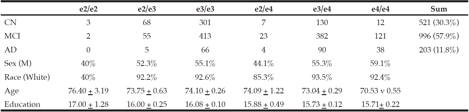
In order to understand the predictive performance of the PRS algorithm above and beyond that which is provided for by APOE status alone, we initially investigate data from 652 CN and MCI subjects selected from ADNI 1, ADNI GO, ADNI 2 and ADNI 3 studies examined between 2005 and 2017 (see Table 1a). Due to the low sample sizes (n≤50) of APOE E2E4 and E4E4 individuals in either CN or MCI groups (see Table 1b) at baseline (bl), further analyses were only carried out in APOE E3 homozygotes and APOE E3/E4 heterozygotes. Therefore, all results shown in this paper were based on 594 CN and MCI subjects who were either carried two copies APOE E3 allele or were APOE E3/E4 heterozygotes (see Table 1c) and had modified PACC score data at entry to the study in addition to having suitable genetic data and at least 5 years’ worth of follow up cognitive testing and imaging scans.
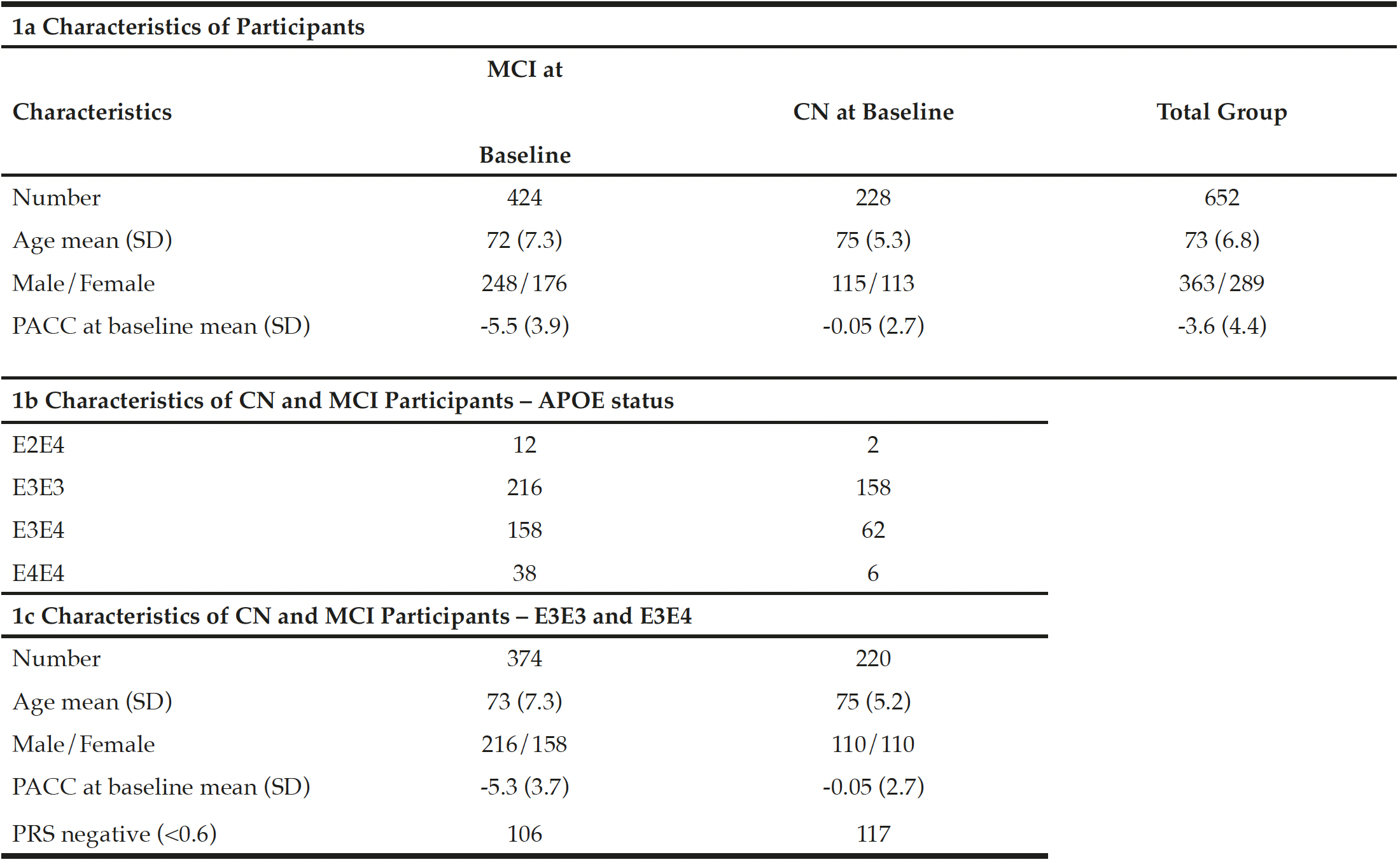
Table 1
Characteristics of participants

Genotyping Procedures and Quality Control

The ADNI samples were genotyped using with Whole Genome Sequencing and/or the Illumina Omni 2.5M BeadChip array. Quality control checks were performed using PLINK software (www.cog-genomics.org/plink/2.0/). Checks included the exclusion of SNPs with missingness greater than 0.02 and minor allele frequency of less than 0.01. SNPs with Hardy-Weinberg equilibrium p-value less than 1 x 10⁻⁶ were also excluded. After such checks 8,990,292 SNPs were left for analysis of which approximately 114,000 were used as part of the polygenic risk scoring algorithm (14).

The ADNI modified PACC score

PACC is a composite score which combines tests that assess episodic memory, timed executive function and global cognition which has been shown to be able to detect the first signs of cognitive decline before clinical signs of MCI manifest (15). In this study, we use a ADNI modified PACC with Digit Symbol Substitution (mPACCdigit) (12, 13) downloaded using R package “adnimerge” (https://adni.bitbucket.io/reference/pacc.html#references).-.
In ADNI, Free and Cued Selective Reminding Test (FCSRT) is not used and has been replaced Delayed Recall test that is included within the Alzheimer’s Disease Assessment Scale (ADAS) as a suitable proxy to be included in the modified PACC score. Furthermore, mPACCdigit score also includes the Digit Symbol Substitution Test (DSST) when available (ADNI1) and mPACCtrailsB uses (log transformed) Trails B as a proxy for DSST. Raw component scores standardized according to the mean and standard deviation of baseline scores of ADNI subjects with normal cognition to create Z scores for each component (Z=(raw – mean(raw.bl))/sd(raw.bl)). The Z scores are reoriented if necessary, so that greater scores reflect better performance. The composite is the sum of these Z scores. At least two components must be present to produce a score. If more than two components are missing, the PACC will be NA.

Calculation of Polygenic Risk Scores

A specifically built, proprietary software called SNPfitRTM was used for all subsequent PRS calculations. The PRS calculations are based on a pre-determined logistic regression model based on the modelling of the association between the incidences of variants within a large panel of SNPs with a known links to AD to the presence of the disease in a substantial cohort of subjects (Escott-Price et al.16). Subject age, sex and APOE status are included as covariates. The software calculates the normalised sum of the individual scores weighted by their effect sizes for each SNP, adds the values for the covariates and derives the predicted risk from the model equation.
Effect sizes were determined from the International Genomics of Alzheimer’s (IGAP) study. The score contribution from SNPs with missing values were imputed based on the population frequency of the effect allele for that SNP.

Statistical Analysis

The polygenic risk scores generated were exported for the analysis presented.
R version 4.0.4 (https://www.r-project.org/) was used to carry out all data processing and analysis. The receiver operating characteristic (ROC) analysis and AUC calculations were performed using R package “pROC”. Modified PACC data were obtained from R package “adnimerge” (https://adni.bitbucket.io/reference/pacc.html#references). T tests were performed in R using the t.test() function to determine whether there is significant different between high and low risk groups (see p-value in Results).
To determine whether applying a PRS approach would provide further accuracy for predicting cognitive decline as measured by a modified PACC, we analysed the cognitively normal APOE E3/E3 and APOE E3/E4 individuals, where both genetics and modified PACC score data were available (n=220, see Table 1c). PRS were calculated and individuals were assigned to either “high risk” (defined as a PRS ≥ 0.6, n=103) or “low risk” (PRS<0.6, n=117) groups (see Table 1c). A similar evaluation was performed on APOE E3/E3 and APOE E3/E4 individuals who entered the study with a diagnosis of MCI and for whom both genetic data and PACC score data were available (n=374 , See Table 1c). PRS were calculated and MCI individuals were assigned to “high risk” (defined as a PRS ≥ 0.6, n=268) or “low risk” (PRS<0.6, n=106) groups (see Table 1c). Note that not all subjects had follow-ups at each time point over the 5 years. Thus, the number of subjects varies at each follow-up check points. A PRS of 0.6 was chosen as a threshold based on an optimal balance between sensitivity and specificity in previous studies (17).


Results

The overall performance for predicting individuals who would decline by at least -1 PACC score within 5 years from a baseline diagnosis of either cognitively normal or mild cognitive impairment was 65.6% (CI:61.3-69.8) area under the curve (AUC), suggesting PRS could be an effective stratification tool to identify patients with a higher likelihood to decline cognitively over a period of 5 years.

PRS to predict early cognitive decline from a cognitively normal baseline

As expected, as measured by modified PACC score, those individuals who carry a copy of the APOE E4 allele are more likely to decline cognitively than those who are APOE E3 homozygotes over a 5-year period (see Figure 1a). The mean change in modified PACC score in APOE E3/E3 after 60 months was just -0.4 points ±4.1 whereas APOEE3/E4 individuals declined, on average, by 1.3 points ±4.7, on the modified PACC score scale after 60 months (see Figure 1a).
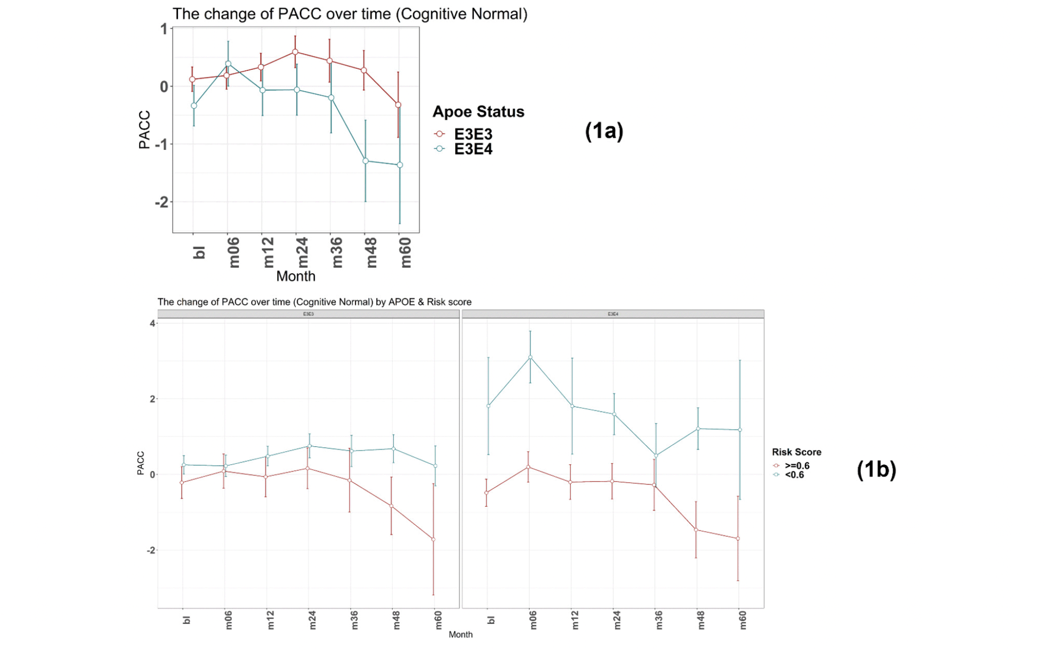
Figure 1
Time-course PACC scores for individuals carrying APOE E3E3 and E3E4 in CN Group: (1a) The change of PACC over time in individuals who entered as cognitively normal over 5-year period grouped by APOE status; (1b) The change of PACC over time in individuals who entered as cognitively normal over 5-year period grouped by risk score
(bl=baseline, m=month)


There was a significant difference in the average change of the modified PACC score approximate to 2 between the two groups observed from as early as 48 months (high risk n=77, low risk n=94; high risk average PACC=-1.2, low risk average PACC=0.7; p-value =0.003, see Table 2). When considering APOE E3 homozygotes alone, the difference in the change of PACC score between the high risk and low risk groups observed was 2 points over 60 months years (high risk n=20, low risk n=51; high risk average PACC=-1.7, low risk average PACC=0.2, p=0.12, see Table 2). Importantly, though sample size is smaller (see Table 2), low PRS risk E3/E4 individuals that entered the study as cognitively normal appeared more likely to remain cognitively stable compared with the high risk group (Figure 1b).
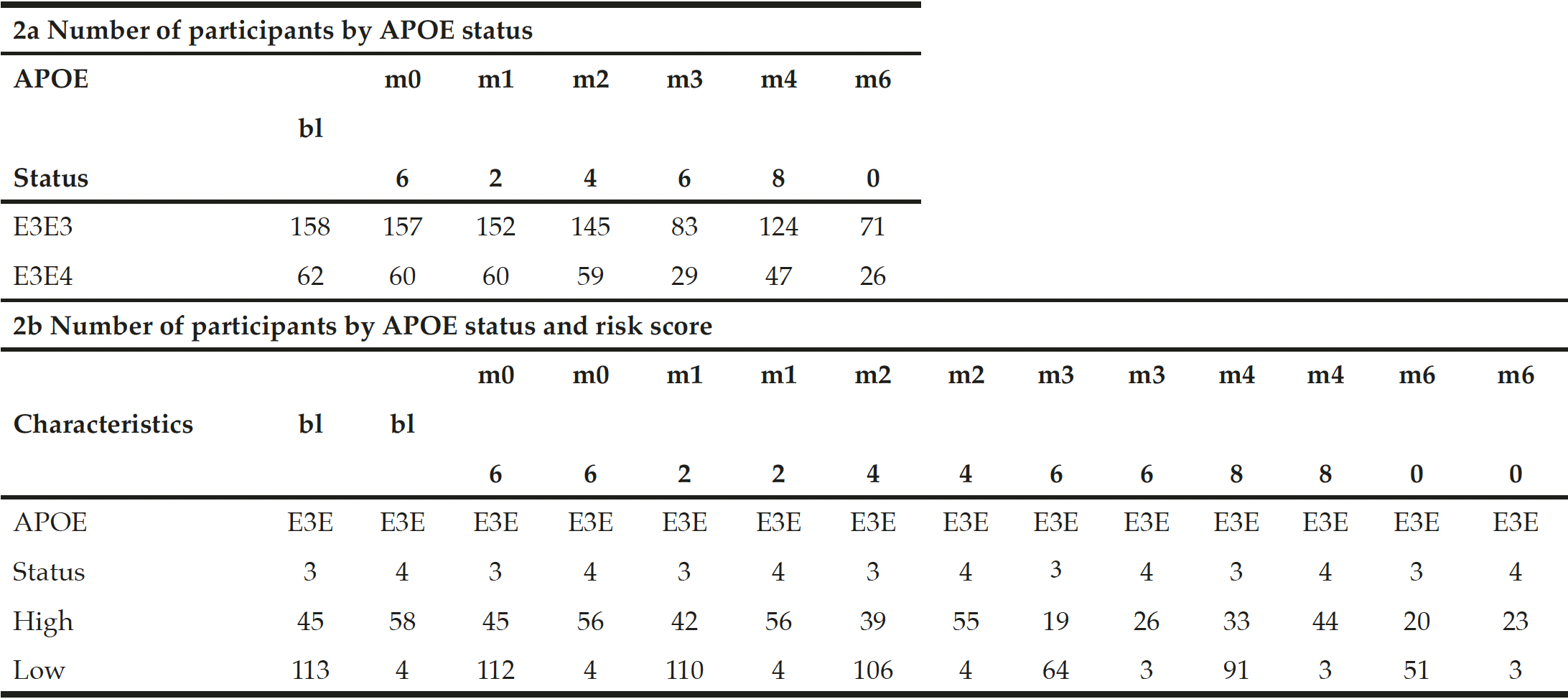
Table 2
Participants carrying APOE E3E3 and E3E4 in CN Group

bl: baseline; m: month. Thus month 6 is represented by m06


PRS to predict early cognitive decline from an MCI baseline

Again, as expected, those individuals carrying an E4 allele demonstrate greater cognitive decline, on average, compared to E3 homozygotes at all timepoints over the 5-year follow-up period (E3/E3 mean PACC change after 60 months -1.4 points ±5.6; E3/E4 mean PACC change after 60 months -8.9 points ±12.2; Figure 2a).
There were no individuals within the APOE E3/E4 MCI cohort (n=158) with a low PRS score (PRS <0.6). This is unsurprising, since these individuals who have already declined cognitively to an MCI diagnosis are likely to have a high PRS. Notwithstanding, this meant that a comparison between low and high PRS risk within the MCI group individuals could not be made. However, the APOE E3 homozygote MCI group contained both high PRS risk (≥0.6, n=110) and low PRS risk (<0.6, n=106) individuals (Figure 2b). Among this group, high PRS risk patients declined, on average, by approximately 1 point more than the low risk group after over 6 months (high risk n=105, low risk n=102; high risk average PACC=-3.9, low risk average PACC=-2.7, p=0.07, see Table 3) and a significant additional 5 points over 60 months (high risk n=59, low risk n=52; high risk average PACC=-8.2, low risk average PACC=-3.2, p-value<0.001, see Table 3) above those calculated as low risk, who did not decline further over the 5 year period studied (Figure 2b).
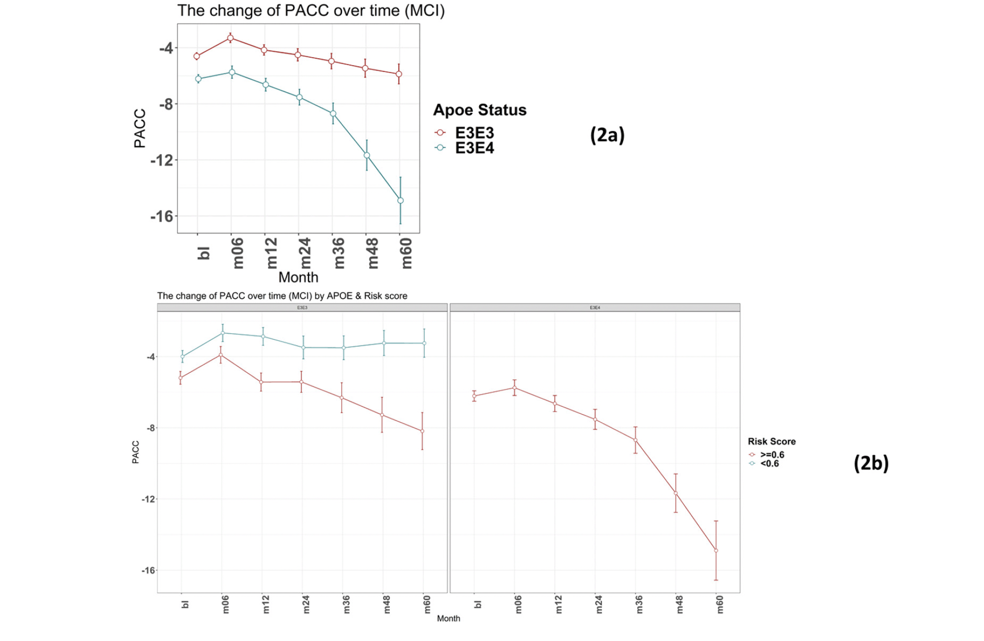
Figure 2
Time-course PACC scores for individuals carrying APOE E3E3 and E3E4 in MCI Group: (2a) The change of PACC over time in individuals who entered as MCI (EMCI or LMCI) over 5-year period grouped by APOE status; (2b) The change of PACC over time in individuals who entered as MCI (EMCI or LMCI) over 5-year period grouped by risk score (bl=baseline, m=month)
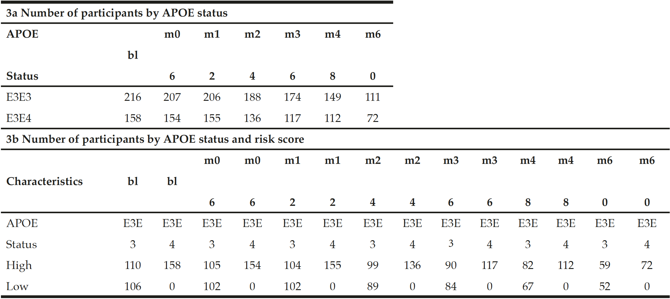
Table 3
Participants carrying APOE E3E3 and E3E4 in MCI group

bl: baseline; m: month. Thus month 6 is represented by m06


Discussion

PRS approaches have demonstrated accuracies of between 75 and 84% for predicting onset of AD when including APOE status, sex and age in addition to PRS (16). In particular, the PRS approach as developed by Escott-Price et al., (14) is built as a sum of the weighted contributed of 10,000s of Single Nucleotide Polymorphisms (SNPs) where the weights are the β-coefficients of each SNP association with the disease. In contrast to other PRS algorithms, where fewer SNPs have been used (for example just 31 SNPs (18)) this approach includes SNPs that are not considered as having genome wide significance in GWAS studies. However, inclusion of this vastly increased number of variants which alone carry sub-threshold significance provides an additive contribution to the overall performance that may be substantive and also reduce risk that performance is not
lost when being applied across different cohorts. Until now the analyses performed using this approach have been carried out to predict those individuals diagnosed with AD or MCI (19) versus those who are cognitively normal, though PRS algorithms have been used to look at a variety of AD pathology and risk by Altmann et al. (20).
Patients who present to clinicians with very mild or subjective cognitive complaints can provide a diagnostic and patient management challenge in terms of decisions on whether to progress to more expensive and/or invasive testing or to discharge. Easier access to risk evaluation data will help better patient management decisions in a cost-efficient manner and provide further basis for dialogue on risk mitigation through lifestyle changes. Furthermore, screening of large pre-symptomatic populations to identify potential clinical trial participants for prevention studies in AD is challenging. Genetic risk prediction can be generated from DNA simply extracted from saliva or blood samples, thus providing a viable route to wide-scale risk stratification to characterise potential clinical trial subjects.
We have previously reported (17) on the performance of a PRS algorithm for predicting those individuals, with a bassline diagnosis of MCI who would decline by at least 15 ADAS-Cog13 points in 4 years with an AUC of 72.8% (CI:67.9-77.7) increasing to 79.1% (CI: 75.6-82.6) when also including those at baseline who were considered cognitively normal. Furthermore, by designating MCI patients as either high or low risk as determined by a PRS threshold of 0.6 it was observed that the high risk group declined, on average, by 1.4 points more on the CDR-SB scale than the low risk group over a period of 4 years. This performance in predicting cognitive decline due to AD was similar to that when defining risk using a pTau/Ab1-42 ratio as measured in a cerebrospinal fluid (CSF) sample.
This study was designed to demonstrate the potential utility of a specific PRS algorithm for identifying individuals at highest risk of developing early or continued cognitive decline from either pre-symptomatic (CN) baseline or a relatively early stage of their disease (MCI). The results show the potential to use a PRS approach to identify those individuals most likely to decline cognitively. Importantly this includes identifying cognitively normal APOE E3 homozygous individuals who are at most risk for early cognitive decline due to AD. This genotype accounts for approximately 60% of the general population and 35% of the AD population but currently would not be considered at higher risk without access to expensive or invasive biomarker testing. PRS could therefore provide a useful tool for identifying individuals within this group who require additional monitoring, investigation or, with future developments, therapeutic intervention.
This study shows that PRS predictions can identify individuals with the highest risk of subtle cognitive decline, as measured by PACC scores, in patients who did not display any measurable symptoms upon entry to the ADNI study. The timeframe of 5 years used for the analysis is relevant in the context of both primary and secondary prevention trials and clinical practice. Furthermore, future work will be conducted to evaluate the predictive performance of our PRS algorithm in order to identify patients during mid-life (40-60 years old) at risk of future cognitive deficits due to AD which can provide a critical strategy for reducing the number. This genetic risk assessment represents an easily accessible intervention with the potential to reduce cost and patient burden through blood or mouth swab testing. Additionally, this genetic risk assessment provides an extremely valuable tool for expanding recruitment into secondary prevention trials which currently are typically limited to recruiting E4 carriers only. Furthermore, as disease modifying drugs enter clinical practice finding an easy to deploy risk prediction test to identify patients most likely to benefit from therapeutic intervention will be critical.
PRS does have its own challenges and limitations. For example, this work considers genetic risk together with age and sex in developing a model for predicting further development of cognitive symptoms but does not consider other risk factors that are known to influence onset and development of disease, for example, lifestyle and environment. Further studies will be required to combine both genetic and lifestyle risk factors to accurately identify those individuals at the most risk of Alzheimer’s disease.

Study Limitations

This study is not without limitations, with sample size being the primary shortcoming. This was particularly relevant in evaluating the APOE E4 carrier sub-group (E2/E4, E3/E4 and E4 homozygous, see Table 1b). Furthermore, studies with larger sample sizes across all diagnostic categories, including those declining from a cognitively normal baseline, will be important to understand broader utility. As with most studies of this nature, observing similar performance in alternative cohorts is important and is critical towards the understanding and confirmation of polygenic risk score assessment for use in clinical trial recruitment and in clinical practice.


*Data used in preparation of this article were obtained from the Alzheimer’s Disease Neuroimaging Initiative (ADNI) database (adni.loni.usc.edu). As such, the investigators within the ADNI contributed to the design and implementation of ADNI and/or provided data but did not participate in analysis or writing of this report. A complete listing of ADNI investigators can be found at: http://adni.loni.usc.edu/wp-content/uploads/how_to_apply/ADNI_Acknowledgement_List.pdf

Conflict of interest: Q. Gao, P. Daunt, A.Gibson and R. Pither are all employees of Cytox Ltd.

Ethical standard: The authors declare that the study was carried out according to all ethical standards.

Acknowledgments: Data collection and sharing for this project was funded by the Alzheimer’s Disease Neuroimaging Initiative (ADNI) (National Institutes of Health Grant U01 AG024904) and DOD ADNI (Department of Defense award number W81XWH-12-2-0012). ADNI is funded by the National Institute on Aging, the National Institute of Biomedical Imaging and Bioengineering, and through generous contributions from the following: AbbVie, Alzheimer’s Association; Alzheimer’s Drug Discovery Foundation; Araclon Biotech; BioClinica, Inc.; Biogen; Bristol-Myers Squibb Company; CereSpir, Inc.; Cogstate; Eisai Inc.; Elan Pharmaceuticals, Inc.; Eli Lilly and Company; EuroImmun; F. Hoffmann-La Roche Ltd and its affiliated company Genentech, Inc.; Fujirebio; GE Healthcare; IXICO Ltd.; Janssen Alzheimer Immunotherapy Research & Development, LLC.; Johnson & Johnson Pharmaceutical Research & Development LLC.; Lumosity; Lundbeck; Merck & Co., Inc.; Meso Scale Diagnostics, LLC.; NeuroRx Research; Neurotrack Technologies; Novartis Pharmaceuticals Corporation; Pfizer Inc.; Piramal Imaging; Servier; Takeda Pharmaceutical Company; and Transition Therapeutics. The Canadian Institutes of Health Research is providing funds to support ADNI clinical sites in Canada. Private sector contributions are facilitated by the Foundation for the National Institutes of Health (www.fnih.org). The grantee organization is the Northern California Institute for Research and Education, and the study is coordinated by the Alzheimer’s Therapeutic Research Institute at the University of Southern California. ADNI data are disseminated by the Laboratory for Neuro Imaging at the University of Southern California. We also acknowledge Prof. Julie Williams, Prof. Valentina Escott-Price, Dr Rebecca Sims and Dr Eftychia Bellou from the University of Cardiff for their advice on adaptation and implementation of the polygenic risk algorithm, and Mr Greg Davidson from Ledcourt Associates Ltd for his contribution on implementation of the algorithm. We thank Dr Simon Flint and Dr Vicky Jones from Cytox Ltd for comments on the manuscript.

Open Access: This article is distributed under the terms of the Creative Commons Attribution 4.0 International License (http://creativecommons.org/licenses/by/4.0/), which permits use, duplication, adaptation, distribution and reproduction in any medium or format, as long as you give appropriate credit to the original author(s) and the source, provide a link to the Creative Commons license and indicate if changes were made.


References

1. Mckhann GM, Knopman DS et al. The diagnosis of dementia due to Alzheimer’s disease: recommendations from the National Institute on Aging-Alzheimer’s Association workgroups on diagnostic guidelines for Alzheimer’s disease. Alzheimers Dement. 2011; 7: 263-269.
2. Wittenberg R, Hu B et al. Projections of older people with dementia and costs of dementia care in the United Kingdom, 2019–2040. Report commissioned by Alzheimer’s Society from the Care Policy and Evaluation Centre (CPEC) at the London School of Economics and Political Science. November 2019.
3. Alzheimer’s Association. Alzheimer disease facts and figures. Alzheimers Dement. 2019; 15: 321-387.
4. Bateman RJ, Xiong C et al. Clinical and biomarker changes in dominantly inherited Alzheimer’s disease. N. Engl. J. Med. 2012; 367: 795-804.
5. Storandt M, Grant EA et al. Rates of progression in mild cognitive impairment and early Alzheimer’s disease. Neurology. 2002 Oct 8; 59(7):1034-41.
6. Hui JS, Wilson RS, et al. Rate of cognitive decline and mortality in Alzheimer’s disease. Neurology. 2003 Nov 25; 61(10):1356-61.
7. Cosentino S, Scarmeas N et al. APOE epsilon 4 allele predicts faster cognitive decline in mild Alzheimer disease. Neurology. 2008 May 6; 70(19 Pt 2):1842-9.
8. Harold D, Abraham R et al. Genome-wide association study identifies variants at CLU and PICALM associated with Alzheimer’s disease. Nat Genet. 2009 Oct; 41(10):1088-93.
9. Lambert JC, Ibrahim-Verbaas CA et al. Meta-analysis of 74,046 individuals identifies 11 new susceptibility loci for Alzheimer’s disease. Nat Genet. 2013 Dec;45(12):1452-8. doi: 10.1038/ng.2802. Epub 2013 Oct 27. PMID: 24162737; PMCID: PMC3896259.
10. Avramopoulos D. Genetics of Alzheimer’s disease: recent advances. Genome Med. 2009 Mar 27; 1(3):34.
11. Stocker H, Möllers T, Perna L, et al, The genetic risk of Alzheimer’s disease beyond APOE ε4: systematic review of Alzheimer’s genetic risk scores. Transl Psych 2018;8;166-174.
12. Donohue MC, et al. The Preclinical Alzheimer Cognitive Composite: Measuring Amyloid-Related Decline. JAMA Neurol. 2014;71(8):961–970. doi: 10.1001/jamaneurol.2014.803. PMID: 24886908; PMCID: PMC4439182.
13. Donohue MC, Sperling RA, Petersen R, Sun C, Weiner MW, Aisen PS, for the Alzheimer’s Disease Neuroimaging Initiative. Association Between Elevated Brain Amyloid and Subsequent Cognitive Decline Among Cognitively Normal Persons. JAMA. 2017;317(22):2305–2316. doi: 10.1001/jama.2017.6669. PMID: 28609533; PMCID: PMC5736301.
14. Escott-Price V, Leonenko G, Sims R et al, Polygenic risk and hazard scores for Alzheimer’s disease prediction. Ann Clin Transl Neurol 2019;6;456-465.
15. Donohue MC, Sperling RA, et al. Australian Imaging, Biomarkers, and Lifestyle Flagship Study of Ageing, Alzheimer’s Disease Neuroimaging Initiative, and Alzheimer’s Disease Cooperative Study (2014) The preclinical Alzheimer cognitive composite: measuring amyloid-related decline. JAMA Neurol 71(8):961–970.
16. Escott-Price V, Sims R, Williams J et al, Common polygenic variation enhances risk prediction for Alzheimer’s disease. Brain 2015;138;3673-3684.
17. Daunt P, Ballard CG, et al. Polygenic Risk Scoring is an Effective Approach to Predict Those Individuals Most Likely to Decline Cognitively Due to Alzheimer’s Disease. J Prev Alzheimers Dis. 2021;8(1):78-83.
18. Desikan RS, Fan CC, Wang Y et al, Genetic assessment of age-associated Alzheimer disease risk: development and validation of polygenic hazard score. PLoS Med 2017 e1002258.
19. Escott-Price V, Myers A, Huentelman AJ, et al, Polygenic risk score analysis of pathologically confirmed Alzheimer’s disease. Ann Neurol 2017;82;311-314.
20. Altmann A, Schott JM, Scelsi et al, A comprehensive analysis of methods for assessing polygenic burden on Alzheimer’s disease pathology and risk beyond APOE. Brain Commun 2020;2; fcz047.

ASSOCIATIONS OF DUAL TASK EXERGAMING WITH COGNITIVE-MOTOR INTERFERENCE IN OLDER ADULTS WITH MILD COGNITIVE IMPAIRMENT: A SINGLE-ARM PILOT STUDY


L. Kannan1, T. Bhatt1


1. Department of Physical Therapy, University of Illinois at Chicago. Chicago, USA

Corresponding Author: Tanvi Bhatt, PT, PhD, Department of Physical Therapy, 1919, W Taylor St, (M/C 898), University of Illinois at Chicago, Chicago, USA 60612, Email – tbhatt6@uic.edu, Phone – +1(312)-355-4443, Fax – +1(312)-996-4583

J Aging Res & Lifestyle 2021;10:54-60
Published online November 16, 2021, http://dx.doi.org/10.14283/jarlife.2021.11


Abstract

Purpose: To examine the feasibility and effectiveness of dual task (DT) exergaming to improve volitional balance control in older adults with mild cognitive impairment (MCI). Methods: Ten older adults with MCI were examined at baseline (week-0) and post-training (week-5) on volitional balance control (maximum excursion of center of gravity, MXE [%]) while performing
cognitive task (auditory clock test or letter number sequencing task) and on the NIH-motor and cognitive toolboxes. DT
exergaming training lasted for 12 sessions which consisted of performing explicit cognitive tasks while playing the Wii-Fit balance games.
Results: From pre- to post-training, MXE improved (p<0.05); however, cognitive accuracy (cognitive task) remained the same (p>0.05).
Improvement in NIH motor and cognitive toolbox tests was observed post-training (p<0.05). Conclusion: DT exergaming was
associated to improvements in balance control under attention-demanding conditions in MCI. Future studies may focus on
examining the efficacy of such training.

Key words: Dual task training, exergaming, mild cognitive impairment, cognitive motor interference.


Introduction

Mild cognitive impairment (MCI), a prodromal stage of dementia, affecting about 15% to 20% of the older adults above the age of 65 in the United States (1) is characterized by substantial memory decline but preserved general intellectual function with subtle balance and gait deficits (2-4). Such deficits are more pronounced during dual tasking (simultaneous performance of cognitive and motor task) resulting in increased cognitive-motor interference (deteriorated performance on either one or both tasks) – a factor likely contributing to the higher fall risk in MCI (3, 5, 6). Therefore, studies have focused on dual task (DT) interventions to enhance and/or preserve the ability to allocate attentional resources to both balance and cognitive tasks when performed concurrently, often needed in daily living (ADLs) (7, 8).
Conventional DT training that involves repeated practice of balance and/or gait activities simultaneously performed with cognitive tasks helps improve motor performance on ADLs but has limited benefits in improving cognitive performance (7, 8). Despite this, barriers such as lack of motivation, adherence to therapy, and limited access to rehabilitation facilities have led to development of alternate therapies involving exergame-based training (9-12). Such training provides real-time biofeedback (visual, auditory, tactile) on movement performance (delivered via low-cost commercial devices – Wii-fit) while implicitly facilitating cognitive domains and is known to be feasible, effective, and highly compliant among MCI (12).
It is known that MCI demonstrate significant structural and functional brain changes associated with executive dysfunction, deteriorated DT performance, and increased fall risk (13, 14). Additionally, exergame-based training may not implicitly address “executive function” (12, 15), and explicit cognitive training may aid in delaying or reversing the apparent cognitive decline. One study revealed promising effects on reducing cognitive-motor interference after 6 weeks of Wii-Fit + cognitive training (DT exergaming) in people with chronic stroke (16). As MCI also show significant cortical pathology, it could be postulated that a similar training may be similarly beneficial (13, 14).
Therefore, this single-arm pilot study aimed to examine the feasibility of a 4-week (12 sessions) DT exergaming intervention among MCI on self-initiated (volitional) balance control tasks under attentional demanding conditions (interference task). We hypothesized that MCI participants would show significant improvement in volitional balance, cognitive accuracy, and improved performance-based motor and cognitive function post-training.


Methods

Participants

Older adults (> 55 years) were recruited from the University of Illinois Hospital Geriatric Clinic and flyers in nearby independent living senior centers and grocery stores. This study was approved by the University of Illinois at Chicago institutional review board. Ten older adults participated in the study after obtaining a written informed consent.

Participants’ eligibility

To be included, participants must score 18-24 out of 30 on the Montreal Assessment on Cognitive Assessment (MoCA). Participants with uncontrolled cardiovascular disease, presence of any neurological condition (e.g., Alzheimer’s disease), and/or severe musculoskeletal diseases that may interfere with the ability to receive the intervention were excluded. Additionally, people with the inability to stand independently without an assistive device for the length of a Wii-Fit game, with a fracture risk heel bone density (measured using Lunar Achilles Insight) T-score < -2.0, and inability to communicate and understand English were excluded.

Research Design

This was a single arm pre-post research design consisting of 4 weeks of DT exergaming sessions. Baseline (at week 0) and post-testing (at 5th week) outcome measures were collected.

Intervention

In total, 12 sessions of individual one-on-one DT exergaming was administered and supervised by a research personnel (physical therapist) in a research facility. Participants wore a gait belt and were supervised during the session. DT exergaming was delivered via Wii-Fit standing balance games which was performed at light intensity (rate of perceived exertion via Borg’s scale with individuals reporting score of 7-11) (17) and was concurrently performed with explicit cognitive games (therapist cued) for 90 minutes/session, 3 times/week. While Wii fit games implicitly addressed cognitive domains like working memory, episodic memory, and visuospatial awareness, explicit cognitive games targeted subdomains of executive function – working memory and attention, and semantic memory, abstract memory. Warm up (step-in-place, trunk twists) and cool down (stretching of lower limb) were performed before and after session, respectively. Refer to supplementary material section for details of protocol.

Assessments

Volitional balance control task: The limits of stability (LOS) test via Balance Master (Equitest® Neurocom) (18) was administered. Participants were secured in a safety harness and asked to stand on the force platform of the Balance Master (Figure 1). Participants were instructed to lean their body either in the forward, backward, left, or right direction to move their center of gravity (COG) projection shown on a screen to the desired direction without losing balance, stepping, or reaching for assistance.

Figure 1
Represents a picture of an individual performing the
limits of stability test on Balance Master (Equitest®
Neurocom) in the forward direction


Cognitive task

Auditory clock test (ACT) (19) and letter number sequencing task (LNS) (20) were administered using the DirectRT EmpirisoftTM (21) software to assess subdomains of executive function (visuo-spatial memory, working memory, attention, and cognitive flexibility). The audio cues were delivered through headphones and responses were recorded through a microphone. The ACT involved responding to different times of the day, “yes” if the hour and the minute hand was on the same side of the clock face and “no” otherwise. The LNS involved sequentially listing alternate letter and number combinations, for example, response to “C5,” was D6, E7, etc.

Interference test

The LOS test (all directions) was performed along with both cognitive tasks mentioned above. Participants began responding to the cognitive cues followed by the LOS task.

NIH Toolbox

An iPad was used to test the motor and cognitive domains. The motor tests include 4-meter gait speed test and 2-minute walk test. The cognitive tests include list sort memory test (working memory), picture sequence memory test (episodic memory), dimensional change card sort test (executive function), flanker inhibitory control and attention test (attention and executive function), and pattern comparison processing speed test (processing speed) (refer supplementary material section).

Outcome measures

Volitional balance control and interference task: Single task (task when performed alone) and performance during interference task was quantified by the movement stability measurement of maximum excursion (MXE, expressed in percentage), which is the maximum ability to shift one’s COG toward the theoretical limit in the desired direction. Higher values indicate better performance.
Cognitive and interference task: Accuracy [(Correct responses)(Total responses)*100] was calculated during single and interference task.
NIH toolbox: Speed (m/sec) for 4-meter gait test and distance covered in 2-minute walk test for endurance was computed. Number of correct responses for list sort memory test and accuracy for the remainder tests was included for analysis.

Statistical analyses

Statistical analyses were performed using SPSS version 24, Chicago, IL, USA. For MXE in each volitional balance control task direction (i.e., forward, backward, left, and right), 2 x 2 repeated measures analysis of variance (ANOVA) was performed to examine the time (pre- to post-training) and task (single vs. interference task) differences on with follow-up post-hoc tests. Similarly, four repeated measures ANOVA for accuracy (cognitive) in ACT and LNS was performed. Paired t-test was conducted for NIH toolbox measures. Refer supplementary material section for details.


Results

Demographics: Demographic characteristics of participants who completed the study are provided in Table 1.

Table 1
Demographics and clinical characteristics of older adults with mild cognitive impairment (MCI). BBS = Berg Balance Scale, MoCA = Montreal Cognitive
Assessment


Volitional balance control and interference task: From pre- to post-training, MXE improved in the forward and left direction (p<0.05) under interference test (Figure 2a-2d). Results of ANOVA and follow-up test are presented in Table 2.

Table 2
Results for balance control task

ST: single task ; ACT: Auditory clock test; LNS: Letter number sequencing; *p<0.05 **p<0.01 ***p<0.001


Cognitive and interference task: Accuracy on ACT (Figure 2e-2h) and LNS (Figure 1i-1l) showed significant improvement only during single task performances (p<0.05), however, no improvement was observed during interference test (p>0.05). Results of ANOVA and follow-up test are presented in Table 3.

Table 3
Results for cognitive task

ST: single task; ACT: Auditory clock test; LNS: Letter number sequencing; *p<0.05 **p<0.01 ***p<0.001


NIH toolbox: A significant increase in gait speed was observed post-training (p<0.05); however, there was no change in the 2-minute walk test distance covered (p>0.05) (Figure 3a). Post-training, significant improvements in NIH cognitive toolbox measures of working memory (p<0.05) (Figure 3b), episodic memory (p<0.01) (Figure 3c), and executive function (p<0.01) (Figure 3d) were observed. However, no improvements were observed in attention (p>0.05) and processing speed (p>0.05).

Figure 2
Association of dual task training with balance control and cognition under single and dual task conditions

Figures a, b, c, & d represent mean and standard deviations (SD) pre- to post-training changes for maximum excursion during volitional balance control under dual task and single task conditions. Figures e, f, g, & h represent mean and SD pre- to post-training cognitive accuracy changes for auditory clock test during dual task and single task conditions. Figures i, j, k, & l represent mean and SD pre- to post-training cognitive accuracy changes for letter number sequencing during dual task and single task conditions


Figure 3
Means and SD for gait speed shown in meters (m) and obtained from the NIH motor toolbox; and b) working memory tested via list sort memory test, (c) episodic memory tested via picture sequence memory test, and (d) executive function via dimensional change card sort obtained from the NIH cognitive toolbox

Greater scores indicate better performance. *, p<0.05


Discussion

As hypothesized, the study results showed a significant improvement in volitional balance control under interference conditions but no improvement in ACT and LNS. Additionally, the intervention resulted in improved gait speed but not endurance for motor function and, similarly, improved working memory, episodic memory, and executive function but not attention and processing speed.
With respect to interference task conditions, the results show improvement in motor performance but no change in ACT and LNS cognitive tasks demonstrating motor prioritization (6). Repeated practice of multidirectional weight shift training that challenged one’s LOS and immediate biofeedback (visual) with knowledge of performance during training could have facilitated the ability to precisely control one’s center of mass (COM) body movement. The LOS test utilizes a significant amount of attentional resources within the dorsolateral prefrontal cortex (DLPFC, associated with executive functions) (22). Post-training, utilization of the shared resources between cognitive and motor areas perhaps improved by channeling available attentional resources to prioritize motor performance – probably due to the CNS’ estimation or perception of the balance task to be more challenging with significant consequences (such as falls) in case of failure. Furthermore, the significantly greater pathology affecting the DLPFC and associative sensorimotor areas (controlling executing function) than the premotor or motor areas (controlling volitional balance) could attribute to motor prioritization (5).
Our study yielded motor benefits in forward and left lean but did not improve backward and right lean. It has been postulated that backward leans are more difficult than forward leans due to directional-specific anatomic constraints and increased reliance on proprioceptive and vestibular systems (over visual system) in older adults (23-25). Aging-induced changes causes impaired integration of these systems and any cognitive decline further depreciates this sensory signal processing, resulting in deteriorated balance control (23, 24). Thus, impaired cognitive-sensory signal processing could explain the poor performance and lowest MXE for backward lean at baseline (single and interference task). Lastly, motor performance was the highest on right lean, which could be related to the dominance/preferred side for performing activities and, therefore, there may have been limited room for improvement in the right lean.
Although there wasn’t an improvement in cognitive performance during interference task, visuo-spatial and working memory (ACT) improved under single task conditions. Apart from the implicit benefits of exergaming (12, 15), and the explicit cognitive training that targeted subdomains of executive function (attention, planning, and working memory) may explain such improvements. However, there were no benefits in cognitive flexibility (i.e., letter number sequencing) despite a positive trend. This task requires simultaneous utilization of attention, processing information, and working memory. Although the training did target these domains, the dosage might not be enough to induce change.
Similar to our study, studies targeting balance training in standing have shown improvements in overground gait speed in MCI (3) and positive transfer to improved mobility (16). This could be attributed to task-specific characteristics of exergames, which involve time limited stepping activities. However, our results showed no improvement in endurance. This could be because the exergames did not incorporate high-intensity training that is known to induce improvement in cardiovascular function in MCI (7, 8). Lastly, due to the implicit (via Wii fit games) and explicit (therapist cued cognitive tasks) cognitive training components of the protocol, improvement in NIH cognitive toolbox measures for executive function, attention, and processing speed were expected and agree with results of previous DT training studies (7, 12).
Despite the positive results, our study has certain limitations. Firstly, there was a small sample size and lack of a control group due to the study’s preliminary nature. Furthermore, the training was limited to 4 weeks, and greater training dosage may yield larger motor and cognitive improvements under DT conditions. While our results demonstrate partial benefits of DT exergaming on balance control (self-initiated), its effects on the primary defense mechanism – reactive balance control – remain to be explored.


Conclusion

Our preliminary study demonstrated that DT exergaming has the potential to improve balance control and, limited benefits in executive function which could potentially have an impact in fall-risk reduction.


Conflict of interest: We have no conflict of interest to declare.

Ethical standards: The study was supported by Midwest Roybal Center for Health Promotion and Translation awarded to Dr. Tanvi Bhatt. This work was carried out after securing approval from the University of Illinois institutional review board protocol 2018-1257 and was registered on clinicaltrials.gov NCT03765398.


References

1. Langa, K.M. and D.A. Levine, The diagnosis and management of mild cognitive impairment: a clinical review. Jama, 2014. 312(23): p. 2551-2561.
2. Sheridan, P.L. and J.M. Hausdorff, The role of higher-level cognitive function in gait: executive dysfunction contributes to fall risk in Alzheimer’s disease. Dementia and geriatric cognitive disorders, 2007. 24(2): p. 125-137.
3. Bahureksa, L., et al., The impact of mild cognitive impairment on gait and balance: a systematic review and meta-analysis of studies using instrumented assessment. Gerontology, 2017. 63(1): p. 67-83.
4. Lopez, O.L., et al., Neuropsychological characteristics of mild cognitive impairment subgroups. Journal of Neurology, Neurosurgery & Psychiatry, 2006. 77(2): p. 159-165.
5. Muir, S.W., K. Gopaul, and M.M. Montero Odasso, The role of cognitive impairment in fall risk among older adults: a systematic review and meta-analysis. Age and ageing, 2012. 41(3): p. 299-308.
6. Plummer, P., et al., Cognitive-motor interference during functional mobility after stroke: state of the science and implications for future research. Archives of physical medicine and rehabilitation, 2013. 94(12): p. 2565-2574. e6.
7. Law, L.L., et al., Effects of combined cognitive and exercise interventions on cognition in older adults with and without cognitive impairment: a systematic review. Ageing research reviews, 2014. 15: p. 61-75.
8. Zhang, W., et al., Interventions to improve gait in older adults with cognitive impairment: a systematic review. Journal of the American Geriatrics Society, 2019. 67(2): p. 381-391.
9. Ben-Sadoun, G., et al., Physical and cognitive stimulation using an exergame in subjects with normal aging, mild and moderate cognitive impairment. Journal of Alzheimer’s Disease, 2016. 53(4): p. 1299-1314.
10. Colombo, M., et al. Virtual reality for persons with dementia: an exergaming experience. in ISARC. Proceedings of the International Symposium on Automation and Robotics in Construction. 2012. IAARC Publications.
11. Ge, S., et al., Technology-based cognitive training and rehabilitation interventions for individuals with mild cognitive impairment: a systematic review. BMC geriatrics, 2018. 18(1): p. 1-19.
12. Zhao, Y., et al., Effectiveness of exergaming in improving cognitive and physical function in people with mild cognitive impairment or dementia: systematic review. JMIR serious games, 2020. 8(2): p. e16841.
13. Crockett, R.A., et al., Resting state default mode network connectivity, dual task performance, gait speed, and postural sway in older adults with mild cognitive impairment. Frontiers in aging neuroscience, 2017. 9: p. 423.
14. Zhang, Y., et al., MRI markers for mild cognitive impairment: comparisons between white matter integrity and gray matter volume measurements. PloS one, 2013. 8(6): p. e66367.
15. Stojan, R. and C. Voelcker-Rehage, A systematic review on the cognitive benefits and neurophysiological correlates of exergaming in healthy older adults. Journal of clinical medicine, 2019. 8(5): p. 734.
16. Kannan, L., et al., Cognitive-motor exergaming for reducing fall risk in people with chronic stroke: A randomized controlled trial. NeuroRehabilitation, 2019. 44(4): p. 493-510.
17. Worley, J.R., S.N. Rogers, and R.R. Kraemer, Metabolic responses to Wii Fit™ video games at different game levels. The Journal of Strength & Conditioning Research, 2011. 25(3): p. 689-693.
18. Jbabdi, M., P. Boissy, and M. Hamel, Assessing control of postural stability in community-living older adults using performance-based limits of stability. BMC geriatrics, 2008. 8(1): p. 1-8.
19. Haggard, P. and J. Cockburn, Dividing attention between cognitive and motor tasks in neurological rehabilitation. Neuropsychological Rehabilitation, 1998. 8: p. 155-170.
20. Mrazik, M., S. Millis, and D.L. Drane, The oral trail making test: effects of age and concurrent validity. Archives of Clinical Neuropsychology, 2010. 25(3): p. 236-243.
21. Jarvis, W., MediaLab (Version 200x. xx)[Computer Software]. Empirisoft Corporation, 2017.
22. Nachev, P., C. Kennard, and M. Husain, Functional role of the supplementary and pre-supplementary motor areas. Nature Reviews Neuroscience, 2008. 9(11): p. 856-869.
23. Berchicci, M., et al., Stepping forward, stepping backward: a movement-related cortical potential study unveils distinctive brain activities. Behavioural brain research, 2020. 388: p. 112663.
24. Carbonneau, E. and C. Smeesters, Effects of age and lean direction on the threshold of single-step balance recovery in younger, middle-aged and older adults. Gait & posture, 2014. 39(1): p. 365-371.
25. Duarte, M. and V.M. Zatsiorsky, Effects of body lean and visual information on the equilibrium maintenance during stance. Experimental brain research, 2002. 146(1): p. 60-69.

EFFECTS OF ONCE-DAILY VERSUS TWICE DAILY DOSING OF CALCIUM FRUCTOBORATE ON KNEE DISCOMFORT. A 90 DAY, DOUBLE-BLIND, PLACEBO CONTROLLED RANDOMIZED CLINICAL STUDY


Z. Pietrzkowski1, A. Roldán Mercado-Sesma3, R. Argumedo1, M. Cervantes1, B. Nemzer2, T. Reyes-Izquierdo1


1. Bioresearch Lab, VDF Futureceuticals Inc.; 23 Peters Canyon Rd, Irvine, CA USA 92606; 2. VDF Futureceuticals Inc.; 2692 N. State Rt. 1-17., Momence, IL, USA 60954; 3. Departamento de Salud-Enfermedad, Centro Universitario de Tonalá, Universidad de Guadalajara. Av. Nuevo Periférico No. 555 Ejido San José Tatepozco, C.P. 45425, Tonalá Jalisco, México.

Corresponding Author: Tania Reyes-Izquierdo, 23 Peters Canyon Rd, Irvine, CA, 92606 USA, Phone +1 949 502 4496, Fax +1 949 502 4987, Email: treyes@futureceuticals.com

J Aging Res Clin Practice 2018;7:31-36
Published online February 22, 2018, http://dx.doi.org/10.14283/jarcp.2018.7


Abstract

Importance: Previous research showed that a twice-daily 108 mg dose of calcium fructoborate (CFB) improved knee discomfort during a 2-week supplementation period. This current double-blind, placebo-controlled randomized study investigates the effects of CFB supplementation on knee discomfort during 90 days of supplementation. Purpose: To evaluate the comparative effects of once-daily and twice-daily dosing of calcium fructoborate on knee joint discomfort for ninety days. Design: 120 participants with self-reported knee discomfort were recruited and randomized into three groups (each N=40). Participants received: 108 mg CFB twice per day (CFB-G1); or, 216 mg CFB in a single dose (CFB-G2); or, placebo. Setting: Subjects were recruited through advertisement in local papers. The researchers assessed intake and within-study levels of knee discomfort by using the McGill Pain Questionnaire (MPQ) and the Western Ontario and McMaster University Arthritis Index (WOMAC). Results: 62 female and 59 male subjects completed the study. Subjects’ average age was (52.84 ± 8.19 years) and average BMI was (26.76 ± 2.50 kg/m2). Statistical differences between groups were calculated using a two-sided, two-sample t-test. Analysis of variance (ANOVA) was used to estimate within-group changes in mean WOMAC and MPQ scores as well as against the control group. When compared to placebo, CFB-G1 showed a significant decrease in reported discomfort on day 14 (P=0.02,) day 30 (P=0.003), day 60 (P<0.0001) and day 90 (P<0.0001) according to WOMAC Scores. A similar decrease was observed for CFB-G2 WOMAC Scores on day 14 (P=0.02), day 30 (P=0.0003), day 60 (P<0.0001) and day 90 (P<0.0001). When compared to placebo, the MPQ score for CFB-G1 group decreased on day 7 (P=0.002), day 14 (P=0.001), day 30 (P<0.0001), day 60 (P<0.0001) and day 90 (P<0.0001). MPQ score decreases were also observed for CFB-G2 group on day 7 (P=0.02), day 14 (P=0.01), day 30 (P<0.0001), day 60 (P<0.0001) and day 90 (P<0.0001). When comparing CFB-G1 and CFB-G2, no significant differences were observed. Importantly, no changes were observed in the WOMAC and MPQ scores within the placebo group. Conclusion: Both CFB groups showed early and significantly improved levels of knee comfort. Knee comfort continued to significantly improve throughout the duration of this 90-day study. No significant differences were observed between the once-daily and the twice–daily doses of CFB.

Key words: Calcium fructoborate, knee discomfort, WOMAC, McGill.


Introduction

Frequent knee pain is a common condition that affects 25 percent of adults. The leading cause of knee pain is a degenerative joint disease, known as degenerative arthritis or osteoarthritis(OA); which is the most prevalent joint disorder in the United States (1). Symptomatic osteoarthritis of the knee affects approximately 10 percent of men and 13 percent of women aged 60 and older (2). The prevalence of osteoarthritis of the knee and other sources of knee pain is increasing (1, 3). Although OA occurs in people of all ages, it is most commonly found in older people. Common risk factors include increasing age, obesity, previous joint injury, overuse of the joint, weak thigh muscles, and genetics.
According to the Arthritis Foundation, rheumatoid arthritis, gout, psoriatic arthritis, lupus, and fibromyalgia, can also cause knee pain (4). Knee pain not only interferes with an individual’s ability to engage in physical exercise, but can also interfere with other basic daily activities. Typically, individuals experiencing knee discomfort have resorted to use of analgesics or non-steroidal anti-inflammatory drugs (NSAIDs) for the relief of symptoms. Unfortunately, NSAIDS have been associated with undesirable side-effects and have been reported to be potentially dangerous for some individuals. Consequently, many active adults prefer a more natural solution for their joint discomfort. Therefore, longer-term use of an effective, natural and safe dietary supplement may be a healthier alternative. Previous research suggests that some nutritional supplements such as vitamins (vitamin C and E, D and B), glucosamine, chondroitin sulfates, trace elements (boron, selenium, zinc and copper) and fish oil can improve symptoms of knee discomfort (5-7). More recently, nutraceuticals have been considered as an alternative to stimulate production of needed components of articular cartilage or by slowing down cartilage damage in people with OA (8).
Calcium fructoborate (CFB) is a nature-identical plant mineral complex (a “borocarbohydrate”) originally found in certain fruits, vegetables, nuts and legumes, and currently produced by a previously described patented process (9). CFB is a non-animal, generally recognized as safe (GRAS), non-genetically modified organism (GMO), water-soluble material that has been reported to be fast-acting and effective at low doses for relief of joint discomfort (10-14). Our previous research showed that calcium fructoborate significantly improved knee comfort 9, 14, 15) and flexibility through a self-reported Western Ontario and McMaster Universities Index (WOMAC) score (16, 17) and McGill Pain Questionnaire (MPQ) index (18-21) during a 2-week supplementation11. This research supported that CFB may provide “fast-acting” relief for joint discomfort if used twice daily at a 108mg dose (11). However, because discomfort associated with many knee problems generally persists longer than the previously-studied two-week time-frame, (e.g., in progressive conditions related to osteoarthritis), further investigation was required to measure the longer-term (sub-chronic) effects of CFB on subjects with knee discomfort. In the present study, we examined the effects of once per day (QD) CFB at 216 mg/dose versus twice per day (BID) CFB at 108 mg/dose versus a placebo during ninety days of supplementation. This paper reveals the results of our investigation.


Materials and Methods

Consent

This study was conducted according to the Declaration of Helsinki guidelines. All procedures involving human subjects were approved by the Institutional Review Board (Comité de Ética en Investigación Biomédica para el Desarrollo de Fármacos, S.A. de C.V., Av. Sebastian Bach No. 5257, Col. La Estancia, C.P. 45030, Zapopan, JAL, Mexico) (IRB: FCE-NCI-16-06-KNN).
After Institutional Review Board protocol approval, subjects were recruited through advertisement in local papers. Three hundred and sixty male and female subjects were prescreened, according to the inclusion and exclusion criteria. All applicants signed an informed consent form. NutraClinical Inc. (San Diego, CA, USA) performed supplement distribution, sample and data collection according to a protocol designed by BioResearch Lab, VDF FutureCeuticals, Inc. (Irvine, CA, USA).

Materials

CFB was provided and standardized by VDF FutureCeuticals, Inc., Momence, IL, USA. Silica oxide and fructose were from (Sigma Chem. Co. St. Louis, MO, USA). Capsules were from Capsuline (Pompano Beach, FL, USA), Nalgene® amber bottles were from Thermo-Fisher Scientific (Waltham, MA, USA).

Inclusion and Exclusion Criteria

Inclusion criteria

Subjects who reported knee discomfort for more than 4 weeks prior to enrollment in the study, and who had an initial McGill Score: >50 – <65 (Average 55.4, SD± 4.05, P=0.64) were included in the study.
Age range: >35 – <65 years; the average age for the subjects included in the study was 52.8 years of age (SD ± 8.19).
Other than reported knee discomfort, subjects were generally healthy with no visible evidence of having respiratory or other infections. Subjects were non-diabetic and free of known allergies to dietary products.
No supplements of any kind were permitted within two weeks prior to and during the study period. Participants were advised to abstain from taking vitamin D, testosterone supplements, and steroid-containing prescription or non-prescription medications for 30 days prior to the study period.
Subjects were not included based on the following criteria: Age: <30 or >65 years, BMI: <21 or >30; pregnant, nursing, or planning to get pregnant; currently enrolled in another study; subjects with cardiovascular diseases; any knee injury, taking medications for pain or non-steroidal anti-inflammatory drugs (NSAIDs), dietary or nutritional supplements, or vitamin D two weeks prior to the start of this trial.

Study description

One hundred and fifty-six subjects who satisfied the inclusion criteria were included in the study, with twelve (12) subjects accounted for each group to replace dropouts in order to complete 120 subjects. Subjects were divided into two groups (78 females and 78 males) and by using simple randomization consisting of 78 tokens containing either a number “1”, “2” or “3”. The researchers matched the tokens to a list containing all the participant names and recorded the codes assigned for every supplementation. In order to maintain a double-blind status, neither the researchers nor the subjects were aware of the contents of the capsules. After the study was completed, all the bottles were collected from the subjects (for compliance) and the data was analyzed.
Baseline assessment on Day 1 included a medical history and physical examination for all subjects. Participants underwent blood collections at baseline and on days 7, 14, 30, 60 and 90. Subjects fasted for at least 12 hours prior to blood collection.
Each participant received two bottles containing white capsules and blue/white capsules along with instructions to take the white capsules in the morning and the blue/white capsules in the afternoon, thirty minutes before meals (breakfast or lunch) and preferably with water. Following a “2-capsule per day” dosage for all groups ensured a comparable perception of all participants being supplemented. Placebo capsules contained 50 mg of silica/80mg fructose for both, white and blue/white capsules. CFB-G1 capsules contained 108mg/capsule of CFB for both white and blue/white capsules. CFB-G2 capsules contained 216mg of CFB in the white capsules and 50mg silica/80mg fructose in the blue/white capsules. On day 1, all subjects received their test products and were instructed to take first white capsule dose immediately after blood collection. McGill and WOMAC Questionnaires were administered at baseline and at 7, 14, 30, 60 and 90 days.

Follow up visits

Each subject received the full number of capsules required for the duration of the study. Subjects received daily telephone calls to assure compliance. As instructed, subjects brought their test bottles to each follow-up visit. During each visit, researchers counted and recorded the remaining number of capsules in the test bottles to ensure compliance.

Rescue medication and concomitant medication

Subjects were allowed to take acetaminophen only in cases where pain exceeded a value of 6 out of 10 on a provided hospital-type pain scale (simple circle drawings of faces depicting increasing levels of pain). Subjects were provided with Acetaminophen 500mg/tablet and asked to take a maximum of 1000mg per day in cases where the pain exceeds tolerability. Participants were instructed to not take this medication within 48 hours of a visit. All the events were recorded in the Rescue Medication Form for every day of the treatment. Subjects were asked to record every Rescue Medication event in the subject’s diary. Subjects were asked to take their diaries to each study visit. A summary on subjects taking the Rescue Medication was generated and included in the final study report. Other concomitant medications were recorded in the Concomitant Medication Log Sheet for every day of the treatment.

Blood Collection

Blood was collected at baseline prior to supplementation and again at days 7, 14, 30, 60 and 90. Samples were always collected under fasting conditions. Two 9 mL blood samples were drawn from an antecubital vein in anticoagulant-free (dry tubes) (BD Vacutainer Franklin Lakes, NJ, USA) in each participant. Immediately after collection, blood samples were allowed to clot. Serum samples were collected upon clot formation after centrifugation. Serum was aliquoted, snap frozen and kept at -70°C until use.

Blood chemistry

Blood chemistry was performed on blood samples after every visit. Serum samples collected from each subject at Day 1, Day 60 and Day 90 underwent analysis to monitor any changes during the trial. Analyses included serum glucose, blood urea, nitrogen, creatine, total bilirubin, alkaline phosphatase, total proteins, albumin, globulin, uric acid, calcium, phosphorus, iron, sodium, potassium, chlorine, CO2, triglyceride, total cholesterol, HDL and LDL. The assays for asparate aminotransferase (AST), alanine aminotransferase (ALT), lactate dehydrogenase (LDH) and gamma-glutamyl transpeptidase (GGTP) were also performed.

McGill Pain Questionnaire

The McGill Pain Questionnaire (MPQ) is a multidimensional pain questionnaire used to quantify the quality and intensity of pain. This scale contains four subscales consisting of 78 words that participants use to indicate feelings of pain. Participants choose seven words from categories of pain description, pain components, evaluation of pain, and a miscellaneous descriptor category. Each chosen word has an associated numerical value, and total scores range from 0 (no pain) to 78 (severe pain). The McGill Pain Questionnaire was administered at baseline, (and used as part of the inclusion/exclusion criteria), and at 7, 14, 30, 60 and 90 days post-supplementation.

Western Ontario and McMaster Universities Arthritis Index

The Western Ontario and McMaster Universities Arthritis Index (WOMAC) is a questionnaire used to assess the physical function of joints. WOMAC consists of 24 items divided into 3 subscales, including pain (5 items; scores range from 0 to 20), stiffness (2 items; scores range from 0 to 8), and functional limitations (17 items; scores range from 0 to 68). Total scores range from 0 (best) to 96 (worst). The WOMAC index was administered at baseline for all subjects included in the study, and again at 7, 14, 30, 60 and 90 days post-supplementation.

Data analysis

Statistical Methods

All statistical analyses were performed using Graphpad Prism version 6.0. A value < 0.05 was taken to indicate statistical significance. Statistical differences in the tested groups were calculated by One-Way ANOVA or using a two-sided, two-sample t-test. To address the a priori hypothesis that the supplements would improve mean described discomfort in study subjects with self-reported knee joint pain (and as further confirmed by the intake criteria), the primary analysis tested the effect of treatment on the mean 7-day and 14-day changes from baseline in WOMAC score and McGill score. Subsequent data analysis was also performed for days 30, 60 and 90. A repeated measures analysis of variance (ANOVA) was used to estimate within-group changes in mean WOMAC and MPQ scores over the duration of the study.


Results

Demographic characteristics of the study population are presented in Table 1. Baseline MPQ values were 55.39 (SD ± 4.05) and WOMAC average values were 46.19 (SD ± 19.2). After randomization, One-way analysis of variance (ANOVA) was performed. The age difference between all three groups was not significant (P=0.47). Since the CFB-G2 group failed the normality test for BMI (P=0.03). Kolmogolov-Smirnoff normality test was performed for both parameters; age and BMI. There were no significant differences at baseline between groups in either MPQ (P=0.64) or WOMAC (P=0.21).
As previously stated, supplements of any kind were not permitted within two weeks prior to and during the study period. Participants were advised to abstain from taking vitamin D, testosterone supplements, and prescription or over-the-counter drugs containing steroids for 30 days prior to the study period. In the placebo group, twenty female and twenty male subjects finished the study, as well as in CF-G1. In CF-G2; twenty-one females and twenty males completed the study.


WOMAC scores are presented in figure 1. When compared to placebo, CFB-G1 showed a significant decrease on day 14 score (95% CI -0.06674 to 17.42) (P=0.02), day 30 (95% CI 3.630-20.77) (P=0.003), day 60 (95% CI 7.254 to 22.85) (P<0.0001) and day 90 (95% CI 17.62 to 32.13) (P<0.0001). This decrease was also observed for CFB-G2 on day 14 (95% CI -0.2749 to 17.10) (P=0.02), day 30 (95% CI 5.654 to 22.69) (P=0.0003), day 60 (95% CI 8.31 to 23.87) (P<0.0001) and day 90 (95% CI 22.03 to 36.45) (P<0.0001). A similar pattern was also observed for MPQ scores. When compared to placebo, the average MPQ score decreased on the CFB-G1 group on day 7 (95% CI 1.516 to 8.334) (P=0.002), day 14 (95% CI 2.936 to 11.86) (P=0.001), day 30 (95% CI 7.341 to 16.31) (P<0.0001), day 60 (95% CI 10.32 to 19.88) (P<0.0001) and day 90 (95% CI 13.39 to 22.46) (P<0.0001). This was also observed for the CFB-G2 group on day 7 (95% CI 0.004905 to 6.780) (P=0.02), day 14 (95% CI 0.5187 to 9.392) (P=0.01), day 30 (95% CI 6.746 to 15.66) (P<0.0001), day 60 (95% CI 9.624 to 19.13) (P<0.0001) and day 90 (95% CI 10.94 to 19.95) (P<0.0001) (Figure 2). When compared to each other, no significant differences were detected between groups CFB-G1 and CFB-G2.

Figure 1
WOMAC Scores by groups from day 0 [D0] to day 90 [D90]. WOMAC scores were significantly reduced after day 14 and continued to be reduced until D90. No significant differences were observed between CFB-G1 and CFB-G2. Data are presented as score values (mean ± SEM). [*] symbol represents statistical significance between Placebo and CFB-G1 and CFB-G2, p<0.05

Abbreviations: CFB, calcium fructoborate; WOMAC, Western Ontario and McMAster Universities Arthritis Index; SEM, standard error of the mean.

Figure 2
MPQ Scores by groups from day 0 [D0] to day 90 [D90]. MPQ scores were significantly reduced at day 7 and continued to be reduced until D90. No significant differences were observed between CFB-G1 and CFB-G2. Data are presented as score values (mean ± SEM). [*] symbol represents statistical significance between Placebo and CFB-G1 and CFB-G2, p<0.05

Abbreviations: CFB, calcium fructoborate; MPQ, McGill Pain Questionnsire; SEM, standard error of the mean.


Blood chemistry analysis at day 0, day 60 and day 90 did not indicate any statistically significant changes of key electrolytes, enzymes, lipids and glucose blood levels. All subjects completed this trial without any indications of unusual side effects.


Discussion

Chronic knee discomfort is a condition that affects the quality of life and impacts mobility. To overcome knee pain, some patients resort to prescription and over-the-counter medications, including opioids or other analgesics to mask pain or steroids to reduce inflammation associated with arthritis (22-24). However, since long-term use of prescription and non-prescription drugs can cause serious side effects (25, 26), the use of dietary supplements has been considered as an alternative in the improvement of knee discomfort while reducing the need for NSAIDs.
Previous research supports the use of supplements containing calcium fructoborate (CFB) for fast-acting joint support (10-12). CFB provides knee discomfort relief in as little as 7 days 11 as measured by WOMAC score and McGill index. This study demonstrates the efficacy of CFB for continuous and increasing relief of knee discomfort over a 90-day period. Data herein is in agreement with and extends the results from our previous research on this supplement (10-12). During our previous research, a twice per day CFB dose was provided at 110mg. In this study, we compared the efficacy of a twice-daily (BID) 108mg dose versus a once-daily (QD) 216mg CFB dose. Our results indicated that both supplementations effectively reduced knee discomfort to a similar extent in WOMAC and McGill scores as compared to placebo. Moreover, no significant differences were observed between both supplemented groups (QD and BID). These results suggest that once-daily dosing may be just as effective as BID and may assure high likelihood of subject compliance. In previous research, calcium fructoborate has not only shown short- and long-term effects on reducing knee discomfort, but also seems to reduce pro-inflammatory and pro-atherogenic markers (27). The effects of CFB on circulating miRNA and on serum biomarkers of inflammation, cartilage and synovium activity are yet to be studied. A future study would help clarify such effects and may help to identify potential mechanisms of action (MOA).


Acknowledgements: We express our gratitude to John Hunter (FutureCeuticals Inc.) for his comments and suggestions in the preparation of this article. We would like to thank Lynn H. for her help in editing the manuscript.

Disclosure Statement: The present study was funded by Futureceuticals, Inc. (Momence IL, USA). All authors declare that they have no conflicts of interest. No competing financial interests exist.

Ethical standards: All procedures performed in this study involving human participants were in accordance with the ethical standards of the institutional review board and with the 1964 Helsinki declaration and its later amendments or comparable ethical standards.


References

1. Nguyen U-SDT, Zhang Y, Zhu Y, et al. Increasing Prevalence of Knee Pain and Symptomatic Knee Osteoarthritis. Annals of internal medicine. 2011; 155: 725-732.
2. Jinks C, Jordan K, Croft P. Measuring the population impact of knee pain and disability with the Western Ontario and McMaster Universities Osteoarthritis Index (WOMAC). Pain. 2002; 100: 55-64.
3. Neugebauer V, Han J, Adwanikar H, Fu Y, Ji G. Techniques for assessing knee joint pain in arthritis. Molecular Pain. 2007; 3: 8.
4. Marks R. Obesity profiles with knee osteoarthritis: correlation with pain, disability, disease progression. Obesity (Silver Spring). 2007; 15: 1867-1874.
5. Wang Y, Prentice LF, Vitetta L, Wluka AE, Cicuttini FM. The effect of nutritional supplements on osteoarthritis. Alternative medicine review. 2004; 9: 275-296.
6. Gregory PJ, Sperry M, Wilson AF. Dietary supplements for osteoarthritis. Am Fam Physician. 2008; 77.
7. Grover AK, Samson SE. Benefits of antioxidant supplements for knee osteoarthritis: rationale and reality. Nutr J. 2016; 15: 1.
8. French TMC, Daniel O. The utility of neutraceuticals in the treatment of osteoarthritis. Current Rheumatology Reports. 2007; 9: 25-30.
9. Miljkovic D, Scorei RI, Cimpoiasu VM, Scorei ID. Calcium fructoborate: plant-based dietary boron for human nutrition. J Diet Suppl. 2009; 6: 211-226.
10. Reyes-Izquierdo T, Phelan, M.J., Keller, R., Shu, C., Argumedo, R., Pietrzkowski, Z. . Short-term efficacy of a combination of glucosamine and chondroitin sulfate compared toa combination of glucosamine, chondroitin sulfate and calcium fructoborate (CFB) on improvement of knee discomfort conditions in healthy subjects. A comparative, double-blind, placebo controlled acute clinical study. J Aging Res Clin Practice. 2014; 3: 223-228.
11. Pietrzkowski Z, Phelan MJ, Keller R, Shu C, Argumedo R, Reyes-Izquierdo T. Short-term efficacy of calcium fructoborate on subjects with knee discomfort: a comparative, double-blind, placebo-controlled clinical study. Clin Interv Aging. 2014; 9: 895.
12. Reyes-Izquierdo T, Nemzer B, Gonzalez AE, et al. Short-term intake of calcium fructoborate improves WOMAC and McGill scores and beneficially modulates biomarkers associated with knee osteoarthritis: a pilot clinical double-blinded placebo-controlled study. Am J Biomed Sci. 2012; 4: 111-122.
13. Scorei ID, Scorei RI. Calcium fructoborate helps control inflammation associated with diminished bone health. Biol Trace Elem Res. 2013; 155: 315-321.
14. Scorei R, Mitrut P, Petrisor I, Scorei I. A double-blind, placebo-controlled pilot study to evaluate the effect of calcium fructoborate on systemic inflammation and dyslipidemia markers for middle-aged people with primary osteoarthritis. Biol Trace Elem Res. 2011; 144: 253-263.
15. Scorei RIR, P. Calcium fructoborate–potential anti-inflammatory agent. Biol Trace Elem Res. 2011; 143: 1223-1238.
16. Bellamy N. Pain assessment in osteoarthritis: experience with the WOMAC osteoarthritis index. Semin Arthritis Rheum. 1989; 18: 14-17.
17. Bellamy N, Buchanan WW, Goldsmith CH, Campbell J, Stitt LW. Validation study of WOMAC: a health status instrument for measuring clinically important patient relevant outcomes to antirheumatic drug therapy in patients with osteoarthritis of the hip or knee. J Rheumatol. 1988; 15: 1833-1840.
18. Melzack R. The McGill Pain Questionnaire: major properties and scoring methods. Pain. 1975; 1: 277-299.
19. Melzack R. Re: Discriminative capacity of the McGill Pain Questionnaire. Pain. 1985; 23: 201-203.
20. Melzack R. The short-form McGill Pain Questionnaire. Pain. 1987; 30: 191-197.
21. Melzack R. The McGill pain questionnaire: from description to measurement. Anesthesiology. 2005; 103: 199-202.
22. James Williams H, Ward JR, Egger MJ, et al. Comparison of naproxen and acetaminophen in a two-year study of treatment of osteoarthritis of the knee. Arthritis & Rheumatology. 1993; 36: 1196-1206.
23. Zhang W, Jones A, Doherty M. Does paracetamol (acetaminophen) reduce the pain of osteoarthritis?: a meta-analysis of randomised controlled trials. Ann Rheum Dis. 2004; 63: 901-907.
24. Towheed T, Maxwell L, Judd M, Catton M, Hochberg MC, Wells GA. Acetaminophen for osteoarthritis. The Cochrane Library. 2006.
25. Chen Y, Jobanputra P, Barton P, et al. Cyclooxygenase-2 selective non-steroidal anti-inflammatory drugs (etodolac, meloxicam, celecoxib, rofecoxib, etoricoxib, valdecoxib and lumiracoxib) for osteoarthritis and rheumatoid arthritis: a systematic review and economic evaluation. 2008.
26. Langman MJ, Jensen DM, Watson DJ, et al. Adverse upper gastrointestinal effects of rofecoxib compared with NSAIDs. JAMA. 1999; 282: 1929-1933.
27. Rogoveanu OC, Mogosanu GD, Bejenaru C, et al. Effects of Calcium Fructoborate on Levels of C-Reactive Protein, Total Cholesterol, Low-Density Lipoprotein, Triglycerides, IL-1beta, IL-6, and MCP-1: a Double-blind, Placebo-controlled Clinical Study. Biol Trace Elem Res. 2015; 163: 124-131.

COMPARISON OF DIETARY INTAKE IN UK ADULTS AGED 50 TO 75 YEARS DURING THE 2020 UK COVID-19 LOCKDOWN COMPARED TO THEIR 2019 INTAKES


E.R. Tuttiett1, B.M. Corfe2, E.A. Williams1


1. Department of Oncology and Metabolism, The Medical School, The University of Sheffield, Sheffield, S10 2RX, England; 2. Human Nutrition Research Centre, Faculty of Medical Sciences, Population Health Sciences Institute, Newcastle University, Newcastle NE2 4HH, UK

Corresponding Author: Esme Tuttiett, University of Sheffield Medical School, Beech Hill Road, S10 2RX, email: ertuttiett1@sheffield.ac.uk, ORCID ID: https://orcid.org/0000-0002-7591-4099, phone: 0114 222 5522

J Aging Res & Lifestyle 2021;10:50-53
Published online September 23, 2021, http://dx.doi.org/10.14283/jarlife.2021.9


Abstract

The lockdown restrictions imposed as a result of COVID-19 impacted on many areas of daily life including dietary behaviours. A cohort of middle-older age adults (n=17), who had previously provided 3-day food diaries in May 2019 were asked to record their 3 day dietary intake in May 2020 when the UK was under lockdown restrictions. Mean (SD) energy intakes were significantly higher by ~750kilojoules in 2020 (8587kJ (1466.9)) compared to 2019 (7837 kJ (1388.9)). This energy increase is equivalent to ~170kcal; approximately 2 slices of bread. Furthermore, recorded meat/meat products, riboflavin, vitamin B6/B12 and iron intakes were all greater in 2020. No other dietary differences were observed between the two timepoints. This was a small, homogenous but well controlled sample, who exhibited a relatively stable diet during lockdown compared with pre-pandemic intakes 12 months earlier. It can be concluded that there was little evidence of food insecurity in this cohort.

Key words: COVID-19, lockdown, diet, food groups.


Introduction

In December 2019, a novel coronavirus (COVID-19), induced by the SARS-CoV-2, emerged. Following its rapid spread, a global pandemic was announced by the World Health Organisation on 11th March 2020 (1) and resulted in a UK national lockdown on 23rd March 2020 (2). Lockdown restrictions consequentially led to lifestyle modifications, including disrupting eating habits, leading to research being undertaken to investigate such changes (3–8).
A cross-national survey was used to compare food dynamics in 1,732 Chinese and 1,547 U.S. households (4). Similar behaviours were recorded by both nationalities and included favouring online shopping and purchasing extra amounts of food when shopping, so fewer trips to buy groceries needed to be made. On the contrary, responses to web-based surveys revealed differing eating behaviours between Spanish and Greek residents (8). Lower restraint eating was reported in Spain, where lockdown regulations were more stringent.
Themes that have emerged globally in the literature regarding changes in diet include the increase in purchasing of: tinned goods, “comfort” foods/confectionary, and baking ingredients (4, 9, 10). More home cooking, including homemade desserts, has been reported during lockdown, mirrored by a decrease in takeaway and ready meal consumption (4, 9, 10). The impact of pre-pandemic health status (11) and socioeconomic status (4) have been implicated as factors that influence dietary behaviours observed during lockdown periods. It is difficult to decipher a common pattern of dietary habits in relation to health emerging as respondents to surveys have often reported a split array of lifestyle behaviours (12).
The majority of the evidence has used web based food frequency questionnaires or surveys that do not always capture accurate dietary intake due to recall bias and missing food items. Furthermore, pre-pandemic dietary intakes in the same population are lacking. In light of this, it was the aim of this research to re-sample a small group of middle-older aged adults who had reported dietary intake using estimated food diaries exactly 12 months before the 1st UK lockdown (13). This demographic is often understudied and there are growing obesity rates in the middle-older adult age group so assessing dietary habits during the lockdown period is of interest. It was hypothesised that lockdown restrictions would have led to changes in dietary behaviours observed in this cohort.


Materials and Methods

Study population and ethics

Twenty-four healthy participants, aged 50-75, who had provided detailed 3-day food diaries in May–July 2019 as part of an unrelated study (13) were re-contacted in May 2020, during UK-wide COVID-19-lockdown restrictions and invited to provide a further 3-day food diary. Prior permission was obtained from all participants in 2019 to be recontacted. Participants were sent a study information sheet, alongside study documents, and implied consent was assumed if documents were returned.
Ethical approval for this study was granted by the University of Sheffield’s ethics committee (ethical approval number: 034260)

Design

This study was a repeated dietary analysis of a convenience sample. The eligibility criteria utilised in the 2019 study (13) dictated participant characteristics. In 2020, participants could either complete the study documentation electronically, and receive it via email, or in paper-version, and receive the documentation in the post. The protocol for completing the 3-day food diary collection (as described elsewhere, (13)) was replicated from the 2019 sampling. In short, participants were asked to record everything they ate and drank during a 24-hour period on 3 occasions during the same week (Monday, Wednesday and Friday). Participants had received previous training for this methodology and utilised a food portion booklet, containing photographs from the Ministry of Agriculture, Food and Fisheries (MAFF) food atlas (Nelson, 1997) to aid with completing this.
Guidance to aid with the return of study documentation was provided and a follow-up discussion between the researcher and the participants was arranged, via a video/telephone call, to check the data for clarity and to obtain further qualitative information about dietary behaviour habits during lockdown. Following completion of all tasks, participants received a £20 voucher to thank them for their participation.

Data analysis

Food diary data was inputted into Dietplan7 nutritional analysis software (Forestfield Software Ltd). This software was used to generate a full report for each participant, containing averages across the three days for energy, macronutrient and micronutrient data, based on UK Composition of Foods tables (14). The report also classified the data into food groups. All statistical analyses were undertaken using SPSS software (version 26.) Data was checked for normality using the Shapiro-Wilk test. Related-Samples Wilcoxon Signed Rank Test analysis was used to assess differences between 2019 (pre-pandemic) and 2020 (lockdown) dietary intakes. A p-value of <0.05 was used to indicate significance.

Table 1
Comparison of energy, macro- and micro-nutrient intakes in the study sample (n=17) on two consecutive years; 2019 vs 2020

Data is presented as average mean (SD) values for all participants (n=17.); p-values denoted Wilcoxon analysis using data collected in 2019 compared to data collected in 2020. Significance was set at p=0.05; kJ= kilojoules; g=grams; mg=milligrams, µg=micrograms


Results

Participant Characteristics

All twenty-four original participants were contacted; twenty agreed to provide a further food diary and four did not respond to the follow up email. One participant dropped out of the research due to time limitations. Two participants (both male) were also removed from the analysis, one who displayed irregular eating behaviour, due to shift working, and one for an incomplete food diary, leaving only female participants remaining (n=17). The mean (SD) age and BMI of the included participants was 61.5 (7.4) years and 23.8 (3.8) kg/m2 respectively.

Energy, Macronutrient and Micronutrient Intakes (table 2)

Mean (SD) energy intakes were 9.6% higher in 2020, compared to 2019; 8587kJ (1466.9) vs 7837 kJ (1388.9). No difference was observed in the dietary intakes of protein, carbohydrate and fat at the two timepoints. In 2020, riboflavin, vitamin B6, Vitamin B12 and iron intakes were significantly higher by an average of 0.5mg, 0.3mg, 3.8µg and 3.5mg respectively. No differences were observed in any other micronutrient.

Table 2
Percentage energy provided by food groups, for all participants (n=17), on two consecutive years; 2019 vs 2020

In this table all participant data has been collated together and averages are presented for all 17 participants, based on their food diary recordings. The information demonstrates the average total amount of energy (kJ) consumed by participants in each food group, per day. Further analysis also demonstrates the percentage of energy each food group contributes to overall energy intakes. p-values are Wilcoxon analysis comparing data collected in 2019 to data collected in 2020. Significance was set at p=0.05. kJ= kilojoules.


Food group analysis (table 2)

No differences were observed at a food group level other than for meat and meat products, which significantly contributed more to the average energy provided as a food group in 2020, compared to 2019 (p=0.003).


Discussion

This research investigated dietary intakes both prior to and during lockdown restrictions in a healthy cohort aged 50-71 years. This study revealed, on average, more kilojoules of energy were consumed by participants in May 2020, compared to the previous year. Intakes of riboflavin, vitamins B6 common B12 and iron were greater in 2020 than 2019. These micronutrients are particularly abundant in meat and exploration of the data at a food group level revealed that meat intakes were significantly greater in 2020.
Overall, the dietary data remained fairly stable across 2019 and 2020, in this population. This would suggest that food security was not an issue for the participants, but caution should be paid to the demographic sampled. Survey analysis revealed that the greatest food insecurity were amongst households in the lowest income categories or had family members who had lost income during the pandemic (4). Overall, from the literature, a split picture has emerged in relation to dietary behaviours as a result of lockdown measures (6, 12), and personal circumstances are likely to be an explanation for these disparities (4).
Analysis of food basket data in Spain suggested that energy intakes rose by an average of 6% (15), a similar finding also observed in this sample. Possible explanations for increased energy consumption could be related to greater intakes of nutritionally sparse but energy-dense foods being consumed, often associated with snacking behaviours. Trends of increased consumption of snacks during lockdown, have also been reported by those responding to surveys (11).
Anecdotally, participants in this research reported more home-cooking in 2020. The evening mealtime was described as an event/social occasion, during lockdown, and even referred to as the “highlight of the day” (data not presented). Similarly, it was reported by individuals in Poland that their consumption of homemade meals increased (3), as did U.S. and Chinese citizens (4). Following further investigation of the food diaries to observe the type of food being documented, it was noted that home-cooked meals were often meat-dominated including casseroles and mince-based dishes, such as bolognaise.
The limitations of this study include the small, homogenous sample. This was a convenience based sample, meaning power calculations were not possible. Collection of further demographic and lifestyle information, including physical activity levels, would have made adjustments for confounding variables possible. A critical strength of this research is that the assessments were undertaken on the same individuals at exactly the same time of year. Furthermore, estimated food diaries were obtained, which provide good estimates of energy, nutrient and food intakes. In contrast, a large proportion of research investigating dietary habits during lockdown have relied upon questionnaires and surveys. These are highly subject to recall bias, consequentially deeming them an inadequate method of dietary assessment. This research should be replicated with larger samples who have provided reliable dietary intake information prior to and during lockdown.


Conclusions

• Diet remained generally stable prior to and during lockdown at nutrient and food group level for this small but well controlled population.
• Capturing information from a variety of backgrounds/SES is an important consideration for future work in order to ascertain the overall implications of lockdown on dietary habits.


Disclosure/conflict of interest: No conflicts of interest.

Ethical standards: Ethical approval for this study was granted by the University of Sheffield’s ethics committee (ethical approval number: 034260).

Acknowledgments: The authors would like to thank the Medical Research Council (MRC) and Versus Arthritis for funding this work. Thanks also go to the participants who engaged with this research.

Funding sources: This work was supported by the Medical Research Council (MRC) and Versus Arthritis as part of the Medical Research Council Versus Arthritis Centre for Integrated Research into Musculoskeletal Ageing (CIMA) [MR/R502182/1]. The sponsors had no role in the design and conduct of the study; in the collection, analysis, and interpretation of data; in the preparation of the manuscript; or in the review or approval of the manuscript.


References

1. WHO Director-General’s opening remarks at the media briefing on COVID-19 – 11 March 2020 https://www.who.int/director-general/speeches/detail/who-director-general-s-opening-remarks-at-the-media-briefing-on-covid-19—11-march-2020 Accessed 14 Decemeber 2020
2. Prime Minister’s statement on coronavirus (COVID-19): 23 March 2020 – GOV.UK https://www.gov.uk/government/speeches/pm-address-to-the-nation-on-coronavirus-23-march-2020 Accessed 14 Decemeber 2020
3. Górnicka M, Drywień ME, Zielinska MA, Hamułka J Dietary and Lifestyle Changes During COVID-19 and the Subsequent Lockdowns among Polish Adults: A Cross-Sectional Online Survey PLifeCOVID-19 Study. Nutrients 2020; 12(8):2324.
4. Dou Z, Stefanovski D, Galligan D, Lindem M, Rozin P, Chen T, et al The COVID-19 Pandemic Impacting Household Food Dynamics: A Cross-National Comparison of China and the U.S. SocArXiv (2020) https://osf.io/preprints/socarxiv/64jwy/
5. Di Renzo L, Gualtieri P, Pivari F, Soldati L, Attinà A, Cinelli G, et al Eating habits and lifestyle changes during COVID-19 lockdown: An Italian survey. J Transl Med 2020; 18(1):229.
6. Deschasaux-Tanguy M, Druesne-Pecollo N, Esseddik Y, Szabo de Edelenyi F, Alles B, Andreeva V, et al Diet and physical activity during the COVID-19 lockdown period (March-May 2020): results from the French NutriNet-Sante cohort study. medRxiv. (2020) https://doi.org/10.1101/2020.06.04.20121855
7. Rodríguez-Pérez C, Molina-Montes E, Verardo V, Artacho R, García-Villanova B, Guerra-Hernández EJ, et al Changes in dietary behaviours during the COVID-19 outbreak confinement in the Spanish COVIDiet study. Nutrients 2020; 12(6):1–19.
8. Papandreou C, Arija V, Aretouli E, Tsilidis KK, Bulló M. Comparing eating behaviours, and symptoms of depression and anxiety between Spain and Greece during the COVID-19 outbreak: Cross-sectional analysis of two different confinement strategies. Eur Eat Disord Rev. 2020; 28(6):836–46.
9. Bracale R, Vaccaro CM. Changes in food choice following restrictive measures due to Covid-19. Nutr Metab Cardiovasc Dis. 2020; 30(9):1423–6.
10. McKevitt F. Grocery growth slows and habits change as UK adapts (2020) English – Kantar Worldpanel https://www.kantarworldpanel.com/en/PR/Grocery-growth-slows-and-habits-change-as-nation-adapts. Accessed 14 December 2020.
11. Robinson E, Boyland E, Chisholm A, Harrold J, Maloney NG, Marty L, et al Obesity, eating behavior and physical activity during COVID-19 lockdown: A study of UK adults. Appetite 2021; 10.1016/j.appet.2020.104853
12. British Nutrition Foundation. BNF survey reveals stress, anxiety, tiredness and boredom are the main causes of unhealthy eating habits in lockdown (2020) https://www.nutrition.org.uk/healthyliving/hewathome/lockdownsurvey.html Accessed 14 December 2020
13. Tuttiett ER, Green DJ, Stevenson EJ, Hill TR, Corfe BM, Williams EA. Short-Term Protein Supplementation Does Not Alter Energy Intake, Macronutrient Intake and Appetite in 50–75 Year Old Adults. Nutrients. 2021; 13(5):1711
14. McCance, R. A., Widdowson, E. M., Institute of Food Research (Great Britain), Public Health England,, & Royal Society of Chemistry (Great Britain). (2015). McCance and Widdowson’s the composition of foods.
15. Batlle-Bayer L, Aldaco R, Bala A, Puig R, Laso J, Margallo M, et al Environmental and nutritional impacts of dietary changes in Spain during the COVID-19 lockdown. Sci Total Environ 2020; 748:141410.

USE OF AN OPEN-SOURCE SOFTWARE TO EXAMINE LOW SKELETAL MUSCLE MASS IN PENILE CANCER PATIENTS: A CROSS-SECTIONAL STUDY


C. Ibilibor, H. Wang, D. Kaushik, R. Rodriguez


Department of Urology, University of Texas Health Science Center at San Antonio, San Antonio, TX, USA

Corresponding Author: Christine Ibilibor, M.D., M.Sc, Department of Urology, University of Texas Health Science Center at San Antonio, 7703 Floyd Curl Drive San Antonio, TX 78229, USA, Phone: 210 567 5676, Fax: 210-567-6868, ibilibor@uthscsa.edu, ORCID: 0000-0002-3432-7883

J Aging Res & Lifestyle 2021;10:45-49
Published online June 28, 2021, http://dx.doi.org/10.14283/jarlife.2021.8


Abstract

Purpose: Low skeletal muscle mass determined radiographically has emerged as an important prognostic marker in penile cancer patients but may be unrecognized in obese patients with a high comorbid disease burden. Moreover, publicly available software for image segmentation are limited. Thus, we describe the prevalence of radiographically low skeletal muscle mass in an obese penile cancer cohort, using an open-source software and examine its association with comorbid disease burden. Methods: This is a cross-sectional study, utilizing retrospective data from patients diagnosed with penile squamous cell carcinoma between October 2009 and December 2019. Available digital files of perioperative computerized tomography were analyzed, using CoreSlicer, an open-source image segmentation software. The correlation between radiographically low skeletal muscle mass, defined as a skeletal muscle index (SMI) less than 55 cm2/m2 and a Charlson Comorbidity Index (CCI) greater than 4 was examined, using logistic and linear regression. Results: Forty two of 59 patients had available digital files. Median SMI and body mass index (BMI) were 54.6cm2/m2 and 30.2kg/m2 respectively for the entire cohort. Of included patients, 54% had radiographically low skeletal muscle mass and a median BMI of 28.9 kg/m2. Radiographically low skeletal muscle mass was associated with a CCI greater than 4 on univariable and multivariable logistic regression with odds ratios of 4.85 (p = 0.041) and 7.32 (p = 0.033), respectively. When CCI was treated as a continuous variable on linear regression, the association between radiographically low skeletal muscle mass and CCI was positive, but not statistically significant with an estimated effect of 1.29 (p = 0.1) and 1.27 (p = 0.152) on univariable and multivariable analysis, respectively. Conclusion: Our data demonstrate that low skeletal muscle mass can be readily assessed with CoreSlicer and is associated with a CCI greater than 4 in obese penile cancer patients.

Key words: Low skeletal muscle mass, obesity, penile cancer, computer software, charlson comorbidity index.


Introduction

Growing examination of the association between body composition and perioperative risk stratification has led to the identification of low skeletal muscle mass (SMM) as an important diagnostic and prognostic marker in patients undergoing major surgery (1). While many different metrics have been put forth to define low SMM as a syndrome of low skeletal muscle quality and quantity, low SMM determined radiographically is marked principally by a low skeletal muscle index (SMI) where SMI is differentiated from SMM by being skeletal muscle surface area at the 3rd lumbar normalized to height (2, 3). Within the oncologic literature, radiographically low SMM has been associated with increased rates of post-operative complications, cancer-specific and all-cause mortality in multiple disease sites, including bladder, gastrointestinal and endometrial cancers (4-7). These poorer oncologic outcomes found in patients with low SMM have been reported to be, at least in part, due to the association between low SMM and higher chronic comorbid disease (4, 8). Increased usage of body composition as a corollary to clinical outcomes in different oncologic sites has led to an increasing number of image analysis platforms available for analytic morphomic. However, software commonly used for image segmentation such as Slice-O-Matic and OsiriX are proprietary and not readily accessible (9). Within the realm of urologic oncology, penile cancer patients have been reported as having the third highest median number of chronic comorbid conditions behind bladder and kidney cancer patients (10). Thus, penile cancer patients represent a population of men with a high comorbid disease burden by virtue of the modifiable patient related factors and such as cigarette smoking and obesity, where the latter is commonly associated with a sedentary lifestyle, that increase the risk of developing of penile cancer (11-13). However, due to the rarity of penile cancer there have been limited studies evaluating the association between low SMM, comorbid disease burden, oncologic and surgical outcomes in this patient population (14-16). We believe the rarity of this cancer type and the given high comorbid disease burden of these men warrants further investigation into the prevalence of low SMM among these patients and its correlation with indices of comorbidity. Within this context, our objective is to investigate the relationship between the presence of radiographically defined low SMM and comorbid disease burden in a penile cancer patient cohort, using an open-source web-based image segmentation software to validate its utility in a clinical population.


Materials & Methods

Patient Population

This is a cross-sectional study, utilizing retrospective data. Thus, after institutional review board approval was obtained (HSC20190555H), patients diagnosed with penile squamous cell carcinoma (PSqCC) between October 2009 and December 2019 were consecutively identified within the University of Texas Health Science Center at San Antonio (UTHSCSA) health system. Among the identified PSqCC patients, only those who had undergone standard of care resection including but not limited to partial penectomy, total penectomy, and ILND were further identified using the following corresponding current procedural terminology (CPT) codes: 54120, 54125, 38765, 38531, and 38760. Approximately 59 patients treated for PSqCC were identified in the UTHSCSA system, of those, 42 had available digital files and were included for analysis. Clinicopathologic data were captured through a retrospective chart review with Charlson Comorbidity Index (CCI) scores calculated from abstracted patient data.

Imaging Analysis

The open-source web-based medical image segmentation platform, CoreSlicer, was used for image analysis. The digital files for perioperative computerized tomography (CT) scans performed within 90 days of the primary tumor resection or ILND were imported from the medical record and anonymized in digital imaging and communications in medicine (DICOM) format and subsequently uploaded into CoreSlicer. Only CT scan imaging obtained prior to ILND were used for patients who underwent this procedure. Within the CoreSlicer user interface, a single cross-sectional image obtained at the level of the 3rd lumbar vertebrae was selected by an investigator (CI) blinded to the patient’s clinical data. Image segmentation at the 3rd lumbar vertebrae was performed using a threshold brush that allows for the demarcation of regions of interest based on attenuation thresholds. Skeletal muscle attenuation thresholds ranged from –29 to +150 Hounsfield units while subcutaneous, visceral, and intramuscular adipose attenuation thresholds ranged from –190 to –30 Hounsfield units. SMI was calculated by normalizing the surface area of skeletal muscle in centimeters squared at the 3rd lumbar vertebrae to height in meters squared. Fat mass index (FMI) was calculated by deriving total fat mass in kilograms from the sum total surface area of adipose in centimeters squared which included subcutaneous, visceral and intramuscular adipose, using the below equation and normalizing it to height in meters squared (17).

Total fat mass (kg) = (Total adipose area at the 3rd lumbar in cm2) × 0.042 + 11.2

Primary Predictor Variables

The primary objective of this study is to determine whether there is an association between radiographically low SMM and a CCI greater than 4; an association between SMI and CCI has been reported previously (8). Low SMM, defined as an SMI less than 55cm2/m2, served as our primary predictor. This cut-off was chosen due to its previously reported use in this patient population (15). Also, we treated low SMM as a categorical variable in our analysis due to its previous use in this patient population and our clinical practice (15). Similarly, obesity defined as an FMI greater than 10 kg/m2, based on the NHANES 2009 criteria, or a body mass index (BMI) greater than 30 kg/m2 served as secondary predictors of elevated CCI (18).

Primary Outcome Variables

The CCI for each patient was calculated by using abstracted patient data and served as the primary outcome variable because it has been previously demonstrated to be an important marker of comorbid disease burden (19, 20). The CCI is based on patient age, and the presence of 16 diseases with varied disease specific severity measures. One point is given for an age between 50-59, and one additional point for each decade above 50 up to 80 years. One point was given for history of myocardial infarction, congestive heart failure, peripheral vascular disease, cerebrovascular accident, dementia, chronic obstructive pulmonary disease, peptic ulcer disease, liver cirrhosis without portal hypertension (3 points for cirrhosis with portal hypertension, with or without a history of variceal bleeding), and history of uncomplicated diabetes mellitus (2 points for diabetes mellitus with end organ damage). Similarly, two points were given for presence of a localized tumor (6 points for metastatic disease), 2 points for leukemia and lymphoma while 6 points were given for the presence of acquired immunodeficiency syndrome. Patients with a CCI greater than 4 points were defined as having a high comorbid disease burden based on previous literature in the penile cancer population (15).

Statistical Analysis

Categorical variables such as low SMM, smoking status and ethnicity were expressed as percentages, while continuous variables such as SMI, and BMI were expressed as medians and interquartile ranges. Predictor variables, low SMM, smoking history, ethnicity, and obesity, were chosen a priori, and used to model our primary outcome variable. Univariable and multivariable linear regression models were used to model the effect of our predictors on CCI as a continuous variable while univariable and multivariable logistic regression models were used to model CCI greater than 4 as a categorical variable. Age was not used as a predictor variable nor included in our regression models because age is incorporated into CCI calculations; thus inclusion of age in any analyses modeling CCI would be over adjusted. All statistical analyses were performed in R Version 4.0.0 (Vienne, Austria). All tests were two-sided and a p-value less than 0.05 was the criterion for statistical significance.


Results

Cohort data. The clinicopathologic characteristics of the 42 included stratified by presence of radiographically low SMM are summarized in Table 1. The 17 patients without available digital files had similar baseline demographics to those of the included patients (data not shown). Median SMI, BMI and FMI for the entire cohort were 54.62cm2/m2, 30.2kg/m2 and 11.49kg/m2 respectively and 54% of patients had radiographically low SMM. Low SMM and normal SMM patients were largely similar based on FMI, BMI, smoking history, ethnicity/race, and inguinal lymph node management with one patient within each group receiving chemotherapy for inguinal and distant metastases at presentation. However, low SMM patients were generally older with a slightly higher median CCI score that approached statistical significance and a slightly higher proportion of pathologic T3 disease that similarly trended toward statistical significance. Patients were further subdivided into four body categories based on BMI, FMI and SMI to determine the proportion of obese patients who had radiographically low SMM. Within the cohort, 28.6% were low SMM obese, 28.6% were normal SMM obese, while 26.2% were low SMM non-obese, and 16.7% were normal SMM non-obese. Figure 1 illustrates the number of patients within each of the four body categories with their representative image after image segmentation in CoreSlicer.

Table 1
Baseline patient characteristics stratified by
the presence of Low Skeletal Muscle Mass

BMI: body mass index, CCI: Charlson Comorbidity Index, FMI: fat mass index, ILN: inguinal lymph node, ILND: inguinal lymph node dissection, IQR: interquartile range, LVI: lymphovascular invasion, SMI: skeletal muscle index, SMM: skeletal muscle mass

Figure 1
Representative CoreSlicer images of four different body compositions

A) normal skeletal muscle mass non-obese, B) low skeletal muscle mass non-obese, C) low skeletal muscle mass obese, D) normal skeletal muscle obese, and with visible differences in the skeletal muscle (red), visceral adipose (yellow) and subcutaneous (blue) surface areas. E) bar graph demonstrating the number of patients in each category. BMI: body mass index, FMI: fat mass index, LS/NO: low skeletal muscle mass/non-obese, LS/O: low skeletal muscle mass /obese, NS/NO: normal skeletal muscle mass/non-obese, NS/O: normal skeletal muscle mass /obese, SMI: skeletal muscle index

Inguinal lymph node dissection subgroup

Twenty-two patients underwent ILND and their clinicopathologic data are shown in Supplemental Table 1. Of note, among the patients classified with radiographically low SMM, a larger proportion underwent only bilateral superficial inguinal lymph node dissection and did not undergo sartorius flap creation. Thus, there was higher percentage of 30-day ILND-related complications in the normal SMM group.
The association between low SMM and CCI

On univariable and multivariable logistic regression analysis, radiographically low SMM had a strong association with CCI greater than 4 while smoking history, Hispanic ethnicity, and obesity defined by FMI and BMI had no association with CCI greater than 4 (Table 2). When CCI was treated as a continuous variable, the estimated effect of low SMM on CCI demonstrated a non-statistically significant positive correlation on univariable and multivariable linear regression (Table 3).

Table 2
Odds ratios for likelihood of having a CCI greater than 4

BMI: body mass index, CCI: Charlson Comorbidity Index, CI: confidence interval, FMI: fat mass index, OR: odds ratio, SMM: skeletal muscle mass

Table 3
Estimated effects of predictors on CCI

BMI: body mass index, CCI: Charlson Comorbidity Index, SE: standard error, SMI: skeletal muscle index, FMI: fat mass index


Discussion

In the present cross-sectional study, we utilize a new web-based open-source image segmentation platform called CoreSlicer in a clinical population which to our knowledge has not be reported previously. This software has been shown to provide image analysis comparable to other proprietary software such as Slice-O-Matic and lends itself to higher ease of use through its wizard format, allowing it to be potentially integrated into a clinical work-flow. CoreSlicer positions itself as a platform for radiomics and the extraction of high-quality data from clinical imaging (9). The clinical value of this open-source software is high given its accessibility; however, it is currently only intended for use in investigative research. Thus, further investigation of CoreSlicer’s use in clinical populations such as the one presented here is warranted.
Here, we also report the prevalence of low SMM in an obese penile cancer population and a nearly 5-fold increased likelihood of high comorbid disease burden in men with low SMM alone and a greater than 7-hold increased likelihood when adjusted for other covariates. A negative linear correlation between CCI and SMI has between reported in a geriatric medical patient population with patients demonstrating a higher CCI and poorer performance status also exhibiting lower SMI values (8). Our study helps confirm this phenomenon in a highly comorbid cancer population.
Penile cancer patients represent a population of men with a high comorbid disease burden owing to the risk factors that are associated with penile cancer development such as cigarette smoking and obesity (11, 12). Thus, the impact of a sedentary lifestyle and overall inactivity on penile cancer risk through its contribution to obesity development, cannot be understated (13). Moreover, obesity represents a modifiable risk factor for penile cancer development (11). In the current study, most men were either obese with a low SMM or obese with a normal SMM, pointing to the prevalence of an elevated BMI in this population. It is currently unclear how lifestyle changes or a change in BMI effect penile cancer development, however further research in this area is warranted.
This study’s limitations include its small sample size and retrospective design, leaving it susceptible to selection bias. However, the baseline demographic data for patients who did not have available digital files for analysis were similar to those in the studied cohort. Also, associations that approached statistical significance in our study may be accentuated in a larger sample size. In addition, our usage of CCI as a categorical variable can create an unbalanced population sample as those above and below our predefined threshold will differ inherently based on age.


Conclusion

In this study, we offer insight into the utilization of a publicly available software for body segmentation and the evaluation for the presence of low SMM in a surgical population with a characteristically high comorbid disease burden.


Conflict of interest: The authors have no conflict of interests to declare, financial or otherwise.

Ethical standard: The authors declare that the study procedures adhere to all ethical standards. Ethics approval for this study was obtained from The University of Texas Health Science Center at San Antonio Instituitional Review Board.

Acknowledgments: Christine Ibilibor has received research support through the University of Texas Health Science Center at San Antonio Cancer Research Training Program supported by the CPRIT Research Training Award (RTA; RP170345).


SUPPLEMENTARY MATERIAL


References

1. Friedman, J., Lussiez, A., Sullivan, J., Wang, S. & Englesbe, M. Implications of sarcopenia in major surgery. Nutr Clin Pract 2015; 30, 175-179, doi:10.1177/0884533615569888.
2. Cruz-Jentoft, A. J. et al. Sarcopenia: revised European consensus on definition and diagnosis. Age Ageing 2019; 48, 16-31, doi:10.1093/ageing/afy169.
3. Dodds, R. & Sayer, A. A. Sarcopenia and frailty: new challenges for clinical practice. Clin Med (Lond) 2016; 16, 455-458, doi:10.7861/clinmedicine.16-5-455 .
4. Kuroki, L. M. et al. Pre-operative assessment of muscle mass to predict surgical complications and prognosis in patients with endometrial cancer. Ann Surg Oncol 2015; 22, 972-979, doi:10.1245/s10434-014-4040-8.
5. Psutka, S. P. et al. Mortality after radical cystectomy: impact of obesity versus adiposity after adjusting for skeletal muscle wasting. J Urol 2015; 193, 1507-1513, doi:10.1016/j.juro.2014.11.088.
6. Simonsen, C. et al. Sarcopenia and Postoperative Complication Risk in Gastrointestinal Surgical Oncology: A Meta-analysis. Ann Surg 2018; 268, 58-69, doi:10.1097/SLA.0000000000002679.
7. Smith, A. B. et al. Sarcopenia as a predictor of complications and survival following radical cystectomy. J Urol 2014; 191, 1714-1720, doi:10.1016/j.juro.2013.12.047 .
8. Gong, G. et al. Correlation between the Charlson comorbidity index and skeletal muscle mass/physical performance in hospitalized older people potentially suffering from sarcopenia. BMC Geriatr 2019; 19, 367, doi:10.1186/s12877-019-1395-5.
9. Mullie, L. & Afilalo, J. CoreSlicer: a web toolkit for analytic morphomics. BMC Med Imaging 2019; 19, 15, doi:10.1186/s12880-019-0316-6.
10. Garg, T. et al. Burden of Multiple Chronic Conditions among Patients with Urological Cancer. J Urol 2018; 199, 543-550, doi:10.1016/j.juro.2017.08.005.
11. Barnes, K. T. et al. Obesity is associated with increased risk of invasive penile cancer. BMC Urol 2016; 16, 42, doi:10.1186/s12894-016-0161-7.
12. Daling, J. R. et al. Penile cancer: importance of circumcision, human papillomavirus and smoking in in situ and invasive disease. Int J Cancer 2005; 116, 606-616, doi:10.1002/ijc.21009.
13. Kushi, L. H. et al. American Cancer Society Guidelines on nutrition and physical activity for cancer prevention: reducing the risk of cancer with healthy food choices and physical activity. CA Cancer J Clin 2012; 62, 30-67, doi:10.3322/caac.20140.
14. Christodoulidou, M. et al. Is sarcopenia a useful prognostic indicator in patients with squamous cell carcinoma of the penis? Journal of Clinical Oncology 2017; 35, 426-426, doi:10.1200/JCO.2017.35.6_suppl.426.
15. Sharma, P. et al. Sarcopenia as a predictor of complications in penile cancer patients undergoing inguinal lymph node dissection. World J Urol 2015; 33, 1585-1592, doi:10.1007/s00345-014-1471-6.
16. Takkamoto, D. et al. A low psoas muscle volume is associated with a poor prognosis in penile cancer. Oncotarget 2020; 11, 3526-3530, doi:10.18632/oncotarget.27719.
17. Mourtzakis, M. et al. A practical and precise approach to quantification of body composition in cancer patients using computed tomography images acquired during routine care. Appl Physiol Nutr Metab 2008; 33, 997-1006, doi:10.1139/H08-075.
18. Kelly, T. L., Wilson, K. E. & Heymsfield, S. B. Dual energy X-Ray absorptiometry body composition reference values from NHANES. PLoS One 2009; 4, e7038, doi:10.1371/journal.pone.0007038.
19. Charlson, M. E., Pompei, P., Ales, K. L. & MacKenzie, C. R. A new method of classifying prognostic comorbidity in longitudinal studies: development and validation. J Chronic Dis 1987; 40, 373-383, doi:10.1016/0021-9681(87)90171-8.
20. Quan, H. et al. Updating and validating the Charlson comorbidity index and score for risk adjustment in hospital discharge abstracts using data from 6 countries. Am J Epidemiol 2011; 173, 676-682, doi:10.1093/aje/kwq433.

EFFECTS OF CONTROLLED WHOLE-BODY VIBRATION TRAINING ON FUNCTIONAL PERFORMANCE AMONG HEALTHY OLDER ADULTS: A 6-WEEK PILOT STUDY


F. Saucedo1, E.A. Chavez2, H.R. Vanderhoof2, J.D. Eggleston2,3


1. Department of Kinesiology, Penn State Altoona, Altoona, PA, USA; 2. Interdisciplinary Health Sciences Doctoral Program, The University of Texas at El Paso, El Paso, TX, USA; 3. Department of Kinesiology, The University of Texas at El Paso, El Paso, TX, USA

Corresponding Author: Fabricio Saucedo, PhD, Department of Kinesiology, Penn State Altoona, 300 Ivyside Park, Rm. 203, Altoona, Pennsylvania 16601, USA, Tel: +1-814-949-5307, E-mail: fns5045@psu.edu

J Aging Res & Lifestyle 2021;10:39-44
Published online June 7, 2021, http://dx.doi.org/10.14283/jarlife.2021.7


Abstract

Background: Falling is the second leading cause of injury-related death worldwide and is a leading cause of injury among older adults. Whole-body vibration has been used to improve fall risk factors in older adults. No study has assessed if vibration benefits can be retained over time. Objectives: The aims of this study were to examine if six-weeks of whole-body vibration could improve fall risk factors and to assess if benefits associated with the training program could be sustained two months following the final training session. Design and Setting: Repeated measures randomized controlled design. Participants: Twenty-four independent living older adults were recruited and were randomly assigned to the WBV or control group. Intervention: Participants performed three sessions of whole-body vibration training per week with a vibration frequency of 20Hz or with only an audio recording of the vibration noise. An assessment of fall risk factors was performed prior to, immediately following, and two-months after the completion of the training program. Main Outcome Measures: Fall risk factors including functional capacity, mobility, strength, and walking speed were assessed pre-training, post-training, and two-months post-training. Results: Seventeen participants completed the study. No improvements (p<0.05) between groups were found in the measures of physical performance. Conclusions: Findings revealed that six weeks of whole-body vibration is not effective in improving fall risk factors or producing benefits post-training.

Key words: Vibration exercise, fall prevention, muscle strength, retention, functional mobility.


Introduction

Falling is the second leading cause of injury-related death worldwide (1) and is a leading cause of injury among older adults. More than one-third of older adults in the United States will fall in a given year (2) and considering the increased prevalence of falls among this age group (i.e. 60 years and older), this presents a significant global healthcare and economic issue (3). Reports have shown that up to 29 million community dwelling older adults experience falls each year, resulting in 7 million injuries requiring medical treatment (4).
Age-related changes in physical performance expose older adults to increased falls risk and high fall-related morbidity (5) and thus, the main focus of interventions should focus on improving performance in areas related to falls. Traditional approaches such as aerobic or resistance training have attempted to improve physical performance to mitigate risk factors associated with increased falls, such as muscle weakness, decreased mobility sensory loss, and deficits in balance (6). However, despite significant efforts, limitations such as physical ability (7) have prevented access and have affected long-term adherence to traditional exercise programs.
Recently, whole-body vibration (WBV) has been utilized to train older adults to improve physical performance (8). Compared to traditional exercise programs, WBV is less strenuous, can be portable and cost-effective, and requires minimal exercise experience (8). Participants stand on the vibration platform and experience low frequency mechanical stimulation, which stimulates the muscle spindles (9). This activates alpha-motor neurons in the central nervous system which elicits tonic muscle contractions in the lower extremities (9). The transmission of vibrations and oscillations to the human body can lead to physiological changes on numerous levels (10). Studies have demonstrated that vibration can nurture coordination and improve muscle strength, which can be an effective method in improving postural control in older adults (11). This has been demonstrated in studies examining six-week training periods, which have demonstrated neuromuscular adaptations and increases in neural activation (12), which in turn might have aid physical performance or lead to acute benefits.
Although several studies have examined the effects of WBV in older adults, only four other studies have assessed if WBV benefits can be retained over time (13–16). These studies assessed performance after a washout period of three weeks, three months, and six months, respectively, and found that participants were not able to sustain WBV training benefits. Therefore, it is not certain if WBV exercise can produce health and performance benefits following the cessation of a training program. Thus, the purposes of this study were to examine if a six-week course of WBV training could improve fall risk factors and to examine if benefits of WBV could be retained over a two-month period after completing the program. It was hypothesized that six weeks of WBV would improve fall risk factors compared to a control (CON) group. Additionally, it was hypothesized that benefits associated with WBV would be sustained over the two-month period following the completion of the WBV program.


Methods

Participants

Twenty-four older adults between the ages of 60-85 with physician clearance, no history of neurological, cognitive, musculoskeletal, cardiovascular, or known gait impairments were recruited for the study (Figure 1). Participants were recruited via advertisements on social media and flyers throughout the Greater El Paso, Texas Region and through our contacts with different institutes and hospitals in the city of El Paso. From the initial 24 recruits, only seventeen participants (13 female and 4 male) ages (70.4 ± 6.2 years) completed the study (Table 1); seven participants did not complete the study due to research restrictions from the COVID-19 global pandemic. Participants were randomly assigned into one of two groups (WBV n=9 or CON n=8) using a random number generator and were briefed on all procedures. Participants were not aware of their assignment into the CON or WBV group and this information was withheld for the duration of the study. Participants provided written informed consent approved by the University’s Institutional Review Board. This was a pilot study utilizing a randomized controlled design and was performed in accordance with the ethical standards as described by the 1964 Declaration of Helsinki.

Figure 1
Study flowchart outlining participant recruitment, randomization, and course of study

Table 1
Group demographic parameters for participants in the whole-body vibration and control group

Values are n, mean ± standard deviation, or as otherwise indicated

Assessment of functional mobility

Functional mobility was evaluated using the timed-up-and-go test (TUG). Participants rose from an armed chair with no use of the arms, walked forward three meters, crossed a marked line on the floor and returned to the original seated position. The test began when the investigator said “go” and ended when the participant returned to the seated position. Participants were instructed to complete the task quickly and safely. The total time taken to complete the task at maximal speed was used for analysis.

Functional capacity was assessed using the two-minute walk test (2MWT). Participants were instructed to walk for two minutes between two cones set 30.48 meters apart. Participants were permitted to rest during the two-minute test but were made aware that the timer would continue to run until time expired. Total distance traveled during the two-minutes was used as a measure of functional capacity.

Assessment of walking speed

Participants performed three walking trials of self-selected normal gait along a 10-meter straight walkway (10MWT). Time to complete each trial in seconds was recorded to the hundredth second. The mean of the of the three trials was used for analysis.

Assessment of muscle strength

Maximum isometric torque of the quadriceps and hamstrings was assessed for all participants. Participants completed a standardized warm-up and test protocol on a motor-driven dynamometer (System 3, Biodex Medical Systems, Inc., Shirley, NY). The knee extension/flexion isometric strength assessment was performed bilaterally, in a seated position on a posterior-inclined (15º) chair. The proximal portion of the leg, pelvis, and shoulders were stabilized with safety belts. The rotational axis of the dynamometer was aligned with the mediolateral knee-joint axis and connected to the distal end of the tibia using an adjustable rigid lever arm. The three-dimensional positions of the rotational axis, the position of the chair, and the length of the lever arm were recorded and were identical for the strength assessment during the other testing sessions (e.g., post-training and two-month follow-up). Each participant performed three repetitions, each lasting seven seconds for both flexion and extension on the dominant and non-dominant leg. Leg dominance was identified by asking the participant which leg would be used to kick a ball. One-minute resting periods were administered between repetitions. The average maximum torque normalized to body mass (Newton-meter/kilogram (Nm/kg)) from the three trials was used for analysis.

Training Intervention

During each training, participants in the WBV group completed one set of vibration training. The training was intermittent with one-minute vibration sessions followed by a one-minute rest, for a total of 10 minutes (Figure 2). To avoid adverse effects or discomfort while on the vibration platform, knee flexion was maintained at 20º (17). To minimize the shoe-dampening effect, participants stood on the platform barefoot. A side-alternating vibration platform (Galileo Med-L, Germany) was used and is depicted in Figure 2. The platform rotated about an anteroposterior axis, so positioning the feet farther from the axis of rotation would result in larger-amplitude vibration. The vibrator provided stimulation at fixed frequency of 20 Hz with the vibration amplitude set to 1.3 mm, a setting designed to stimulate the stretch reflex and promote muscle function (Galileo Med-L, German). This vibration frequency was selected to maximize comfort and retention in the protocol and to reduce the risk of excess stimulation or resonating of the physiological systems (18). The assessments for physical performance described previously were performed in the morning hours prior to training (pre), immediately following the completing of the six-week WBV program (post), and two months after the completion of the protocol for retention (rtn). All sessions were performed in the laboratory at The University of Texas at El Paso all assessors were not blinded to participant group allocation.

Schematics of (a) the whole-body vibration timeline and protocol breakdown and (b) participant set-up on the side-alternation vibration platform. Vibrations were delivered intermittently at a frequency of 20 Hz and a vibration amplitude of 1.3mm


The CON group completed an identical program with no vibration. An audio recording of the vibrator motor was played during the session to mimic the sound of the WBV protocol (19).
Training sessions occurred three times per week, for six weeks. At least 24 hours were observed between consecutive training sessions. Successful completion of the programs occurred when each participant completed 18 sessions. Training sessions were supervised and conducted individually to monitor participant status and note any adverse mild effects potentially associated with training (itching, edema of the legs, soreness) (20). Participants were instructed to hold a stability bar attached to the vibration platform to minimize any fall risk.

Statistical analyses

An a priori sample estimate of 32 participants was calculated in G-Power 3.1 with a critical alpha-level set at 0.05, a large effect size (d= 1.03), and power of 0.80. Analyses were performed using SPSS software version 24 (IBM, Armonk, New York). A Chi-Square Test was conducted to assess between group differences in baseline characteristics and Fisher’s Exact Test was used to denote significance. Repeated measures analysis of variance (ANOVA) was used to identify the effect of WBV training on muscle strength, and performance on the TUG, 2MWT, and 10MWT. The within subject factor was the time instances (pre vs. post vs. rtn) while group (WBV vs. CON) served as the between subject factor. An alpha level of p <0.05 was used to determine statistical significance.

Results

Baseline characteristics are presented in Table 1. The Chi-Square Test revealed no differences in gender
between groups and no differences were identified between groups in age (yrs.), height (m), or mass (kg). No significant time by group 2-way interaction was detected for any of the variables (Table 2), however, isometric extension of the left leg approached significance (p =0.090) (Figure. 4b). The ANOVA revealed a significant main effect of time for the 10MWT (p =0.033) (Figure. 3a) and the 2MWT (p =0.013) (Figure. 3b), but not for the TUG test (Figure. 3c) or the right and left measures of leg strength (Figure. 4a-b). Mean and standard error values are displayed in Figures 3 and 4.

Table 2
Performance outcomes for the control and whole-body vibration group for all testing periods

TUG: Timed Up and Go test; 2MWT: Two-minute walking test; 10MWT: 10-meter walking test. p-value reflects between-group differences

Figure 3
Group means and standard error bars for (a)10MWT, (b) 2MWT, and (c) TUG Test for the pre-test (Pre), post-test (Post), and two-month retention (Rtn). Asterisk (*) indicates within group differences p<0.05 for the duration of the study

Figure 4
Group means and standard error bars for the (a-b) right and left max extensor torque and (c-d) right and left max flexor torque for the pre-test (Pre), post-test (Post), and two-month retention (Rtn)


Discussion

The aims of this study were to examine if six-weeks of WBV training could improve fall risk factors in older adults and to examine whether WBV benefits could be retained at least two months after completion of the WBV program. It was hypothesized that participants in the WBV group would improve in all fall risk factors. Additionally, it was hypothesized that all performance benefits associated with WBV would be retained in participants after two months. Based on the study findings, the hypotheses were not supported.
Other studies have also shown that WBV is no more effective than placebo conditions or traditional methods of intervention (21, 22). The findings from these studies as well as our study contrast those reported previously (23, 24). The study by Kawanabe and colleagues (2007) found that incorporating WBV training into a conventional regimen consisting of lower extremity strength exercises significantly improved walking speed in the 10MWT compared to the exercise-only group. Simão et al., (2012) determined that WBV signifcanlty improved distance walked during the 6MWT (similar to our 2MWT), and walking speed in the 10MWT. However, much like the study by Kawanabe et al., (2007), the participants in the study underwent a combination of WBV and squat therapy (24). Several other studies have reported performance improvements associated with WBV, but these too have combined exercise with a WBV regimen (19, 25).

Few studies conducted previously have implemented protocols similar to our study. One study reported significant between group differences with participants in the WBV group showing greater increases in muscle strength and muscle hypertrophy compared to the control group (26). Another study reported significant improvements in participants who underwent 8-weeks of WBV. Participants experienced improvements in isometric knee extensor and flexor strength (8). This study did not include a control group, as our study, limiting their ability directly link performance benefits to the intervention.
Our study did not provide any evidence indicating any performance improvements linked to WBV. One possible reason for this outcome may relate to the duration of the study intervention. Other studies have commonly implemented WBV training periods lasting between three and eight months (25, 27). While the six-week period that we chose may be sufficient to yield neuromuscular adaptations or increase neural activation (12), which in turn might have aided physical performance or lead to acute benefits, it may be possible that six weeks does not suffice to obtain benefits from WBV. Other studies have implemented six-week WBV interventions in older adults and have reported improved balance and mobility/walking scores (19, 28). With the exception of the findings reported by Sitja-Rabert et al., (22), which found no improvements in physical performance in older adults after a six-week vibration intervention, studies implementing six-week interventions have shown improvements in fall risk factors and therefore other explanations for the findings in our study must be considered.
We acknowledge several limitations in this study. One possible limitation is that the training intensity and frequency were not adequate to elicit physiological changes linked to the improvements in the fall risk factors. The vibration frequency selected for the current study was 20 Hz and it was delivered intermittently for 60-seconds for a total of five-minutes, three times weekly. Vibration frequencies ranging from 12.5 to 20 Hz have typically been classified as low-intensity, while frequencies from 30-50 Hz have been classified as high-intensity (29). In theory, higher vibration frequencies elicit greater responses from the proprioceptors of the lower-extremities, however, many studies have shown that WBV interventions utilizing 20 Hz still result in improved performance outcomes (i.e. lower falls risk) (8). Another limitation that might have resulted in lack of significant findings in the present study is potentially attributed to the small sample size. Based on the a priori sample size estimation, a total of 32 participants was required to achieve sufficient statistical power. A posteriori power-analysis revealed that with the 17 total participants that were recruited, the present study only yielded a statistical power of 0.22 at the 0.05 alpha level, thereby increasing the likelihood of type 2 error. Considering the smaller sample size composed of generally healthy and high-functioning participants, a ceiling effect could have resulted. The COVID-19 global pandemic impacted the study sample size, but future studies will aim to increase the study sample size to increase study power. One final limitation may be linked to the methodology that was utilized in the study. Isokinetic dynamometry was used to assess leg strength, specifically knee flexion and extension torque and no significant findings were found. While WBV can be effective in stimulating proprioception of the lower extremity, the vibratory stimulation is mainly targeted distally at the ankle joint because this is where the majority of the signal is dampened. Therefore, it would be more appropriate for future studies to examine ankle plantarflexion and dorsiflexion. This may also be beneficial as the plantarflexion and dorsiflexion play a vital role in the ankle mechanism for postural control.
The overall conclusion from this study was that six-weeks of WBV was not effective in improving physical performance on fall risk assessments among healthy older adults. While the findings from this study did not reveal statistically significant findings, there is one key strength which should be emphasized. This study represents the only one of a few studies to have looked at possible retention of benefits in older adults. Although not significant, this study could potentially be utilized in the scientific community to modify and design future protocols to examine the effects of WBV on risk factors associated with falls and retention among older adults. Future studies are required to identify the full benefits of WBV on improving performance fall risk factors in older adults.


Conflict of interest: None declared.

Acknowledgments: The authors thank Bianca Tovar, Alyssa Olivas, Pearl Quintero, and Christian Sanchez for their assistance

Funding sources: This study was funded The University of Texas at El Paso Dodson Research Grant, the Texas American College of Sports Medicine Student Research Development Award, and a generous contribution from the Virtual Reality and Motor Control Research Laboratory directed by Dr. Jason Boyle.


References

1. World Health Organization, “Falls.” https://www.who.int/news-room/fact-sheets/detail/falls (accessed Oct. 29, 2020).
2. W. P. Berg, H. M. Alessio, E. M. Mills, and C. Tong, “Correlates of Recurrent Falling in Independent Community-Dwelling Older Adults,” J. Mot. Behav. 1997; 29 (1): 5-16.
3. M. E. Tinetti, “Preventing falls in elderly persons,” N. Engl. J. Med. 2003; 348 (1): 42-49.
4. A. Dellinger, “Older Adult Falls: Effective Approaches to Prevention,” Current Trauma Reports. 2017; 3 (2): 118-123.
5. C. S. Florence, G. Bergen, A. Atherly, E. Burns, J. Stevens, and C. Drake, “Medical Costs of Fatal and Nonfatal Falls in Older Adults,” J. Am. Geriatr. Soc. 2018; 66 (4): 693-698.
6. J. Woo, A. Hong, E. Lau, and H. Lynn, “A randomised controlled trial of Tai Chi and resistance exercise on bone health, muscle strength and balance in community-living elderly people,” Age Ageing. 2007; 36 (3): 262-268.
7. J. H. Rimmer, “Barriers associated with exercise and community access for individuals with stroke,” J. Rehabil. Res. Dev. 2008; 45 (2): 315-322.
8. F. Yang, G. A. King, L. Dillon, and X. Su, “Controlled whole-body vibration training reduces risk of falls among community-dwelling older adults,” J. Biomech. 2015; 48 (12): 3206-3212.
9. A. Bogaerts, C. Delecluse, A. L. Claessens, W. Coudyzer, S. Boonen, and S. M. P. Verschueren, “Impact of whole-body vibration training versus fitness training on muscle strength and muscle mass in older men: A 1-year randomized controlled trial,” Journals Gerontol. – Ser. A Biol. Sci. Med. Sci. 2007; 62 (6): 630-635.
10. K. H. Madou and J. B. Cronin, “The effects of whole body vibration on physical and physiological capability in special populations.,” Hong Kong Physiother. J. 2008; 26: 24-38.
11. A. Bogaerts, S. Verschueren, C. Delecluse, A. L. Claessens, and S. Boonen, “Effects of whole body vibration training on postural control in older individuals: A 1 year randomized controlled trial,” Gait Posture. 2007; 26 (2): 309-316.
12. B. M. Baroni, R. Rodrigues, R. A. Franke, J. M. Geremia, D. E. Rassier, and M. A. Vaz, “Time course of neuromuscular adaptations to knee extensor eccentric training,” Int. J. Sports Med. 2013; 34 (10): 904-911.
13. P. J. Marín et al., “Effects of vibration training and detraining on balance and muscle strength in older adults,” J. Sport. Sci. Med. 2011; 10 (3): 559-564.
14. S. Y. Tseng, C. L. Lai, K. L. Chang, P. S. Hsu, M. C. Lee, and C. H. Wang, “Influence of whole-body vibration training without visual feedback on balance and lower-extremity muscle strength of the elderly,” Med. (United States). 2016; 95 (5).
15. R. D. Pollock, F. C. Martin, and D. J. Newham, “Whole-body vibration in addition to strength and balance exercise for falls-related functional mobility of frail older adults: A single-blind randomized controlled trial,” Clin. Rehabil. 2012; 26 (10): 915-923.
16. J. M. Crielaard et al., “Evaluation of the impact of 6-month training by whole body vibration on the risk of falls among nursing home residents, observed over a 12-month period: a single blind, randomized controlled trial,” Aging Clin. Exp. Res.. 2014; 26 (4): 369-376.
17. M. Mikhael, R. Orr, F. Amsen, D. Greene, and M. A. F. Singh, “Effect of standing posture during whole body vibration training on muscle morphology and function in older adults: A randomised controlled trial,” BMC Geriatr. 2010; 10 (1): 74.
18. C. Rubin, M. Pope, J. C. Fritton, M. Magnusson, T. Hansson, and K. McLeod, “Transmissibility of 15-Hertz to 35-Hertz Vibrations to the Human Hip and Lumbar Spine: Determining the Physiologic Feasibility of Delivering Low-Level Anabolic Mechanical Stimuli to Skeletal Regions at Greatest Risk of Fracture because of Osteoporosis,” Spine. 2003; 28 (23): 2621-2627.
19. I. Bautmans, E. Van Hees, J. C. Lemper, and T. Mets, “The feasibility of whole body vibration in institutionalised elderly persons and its influence on muscle performance, balance and mobility: A randomised controlled trial [ISRCTN62535013],” BMC Geriatr. 2005; 5 (1): 17.
20. F. M. H. Lam, R. W. K. Lau, R. C. K. Chung, and M. Y. C. Pang, “The effect of whole body vibration on balance, mobility and falls in older adults: A systematic review and meta-analysis,” Maturitas. 2012; 72 (3): 206-213.
21. E. Rocha-Vieira et al., “The Effect of Adding Whole-Body Vibration to Squat Training on the Functional Performance and Self-Report of Disease Status in Elderly Patients with Knee Osteoarthritis: A Randomized, Controlled Clinical Study,” J. Altern. Complement. Med. 2011; 17 (12): 1149-1155.
22. M. Sitjà-Rabert, M. J. Martínez-Zapata, A. F. Vanmeerhaeghe, F. R. Abella, D. Romero-Rodríguez, and X. Bonfill, “Effects of a Whole body vibration (WBV) exercise intervention for institutionalized older people: A randomized, multicentre, parallel, clinical trial,” J. Am. Med. Dir. Assoc. 2015; 16 (2): 125-131.
23. K. Kawanabe, A. Kawashima, I. Sashimoto, T. Takeda, Y. Sato, and J. Iwamoto, “Effect of whole-body vibration exercise and muscle strengthening, balance, and walking exercises on walking ability in the elderly,” Keio J. Med. 2007; 56 (1): 28-33.
24. A. P. Simão et al., “Functional performance and inflammatory cytokines after squat exercises and whole-body vibration in elderly individuals with knee osteoarthritis,” Arch. Phys. Med. Rehabil. 2012; 93 (10): 1692-1700.
25. A. Bogaerts, C. Delecluse, S. Boonen, A. L. Claessens, K. Milisen, and S. M. P. Verschueren, “Changes in balance, functional performance and fall risk following whole body vibration training and vitamin D supplementation in institutionalized elderly women. A 6 month randomized controlled trial,” Gait Posture. 2011; 33 (3): 466-472.
26. A. Machado, D. García-López, J. González-Gallego, and N. Garatachea, “Whole-body vibration training increases muscle strength and mass in older women: A randomized-controlled trial,” Scand. J. Med. Sci. Sport. 2010; 20 (2): 200-207.
27. K. S. Leung, C. Y. Li, Y. K. Tse, T. K. Choy, P. . Leung, and W. H. Cheung, “Effects of 18-month low-magnitude high-frequency vibration on fall rate and fracture risks in 710 community elderly – A cluster-randomized controlled trial,” Osteoporos. Int. 2014; 25 (6): 1785-1795.
28. O. Bruyere et al., “Controlled whole body vibration to decrease fall risk and improve health-related quality of life of nursing home residents,” Arch. Phys. Med. Rehabil. 2005; 86 (2): 303-307.
29. J. Muir, D. P. Kiel, and C. T. Rubin, “Vibration Exercise Devices To Standing Adults,” Sport Med. Aust. 2013; 16 (6): 526-531.


NUTRITIONAL ASSESSMENT IN PATIENTS WITH EARLY-ONSET AUTOSOMAL DOMINANT ALZHEIMER’S DISEASE DUE TO PSEN1- E280A GENETIC VARIANT: A CROSS-SECTIONAL STUDY


M. Gómez-Vega1,2, E. Garcia-Cifuentes2,3, D. Aguillon1,2, J.E. Velez2, A. Jaramillo-Jimenez1,2,4,5, D. Vasquez2,6, C. Gómez-Henck2, C. Andrés Tobon1, G.C. Deossa Restrepo7, F. Lopera2


1. Grupo Neuropsicología y Conducta, Facultad de Medicina, Universidad de Antioquia, Institución Prestadora de Servicios de Salud – IPS Universitaria, Medellín, Colombia; 2. Grupo Neurociencias de Antioquia, Facultad de Medicina, Universidad de Antioquia, Medellín, Colombia; 3. Semillero de Neurociencias y Envejecimiento, Facultad de Medicina, Instituto de Envejecimiento, Pontificia Universidad Javeriana, Bogotá, Colombia; 4. Centre for Age-Related Medicine (SESAM), Stavanger University Hospital. Stavanger, Norway; 5. Faculty of Health Sciences, University of Stavanger. Stavanger, Norway; 6. Grupo de investigación en Epidemiología y Bioestadística, Universidad CES, Medellín, Colombia; 7. Escuela de nutrición y dietética, Universidad de Antioquia, Medellín, Colombia.ORCID digit: Manuela Gómez-Vega: 0000-0002-2000-4901; Elkin Garcia-Cifuentes: 0000-0003-4214-0266; David Aguillon: 0000-0003-2283-536X; Juan Esteban Velez: 0000-0002-0800-5709; Alberto Jaramillo-Jimenez: 0000-0001-5374-6410; Daniel Vasquez: 0000-0002-2586-162X; Clara Gómez Henck: 0000-0003-0848-8330; Carlos Andrés Tobon: 0000-0002-5787-6279; Gloria Cecilia Deossa Restrepo: 0000-0002-1635-1601; Francisco Lopera: 0000-0003-396-1484

Corresponding author: Manuela Gómez Vega, Grupo Neuropsicología y Conducta, Facultad de Medicina, Universidad de Antioquia, Institución Prestadora de Servicios de Salud – IPS Universitaria, Medellín, Colombia E:mail: manugomezvega@gmail.com; Tel: (+57) 319 5245304; Fax: (+574) 219 6444

J Aging Res & Lifestyle 2021;10:32-38
Published online June 6, 2021, http://dx.doi.org/10.14283/jarlife.2021.6


Abstract

Background: Weight loss and malnutrition are frequent findings in late-onset and sporadic presentations of Alzheimer’s Disease (AD). However, less is known about nutritional status in Early-Onset Autosomal Dominant AD (EO-ADAD). Objective: To analyze the association between nutritional status and other clinical and sociodemographic characteristics in individuals with a genetic form of EO-ADAD. Design, settings, and participants: Cross-sectional study with 75 non-institutionalized participants from a cohort of Autosomal Dominant AD (13 with mild cognitive impairment and 61 with dementia, ages from 38 to 67 years) underwent a structured clinical assessment with emphasis on nutritional status. Measurements: Primary outcome was nutritional status and it was measured using the Mini Nutritional Assessment (MNA). Patients were categorized according to MNA total score, as undernourished (MNA ≤23.5) and well-nourished (MNA ≥ 24). Sociodemographic and clinical variables identified as potential predictors or confounders of nutritional status were also collected. Results: Undernourishment by MNA was present in 57.3% of the sample. Forty-two percent of participants had abnormal BMI values considered lower than 18.5 or higher than 24.9 kg/m2. Total BMI values were similar in well and undernourished patients (median 24.2 IQR 3.59 and median 23.9 IQR 4.42, respectively, p=0.476). When comparing well and undernourished groups, we found statistically significant differences for variables: severity of dementia (p=0.034), frailty (p=0.001), multimorbidity (p=0.035) and, polymedication (p=0.045). Neither adjusted logistic regression nor the Poisson regression showed that any clinical or sociodemographic variables explained undernourishment. Conclusions: Undernourishment was a frequent finding in our sample of EO-ADAD, especially in later stages of the disease. Patients with polymedication, multimorbidity, frailty and severe dementia show differences in their nutritional status with a tendency to be more frequently undernourished. Further studies with larger sample sizes are needed to establish this association.

Key words: Autosomal Dominant Alzheimer’s Disease, Mini Nutritional Assessment, malnutrition.


Introduction

Alzheimer’s Disease (AD) is the main cause of dementia worldwide (1). Less than 1% of all AD cases are due to a genetic variant with familial aggregation. However, these forms of dementia are usually more severe and have an earlier onset (before age 65) (2). As a case in point, the Group of Neurosciences of Antioquia (GNA by its name in Spanish: Grupo de Neurociencias de Antioquia) in Colombia, has longitudinally followed around 6000 individuals at risk of Early-Onset Autosomal Dominant AD (EO-ADAD), 20% of them potentially carrying a single genetic variant, E280A (Glu280Ala) in Presenilin 1 (PSEN1), responsible for the disease (3). In this population, the mean age of onset for Mild Cognitive Impairment (MCI) is 44 years of age and 49 years of age for dementia (4), which is approximately 20 years younger than in late-onset AD (2).
Changes in nutritional status and weight loss have been widely studied before the onset and during the course of sporadic AD (5) representing a mortality predictor (6). Some mechanisms involved are neurodegeneration of specific brain regions (7), inflammatory processes (8) and, olfactory and taste dysfunction (9). Besides, some dementia-specific symptoms such as executive function and planning impairments, amnesia, behavioral and neuropsychiatric disorders (10, 11) dysphagia, side effects of pharmacotherapy (12), among others, lead to reduced dietary intake and malnutrition.
Malnutrition is related to modification of many epigenetic markers, resulting in the development of complex systemic disorders such as diabetes, obesity and hypertension: all related to higher cardiovascular risk and greater progression of AD (13–15). Malnutrition increases mortality rates in adults with dementia, causes reduced muscle mass, loss of autonomy, increased falls, decubitus ulcers, systemic infections (11) and rapid cognitive decline (16).
Approximately 44% of cognitively impaired elderly subjects are at risk of malnutrition and 15% suffer from it, albeit with variations in measurement from one region to another (17). Despite wide availability of information on malnutrition in late and sporadic forms of AD, only a few studies have assessed nutritional variables either in early-onset AD or in preclinical stages of ADAD (18–22). Studying the relationship between nutrition and EO-ADAD may provide a basis for effective preventive strategies as a public health priority against malnutrition, as well as a better understanding of morbimortality risk, due more to dementia than aging itself.
The purpose of the current study is to analyze the association between nutritional status in individuals with a genetic form of EO-ADAD and some clinical and sociodemographic potential determinants of malnutrition.


Methods

Study design and population

This is a cross-sectional study with a convenience sample of 75 individuals. The study population consisted of a sub-group of the longitudinal cohort of participants with EO-ADAD due to a genetic variant in PSEN1 (E280A) followed by the GNA. Major eligibility criteria included: diagnosis of cognitive impairment provided by an expert neurologist based on clinical and neuropsychological aspects (23, 24), carrier status of the PSEN1-E280A genetic variant and written informed consent from the participant or authorized proxy. Exclusion criteria were: functional limitation defined as a Global Deterioration Scale ≥6 (25) and clinical diagnosis of cerebrovascular disease.
Data was collected from records of 75 non-institutionalized patients (13 with MCI and 62 with dementia) participating in the ongoing project of the GNA called “Characterization of frailty syndrome in a population with early-onset Alzheimer’s Disease due to a genetic variant in PSEN1-E280A, using the evaluation methodology: Multimodal Approach for the Patient with Alzheimer’s and other Dementias (AMPAD)”, in Spanish: “Caracterización del síndrome de fragilidad en población con Enfermedad de Alzheimer de inicio precoz por variante genética PSEN1- E280A usando la metodología de evaluación: Abordaje Multimodal al Paciente con Alzheimer y otras Demencias (AMPAD)”. The genotypification of PSEN1-E280A variant is regularly conducted by the GNA using the molecular method PCR-RFLP (26), in all the members of the kindred that is being longitudinally followed since the 1990s (3).
Written informed consent was obtained from participants and their caregivers before study enrollment. The study was approved by the ethics committee of the Institute of Medical Research – School of Medicine of the University of Antioquia act 005 /2020.

Clinical assessment

Demographic and clinical data were obtained by a trained physician with a simultaneous interview of both participants and their caregivers. Polymedication was defined as taking ≥ 3 medications (24) and multimorbidity as having ≥ 2 chronic conditions (29). Hypertension, dyslipidemia and diabetes mellitus 2 were assessed by having it recorded in a previous medical record.
Frailty status was assessed through the Short Physical Performance Battery (SPPB) and the Timed Up and Go Test following standardized methods. The SPPB classifies individuals as non-frail or pre-frail/frail with a total score > 9, and ≤ 9, respectively (30). Participants who could not complete the SPPB because of major cognitive impairment were assessed using gait speed, classifying those with speed on a 6 meter walk < 1m/s as pre-frail/frail (31).
Anthropometric measures included brachial, calf and abdominal circumference obtained with a measuring tape SECA 101 (sensibility 0.1 cm). The abdominal perimeter was measured at the midpoint between the last rib and the upper border of the iliac crest and was categorized as normal or at risk of metabolic syndrome (women ≥ 80, men ≥90 cm) (32). Weight was measured in light clothing to the nearest 0.1 kg using SECA 813 electronic scale (sensibility 0.1 kg) and height using a wall-mount SECA 206 stadiometer. World Health Organization`s classification was used for Body Mass Index (BMI) (body weight [kg] divided by body height squared [m2]): underweight <18.5, normal weight 18.5 – 24.9, overweight 25 -29.9 and obesity ≥30 (33).

Main outcome measure

Main outcome for nutritional status was the total score on the Mini Nutritional Assessment® Guideline (MNA) questionnaire (including both screening and assessment). MNA has been validated for the evaluation of nutritional status of frail elderly including those with AD (17, 34). It has shown high sensitivity, specificity, and positive predictive value (96%, 98% and, 97% respectively) (35). MNA has also been used in younger populations (36). Brachial and calf circumferences were measured as described in the MNA guideline (37). It classifies patients into well-nourished (MNA score >23.5), at risk of malnutrition (MNA score=17.0–23.5), or malnourished (MNA score <17).

Neuropsychological assessment

All participants evaluated in the longitudinal follow-up of the Group of Neurosciences of Antioquia undergo a standardized neuropsychological assessment by a trained clinician based on the research group protocol (38). For the dementia stage grading, we collected neuropsychological data from visits in the prior three months to the clinical evaluation. We included: Mini-Mental State Examination from 0 to 30, where higher values indicate a better cognitive function (39), Global Deterioration Scale used to establish cognitive and functional impairment ranging from 1 to 7, and Barthel Index (40) for impairment in basic activities of daily living, lower scores indicating greater dependency.

Statistical analysis

Absolute and relative measures of demographic and clinical variables were obtained for qualitative data, central tendency and dispersion measures were evaluated using median and interquartile range. For statistical purposes, we categorized subjects according to the MNA total score into well-nourished (MNA score >23.5), and undernourished (MNA score ≤23.5) groups. Therefore, the undernourished group consisted of both: subjects at risk of malnutrition (n = 37) as well as those classified as malnourished (n = 6) according to the MNA total score.
To compare demographic and clinical variables between the well-nourished and undernourished groups, group differences in demographic (i.e. age, gender, and years of education) and clinical variables (i.e. severity of dementia, frailty, multimorbidity, polymedication, BMI, diagnosis of diabetes mellitus type 2, hypertension, dyslipidemia and risk of metabolic syndrome) were evaluated with independent samples Mann-Whitney U or T-test (depending on normal distribution according to Shapiro Wilk test) and Chi-square test for continuous and categorical variables, respectively.
To analyze the association between nutritional status and clinical and demographic characteristics, we performed a bivariate and multivariate analysis using logistic regression models. Unadjusted models included each clinical and demographic variable as explicative variables and nutritional status as a binary outcome (i.e. well-nourished and undernourished categories). The variables included in the bivariate analysis were used to adjust the estimators in the multivariate analysis and all of them had Variance Inflation Factors (VIF) values smaller than 2. Thus, an adjusted model included relevant demographic and clinical predictors mutually adjusted, and nutritional status as a binary outcome. Besides, these results were also verified by estimating the Prevalence Ratios (instead of Odds ratios) for undernutrition and well-nourishment groups, thus, Poisson regressions with robust variance estimation using the White’s estimator with an Omega value of 1 (Supplementary material 1), following previously published recommendations for cross-sectional designs with a binary outcome (41, 42). However, no major differences were evidenced between both methods. All the hypothesis tests were performed using an alpha value of 0.05 and a confidence interval of 0.95. The statistical analysis was performed using R software (version 3.6.1) (43).


Results

The demographic and clinical characteristics of the sample are shown in Table 1. Ages varied from 38-67 years with a median of= 49 years of age, IQR=8. We found an overall frequency of undernutrition of 57.3% (n=43) in patients with EO-ADAD by MNA. Of those in the undernourished group, 67.5% (n=29) were in later stages of AD (moderate and severe dementia). Forty-two percent (n=32) of the sample had abnormal BMI values distributed as underweight n=2, overweight n=25, obesity n=5. BMI values were similar in well and undernourished patients (median =24.2 IQR = 3.59 and median=23.9 IQR= 4.42, respectively, p=0.476).

Table 1
Demographic and clinical characteristics of the sample according to nutritional status

BMI: Body Mass Index; MCI = Mild Cognitive Impairment, IQR=Interquartile Range. *p-Values for differences between Well-nourished and Undernourished groups using independent samples T-test, and Chi-squared, multiple comparisons for severity of dementia were adjusted with Tukey test. ‡ BMI classification using the World Health Organization reference values. ×Risk of metabolic syndrome was defined as an abdominal perimeter >80 cm for women and >90 cm for men.


There were statistically significant differences for the variables: severity of dementia, frailty, multimorbidity and, polymedication when comparing well and undernourished groups. Undernourished patients tend to be more commonly frail (n=22, 52.1%) than well-nourished patients (n=5, 15.6%) p=0.001. Likewise, the differences between the groups regarding severity of dementia were significant (p=0.034), particularly when comparing MCI vs. severe dementia (p = 0.035). For the other variables, we found no statistically significant differences (see Table 1).

Associations between clinical variables and nutritional status

In the unadjusted bivariate analysis, the variables moderate or severe dementia, frailty, polymedication, and multimorbidity were associated with the undernourished category. However, after adjusting for all clinical variables included in the analysis, there was no significant association for any of the variables (Table 2).

Table 2
Associations between clinical and sociodemographic variables with undernourishment

OR = Odds Ratio; 95%CI = 95% confidence interval. * All the variables included in the bivariate analysis were used to adjust the estimators in the multivariate analysis.


Discussion

Our study analyses the association between nutritional status in individuals with a genetic form of EO-ADAD (median age 49 years) and some clinical and sociodemographic characteristics. Overall, we found undernutrition to be present in 57.3% (n=43) of the sample. We did not find other studies that use MNA in any kind of early-onset dementia. Nevertheless, these results are comparable to previous studies conducted in elderly adults with cognitive impairment due to AD, which show a similar frequency of undernutrition by MNA score similar to the one found: in Korea (46%) (44), Japan (57%) (10), Netherlands (14.1% ) (45), in France (21-25%) (17,34), and as high as 96% in Italy (46). These differences might be explained by disease duration, clinical-stage, study design and culture. However, considering that dementia is a common variable between these studies and our study, we suggest that undernutrition might be present independently from age of onset, and perhaps it is attributable by different mechanisms to the dementia syndrome.
Thirty-two patients (42.6%) had abnormal BMI values (including under/overweight and obesity), which is consistent with a study of cardiovascular risk factors of a French cohort of early-onset AD (18). Furthermore, it is remarkable that n=13 (40%) of these patients with abnormal BMI values were part of the well-nourished group, which could be probably explained because the BMI cut-off values of the MNA differ from the WHO reference values for middle-aged adults. After analyzing BMI as a continuous variable, we did not observe any differences between the well-nourished and undernourished groups. Likewise, we did not find any differences when comparing brachial, calf and abdominal circumferences. Therefore, despite the common use of BMI and other anthropometric variables to classify nutritional status, these results suggest, that anthropometric variables (as an independent measure) are not well related to nutritional status according to MNA in this population of middle-aged adults with dementia. For clinical purposes, we suggest, the importance of complementing the nutritional evaluation with both MNA and BMI in this population. Future studies are needed to address results adapting thresholds of BMI, brachial, and calf circumferences on the MNA in early-onset dementia, to increase its sensibility.
The tendency of frail patients to be more likely in the undernourished group found in our study has been previously proven in older adults, leading to the consideration of MNA as an appropriate tool to measure frailty (47). Likewise, multimorbidity and therefore polypharmacy have been postulated, by different mechanisms, to exert a negative impact on nutrition, due to disorders in food intake, insufficient absorption of nutrients and indirect metabolic effects (21, 48). Nevertheless, in the multivariate analysis, no association was found when adjusting for all variables included. This result could be partly attributable to the underestimation of any significant difference caused by grouping participants “at risk of malnutrition” and “malnutrition” as one variable, since those “at risk of malnutrition” could be differentiated to a lesser extent with the comparison group “well-nourished”. Another explanation may be given by other variables not included in our analysis that have been proven to have a relation with malnutrition in dementia, such as nutrient intake, Apolipoprotein E status and inflammation (49, 50) or due to the sample size.
The frequency of multimorbidity, polymedication, and frailty in our sample was consistent with the prevalence of the same geriatric syndromes in older patients (51). An in-vitro study of our group in carriers of the same E280A genetic variant found that cholinergic neurons (differentiated from multipotent mesenchymal cells) display high levels of reactive oxygen species, loss of mitochondrial membrane potential and DNA fragmentation unlike other mesenchymal-derived cells (52). Therefore, to date, we cannot attribute the similarities between the geriatric syndromes in our middle-aged adults and older people only to the genetic variant. On the other hand, neuropathology that leads to cognitive impairment has been proven to directly affect indicators of frailty, which could be in our case, a possible explanation for our findings (53). Thus, the novelty of our study lies in the high frequency of geriatric syndromes, including undernutrition, in a group of patients with EO-ADAD, raising the importance of a complete clinical assessment with emphasis on nutrition of middle-aged adults with dementia.

Strengths and limitations

We analyzed a convenience sample (N=75) which causes a lack of adequate statistical power. The sampling methodology and cross-sectional design do not allow us to establish causality and therefore, results and conclusions should be read with precaution for other populations. Nevertheless, we acknowledge the value of our findings given the infrequent presentation of EO-ADAD. Healthy control groups should be desirable to contrast our hypothesis in further studies. Other forms of evaluation such as food-frequency and food-security questionnaires and the measurement of plasma levels of nutrients should be applied in future research. Some limitations of the MNA in our context, are the questionable reliability and validity of responses from participants with dementia regarding self-perception, and the applicability of the thresholds of anthropometric variables validated in elderly populations but not in middle-aged adults. The MNA is a useful tool for grading nutritional status, but adaptations in anthropometric and self-graded parameters may be required for its use in EO-ADAD.


Conclusion

Undernourishment, determined by MNA score, is a frequent finding in patients with EO-ADAD, especially in those at later stages of the disease or those who are frail. However, BMI, brachial and calf circumferences as independent measures are not different between well and under-nourished groups. Polymedication and multimorbidity seem to have a direct relationship with an altered nutritional status. Likewise, frail patients with ADAD and those with severe dementia are more likely to be undernourished. Understanding the specific aspects of nutritional status that better describe this population may potentially improve diagnosis, treatment, and prognosis, slow cognitive decline, reduce comorbidity and impact quality of life and caregiver burden.


Author contributions: MGV was responsible for conducting the literature search, designing the research protocol, evaluating participants, interpreting results, writing the protocol and manuscript. EGC was responsible for designing the research protocol, evaluating participants, extracting and analyzing data, and interpreting results. DA was responsible for designing the protocol, evaluating participants, and interpreting results. CGH was responsible for conducting the literature search and writing the report. DV was responsible for extracting and analyzing data and creating the tables and figures. AJ contributed to the design of the research protocol, clinical evaluation of participants, and extraction of data. JEV contributed to the research protocol, evaluated participants, and extracted data. GDR was responsible for methodological review of the protocol, literature search, interpreting results and conclusions. CAT contributed to interpreting results and reviewing and editing the report. FL helped interpreting the results and provided feedback on the report.

Conflict of interest: Dr. Gomez Vega, Dr. Aguillon and Dr. Tobon report grants from Ministerio de Ciencia, Tecnología e Innovación-Minciencias, during the conduct of the study; Dr. Lopera reports grants from Minciencias, during the conduct of the study; grants from API COLOMBIA, outside the submitted work; Dr. Garcia-Cifuentes, Dr. Velez, Dr. Jaramillo-Jimenez, Dr. Vasquez, Dr. Gomez Henck, and Dr. Deossa Restrepo have nothing to disclose.

Ethical standards: Written informed consent was obtained from participants and their caregivers before study participation and publication. The study was approved by the ethics committee of the Institute of Medical Research – School of Medicine of the University of Antioquia act 005 /2020. All procedures followed were following the ethical standards of the Helsinki Declaration.

Acknowledgments: We thank the participants and family members enrolled in the longitudinal follow-up by the Group of Neurosciences of Antioquia for their kind contribution to science during the last 30 years. We also thank the Ministry of Sciences of Colombia and Colciencias for their support in providing research funding awarded to MGV in the Joven Investigador Profesional de Colciencias call (JIC-14-2019).

Funding sources: This research was supported by the Ministry of Sciences of Colombia and Colciencias, immersed in the project: “Identificación de biomarcadores preclínicos en enfermedad de Alzheimer a través de un seguimiento longitudinal de la actividad eléctrica cerebral en poblaciones con riesgo genético” executed by the Group of Neurosciences of Antioquia and Group Neuropsychology and Conduct (Project number: 111577757635). The sponsors had no role in the design and conduct of the study; in the collection, analysis, and interpretation of data; in the preparation of the manuscript; or the review or approval of the manuscript.

Availability of data and material: All authors have full access to the data.


SUPPLEMENTARY MATERIAL1


References

1. Querfurth HW, Laferla FM. Alzheimer’s Disease. N Engl J Med. 2010;362(4):329–44.
2. Acosta-Baena N, Sepulveda-Falla D, Lopera-Gómez CM, Jaramillo-Elorza MC, Moreno S, Aguirre-Acevedo DC, et al. Pre-dementia clinical stages in presenilin 1 E280A familial early-onset Alzheimer’s disease: A retrospective cohort study. Lancet Neurol. 2011;10(3):213–20.
3. Lopera F, Ardilla A, Martínez A, Madrigal L, Arango-Viana JC, Lemere CA, et al. Clinical features of early-onset Alzheimer disease in a large kindred with an E280A presenilin-1 mutation. J Am Med Assoc. 1997;277(10):793–9.
4. Ryman DC, Acosta-Baena N, Aisen PS, Bird T, Danek A, Fox NC, et al. Symptom onset in autosomal dominant Alzheimer disease. Neurology. 2014;83(3):253–60.
5. García-Ptacek S, Faxén-Irving G, Čermáková P, Eriksdotter M, Religa D. Body mass index in dementia. Eur J Clin Nutr. 2014;68(11):1204–9.
6. White H, Pieper C, Schmader K. The Association of Weight Change in Alzheimer’s Disease with Severity of Disease and Mortality: A Longitudinal Analysis. J Am Geriatr Soc. 1998;46(10):1223–7.
7. Grundman M, Corey-Bloom J, Jernigan T, Archibald S, Thal LJ. Low body weight in Alzheimer’s disease is associated with mesial temporal cortex atrophy. Neurology. 1996;46(6):1585–91.
8. Engelhart MJ, Geerlings MI, Meijer J, Kiliaan A, Ruitenberg A, Van Swieten JC, et al. Inflammatory Proteins in Plasma and the Risk of Dementia: The Rotterdam Study. Arch Neurol. 2004;61(5):668–72.
9. Stanciu I, Larsson M, Nordin S, Adolfsson R, Nilsson LG, Olofsson JK. Olfactory Impairment and Subjective Olfactory Complaints Independently Predict Conversion to Dementia: A Longitudinal, Population-Based Study. J Int Neuropsychol Soc. 2014;20(2):209–17.
10. Kimura A, Sugimoto T, Kitamori K, Saji N, Niida S, Toba K, et al. Malnutrition is Associated with Behavioral and Psychiatric Symptoms of Dementia in Older Women with Mild Cognitive Impairment and Early-Stage. Nutrients. 2019;11(8):1–14.
11. Tamura B, Masaki K, Blanchette P. Weight Loss in Patients with Alzheimer’s Disease. J Nutr Elder. 2008;26(3–4):21–38.
12. Little MO. Updates in nutrition and polypharmacy. Curr Opin Clin Nutr Metab Care. 2018;21(1):4–9.
13. Magierski R, Kłoszewska I, Sobow T. Evaluation of the influence of metabolic processes and body composition on cognitive functions: Nutrition and Dementia Project (NutrDem Project). Eur J Clin Nutr. 2014;68(11):1200–3.
14. Lefèvre-Arbogast S, Wagner M, Proust-Lima C, Samieri C. Nutrition and Metabolic Profiles in the Natural History of Dementia: Recent Insights from Systems Biology and Life Course Epidemiology. Curr Nutr Rep. 2019;8(3):256–69.
15. Nicolia V, Lucarelli M, Fuso A. Environment, epigenetics and neurodegeneration: Focus on nutrition in Alzheimer’s disease. Exp Gerontol J. 2014;1–5.
16. Soto ME, Secher M, Gillette-Guyonnet S, Van Kan GA, Andrieu S, Nourhashemi F, et al. Weight Loss and Rapid Cognitive Decline in Community-Dwelling Patients with Alzheimer’s Disease. J Alzheimer’s Dis. 2012;28(3):647–54.
17. Vellas B, Lauque S, Gillette-Guyonnet S, Andrieu S, Cortes F, Nourhashemi F et al. Impact of nutritional status on the evolution of Alzheimer’s disease and on response to acetylcholinesterase inhibitor treatment. J Nutr Heal Aging. 2005;9(2):75–80.
18. Chen Y, Sillaire AR, Dallongeville J, Skrobala E, Wallon D, Dubois B, et al. Low Prevalence and Clinical Effect of Vascular Risk Factors in Early-Onset Alzheimer’s Disease. J Alzheimer’s Dis. 2017;60(3):1045–54.
19. Gu Y, Scarmeas N, Cosentino S, Brandt J, Albert M, Blacker D, et al. Change in Body Mass Index Before and After Alzheimer’s Disease Onset. Curr Alzheimer Res. 2014;11(4):349–56.
20. Muñoz AM, Agudelo GM, Lopera FJ. Diagnóstico del estado nutricional de los pacientes con demencia tipo Alzheimer registrados en el Grupo de Neurociencias, Medellín, 2004. Biomédica. 2006;26(1):113–25.
21. Gerritsen AAJ, Bakker C, Verhey FRJ, de Vugt ME, Melis RJF, Koopmans RTCM, et al. Prevalence of Comorbidity in Patients With Young-Onset Alzheimer Disease Compared With Late-Onset: A Comparative Cohort Study. J Am Med Dir Assoc. 2016;17(4):318–23.
22. Panegyres PK, Chen HY. Differences between early and late onset Alzheimer’s disease. Am J Neurodegener Dis. 2013;2(4):300–6.
23. Petersen RC, Doody R, Kurz A, Mohs RC, Morris JC, Rabins P V., et al. Current Concepts in Mild Cognitive Impairment. Arch Neurol. 2001;58(12):1985–92.
24. McKhann G, Knopman DS, Chertkow H, Hyman BT, Jack Jr CR, Kawas CH, et al. The diagnosis of dementia due to Alzheimer’s disease: Recommendations from the National Institute on Aging- Alzheimer’s Association workgroups on diagnostic guidelines for Alzheimer’s disease. Alzheimers Dement. 2012;7(3):263–9.
25. Reisberg B, Ferris S, De Leon M, Crook T. The Global Deterioration Scale for Assessment of Primary Degenerative Dementia. Am J Psychiatry. 1982;139(9):1136–9.
26. Clark RF, Hutton M, Fuldner M, Froelich S, Karran E, Talbot C et al. The structure of the presenilin 1 (S182) gene and identification of six novel mutations in early onset AD families. Nat Genet. 1995;11(2):219–22.
27. Barbe C, Jolly D, Morrone I, Wolak-Thierry A, Dramé M, Novella J-L, et al. Factors associated with quality of life in patients with Alzheimer’s disease. BMC Geriatr. 2018 Jul;18(1):159.
28. Berndt S, Maier C, Schütz HW. Polymedication and medication compliance in patients with chronic non-malignant pain. Pain. 1993 Mar;52(3):331–9.
29. Navickas R, Petric V-K, Feigl AB, Seychell M. Multimorbidity: What Do We Know? What Should We Do? J Comorbidity. 2016;6(1):4–11.
30. da Câmara SMA, Alvarado BE, Guralnik JM, Guerra RO, Maciel ÁCC. Using the Short Physical Performance Battery to screen for frailty in young-old adults with distinct socioeconomic conditions. Geriatr Gerontol Int. 2013;13(2):421–8.
31. Savva GM, Donoghue OA, Horgan F, O’Regan C, Cronin H, Kenny RA. Using Timed Up-and-Go to identify frail members of the older population. J Gerontol A Biol Sci Med Sci. 2013 Apr;68(4):441–6.
32. Alberti KGMM, Eckel RH, Grundy SM, Zimmet PZ, Cleeman JI, Donato KA, et al. Harmonizing the metabolic syndrome: A joint interim statement of the international diabetes federation task force on epidemiology and prevention; National heart, lung, and blood institute; American heart association; World heart federation; International . Circulation. 2009;120(16):1640–5.
33. World Health Organization. El estado físico: Uso e interpretación de la antropometría [Internet]. OMS, Serie de Informes Técnicos. 1993. Available from: http://whqlibdoc.who.int/trs/WHO_TRS_854_spa.pdf?ua=1
34. Guerin O, Soto ME, Brocker P, Robert PH, Benoit M, Vellas B et al. Nutritional status assessment during Alzheimer’s disease: results after one year (the REAL French Study Group). J Nutr Heal Aging. 2005;9(2):81—84.
35. Vellas B, Guigoz Y, Garry PJ, Nourhashemi F, Bennahum D, Lauque S, et al. The Mini Nutritional Assessment (MNA) and Its Use in Grading the Nutritional State of Elderly Patients. Nutrition. 1999;15(2):116–22.
36. Demirel B, Atasoy BM. Comparison of Three Nutritional Screening Tools to Predict Malnutrition Risk and Detect Distinctions Between Tools in Cancer Patients Receiving Radiochemotherapy. Nutr Cancer. 2018;70(6):867–73.
37. Nestle Nutrition Institute. Cribado Nutricional. Guía para rellenar el formulario Mini Nutricional Assessment [Internet]. Nestle Nutr Inst Workshop Ser. 2015. p. 20. Available from: http://www.mna-elderly.com/forms/mna_guide_spanish.pdf
38. Torres VL, Vila-Castelar C, Bocanegra Y, Baena A, Guzmán-Vélez E, Aguirre-Acevedo DC, et al. Normative data stratified by age and education for a Spanish neuropsychological test battery: Results from the Colombian Alzheimer’s prevention initiative registry. Appl Neuropsychol. 2019;0(0):1–15.
39. Folstein MF, Folstein SE, McHugh PR. “Mini-Mental State” A practical method for grading the cognitive state of patients for the clinician. J psychiat Res. 1975;12:189–98.
40. Mahonei FI, Barthel DW. Functional Evaluation: The Barthel Index. Md State Med J. 1965;14:61–5.
41. Zou G. A Modified Poisson Regression Approach to Prospective Studies with Binary Data. Am J Epidemiol. 2004;159(7):702–6.
42. Barros AJD, Hirakata VN. Alternatives for logistic regression in cross-sectional studies: An empirical comparison of models that directly estimate the prevalence ratio. BMC Med Res Methodol. 2003;3:1–13.
43. R Core Team. R: A language and environment for statistical computing. R Foundation for Statistical Computing [Internet]. Viena, Austria; 2019. Available from: https://www.r-project.org/.
44. Jang JW, Kim Y, Choi YH, Lee JM, Yoon B, Park KW, et al. Association of nutritional status with cognitive stage in the elderly Korean population: The Korean brain aging study for the early diagnosis and prediction of alzheimer’s disease. J Clin Neurol. 2019;15(3):292–300.
45. Droogsma E, Van Asselt DZB, Scholzel-Dorenbos CJM, Van Steijn JHM, Van Walderveen PE, Van Der Hooft CS. Nutritional status of community-dwelling elderly with newly diagnosed Alzheimer’s disease: Prevalence of malnutrition and the relation of various factors to nutritional status. J Nutr Heal Aging. 2013;17(7):606–10.
46. Tombini M, Sicari M, Pellegrino G, Ursini F, Insardá P, Di Lazzaro V. Nutritional Status of Patients with Alzheimer’s Disease and Their Caregivers. J Alzheimer’s Dis. 2016;54(4):1619–27.
47. Abellan Van Kan G, Vellas B. Is the mini nutritional assessment an appropriate tool to assess frailty in older adults? J Nutr Heal Aging. 2011;15(3):159–61.
48. Zadak Z, Hyspler R, Ticha A, Vlcek J. Polypharmacy and malnutrition. Curr Opin Clin Nutr Metab Care. 2013;16(1):50–5.
49. Olde Rikkert MGM, Verhey FR, Sijben JWC, Bouwman FH, Dautzenberg PLJ, Lansink M, et al. Differences in nutritional status between very mild Alzheimer’s disease patients and healthy controls. J Alzheimer’s Dis. 2014;41(1):261–71.
50. Volkert D, Chourdakis M, Faxen-Irving G, Frühwald T, Landi F, Suominen MH, et al. ESPEN guidelines on nutrition in dementia. Clin Nutr. 2015;34(6):1052–73.
51. Kane RL, Shamliyan T, Talley K, Pacala J. The association between geriatric syndromes and survival. J Am Geriatr Soc. 2012;60(5):896–904.
52. Soto-Mercado V, Mendivil-Perez M, Velez-Pardo C, Lopera F, Jimenez-Del-Rio M. Cholinergic-like neurons carrying PSEN1 E280A mutation from familial Alzheimer’s disease reveal intraneuronal sAPPβ fragments accumulation, hyperphosphorylation of TAU, oxidative stress, apoptosis and Ca2+ dysregulation: Therapeutic implications. PLoS One [Internet]. 2020;15(5):1–29. Available from: http://dx.doi.org/10.1371/journal.pone.0221669
53. Robertson DA, Savva GM, Kenny RA. Frailty and cognitive impairment-A review of the evidence and causal mechanisms. Ageing Res Rev [Internet]. 2013;12(4):840–51. Available from: http://dx.doi.org/10.1016/j.arr.2013.06.004

A CROSS-SECTIONAL ANALYSIS OF APOE GENE POLYMORPHISM AND THE RISK OF COGNITIVE IMPAIRMENTS IN THE ALZHEIMER’S DISEASE NEUROIMAGING INITIATIVE STUDY


G. Wang1, D.E. Vance2, W. Li3, for the Alzheimer’s Disease Neuroimaging Initiative*


1. Tongji Hospital, Tongji Medical College, Huazhong University of Science and Technology, Wuhan, China; 2. Office of Research and Scholarship, School of Nursing, University of Alabama at Birmingham, Birmingham, Alabama; 3. Physician Assistant Studies, School of Health Professions, University of Alabama at Birmingham, Birmingham, Alabama

Corresponding Author: Dr. Wei Li, SHPB 485, 1720 2nd Avenue South, Birmingham, AL 35294, USA, Phone: 205-996-2656, Fax: 205-975-7302, Email: wli@uab.edu

J Aging Res & Lifestyle 2021;10:26-31
Published online April 26, 2021, http://dx.doi.org/10.14283/jarlife.2021.5


Abstract

Background: It is inconclusive on how apolipoprotein epsilon (APOE) gene polymorphism is associated with the risk of having mild cognitive impairment (MCI) or Alzheimer’s disease (AD). Objectives: To investigate how APOE genotype is associated with the risk of MCI or AD using the data collected from the Alzheimer’s Disease Neuroimaging Initiative (ADNI) participants. Methods: A cross-sectional design was used to analyze the baseline data collected from the 1,720 ADNI participants. APOE gene polymorphism was analyzed on how they are related to the risk of cognitive impairments of either MCI or AD using a percent yield (PY) method. Then cognitive functions were compared among six different APOE genotypes using a two-way ANCOVA by controlling possible confounding factors. Results: The prevalence of six APOE genotypes in 1,720 participants is as following: e2/e2 (0.3%), e2/e3 (7.4%), e3/e3 (45.4%), e2/e4 (2%), e3/e4 (35%) and e4/e4 (9.9%). The e2/e2 and e4/e4 genotypes were associated with the lowest and the highest risk respectively for cognitive impairments of either MCI or AD. Further, a worse cognitive diagnosis was associated with an increasing number of APOE e4 allele in a dose dependent manner. Participants with genotype e3/e3 had a better memory measure than those with the genotype of e3/e4. Conclusions: APOE gene polymorphism is associated with different level of risks for cognitive impairments. The heterozygous genotype e3/e4 is associated with a worse memory function compared to the genotype of e3/e3. Further investigations are needed to intervene the cognitive deteriorations in those with at risk APOE genotypes.

Key words: Alzheimer’s disease (AD), apolipoprotein epsilon (APOE), mild cognitive impairment (MCI), aging, lifestyle.


In 1993, the apolipoprotein epsilon (APOE) gene polymorphism was reported to be associated with the risk of late-onset Alzheimer’s disease (AD) (1). APOE, the major susceptibility gene for late-onset, sporadic AD, is located on chromosome 19q13.2 (2). There are three common APOE alleles: e2, e3, and e4, which are defined by two single nucleotide polymorphisms in APOE (rs429358/e4, rs7412/e2) (3). As a result, there are six different APOE genotypes: e2/e2, e2/e3, e2/e4, e3/e3, e3/e4, and e4/e4. Three of them are homozygous (e2/e2, e3/e3, and e4/e4) and the remaining three are heterozygous (e2/e3, e2/e4, and e3/e4) genotypes. In 1994, allele e2 was demonstrated to have protective effects against AD (4). By contrast, allele e4 could increase the risk of sporadic AD in a dose dependent manner (4). In 2003, one study confirmed the association between allele e4 and the risk of AD (5). In 2018, AD risk was shown to be increased with APOE genotype varying from e2/e3 to e2/e4 to e3/e3 to e3/e4 to e4/e4 in a population-based cohort study (6). However, it is still not clear how APOE genotype is associated with the risk of mild cognitive impairment (MCI) and memory function. Using the data collected from the Alzheimer’s Disease Neuroimaging Initiative (ADNI), the purpose of this secondary data analysis study was to categorize how APOE genotype is associated with: 1) the risk of MCI or AD; and 2) cognitive performance. Interestingly, the APOE gene polymorphism was shown to correlate with the outcome in 58 individuals with mild-to-moderate AD after a 10-weeks-long multidimensional stimulation therapy (7). The ADNI participants represent an elderly group who are or were living a relatively healthy lifestyle. The potential findings of our study would be meaningful to direct the care for those with cognitive impairments based on the information of APOE gene polymorphism and cognitive status (MCI or AD). For example, besides living a healthy lifestyle, the clinical management of individuals with cognitive impairments can be optimized by considering the current cognitive diagnosis and the risk of cognitive deterioration associated with a certain APOE genotype.


Methods

ADNI

Data were downloaded from the ADNI database (adni.loni.usc.edu) on October 6, 2019. As an ongoing project, ADNI was launched in 2003 and have been sponsored by the following agencies: National Institute on Aging (NIA), National Institute of Biomedical Imaging and Bioengineering (NIBIB), Food and Drug Administration (FDA), private pharmaceutical companies, and non-profit organizations. The primary goal of the ADNI has been to test whether serial magnetic resonance imaging (MRI), positron emission tomography (PET), biomarkers, and clinical and neuropsychological assessment can be combined to measure the progression of MCI and early AD (8). In the first three phases (1, GO, and 2), the ADNI recruited over 1,700 adult participants from over 50 sites across the United States and Canada. The participants were people (55 to 90 years old), and they consisted of people with different cognitive diagnosis at the baseline visit. Further information about this parent study can be found at http://www.adni-info.org/ and in previous reports (8-13).

APOE Genotyping

APOE genotyping was done using DNA from blood samples collected from ADNI participants. For ADNI-1 participants, APOE genotyping was done through polymerase chain reaction (PCR) amplification, Hhal restriction enzyme digestion, and subsequent standard gel resolution processes (14, 15). For ADNI-GO and ADNI-2 participants, genotyping was carried out by Prevention Genetics and LGC Genomics. Prevention Genetics employed array processing using allele-specific PCR with universal molecular beacons (16, 17). At LGC, assays were performed using competitive allele-specific PCR, enabling bi-allelic scoring of single nucleotide polymorphisms. Genotypes were called and returned to the ADNI Genetics Core after manual quality control.

Baseline Cognitive Diagnosis

For ADNI phase 1, participants were recruited with three cognitive diagnoses at baseline: healthy control (HC), MCI, and AD. The recruitment criteria for HC participants included MMSE scores between 24-30 (inclusive), a clinical dementia rating (CDR) of 0, non-depressed, no diagnosis of either MCI or dementia. The recruitment criteria for participants with MCI included Mini Mental Status Examination (MMSE) scores between 24-30 (inclusive), a memory complaint, have objective memory loss measured by education adjusted scores on Wechsler Memory Scale Logical Memory II, a CDR of 0.5, absence of significant levels of impairment in other cognitive domains, essentially preserved activities of daily living, and an absence of dementia. The recruitment criteria for participants with AD included MMSE scores between 20-26 (inclusive), CDR of 0.5 or 1.0, and meeting NINCDS/ADRDA criteria for probable AD.
For phases GO and 2, the diagnosis of MCI was separated into early MCI (EMCI) and late MCI (LMCI). The criteria for EMCI were: MMSE scores between 24-30 (inclusive), a memory complaint (reported by subject or informant), must have objective memory loss measured by education adjusted scores on delayed recall of one paragraph from Wechsler Memory Scale Logical Memory II (between approximately 0.5 and 1.5 SD below the mean of Cognitively Normal), a CDR of 0.5, absence of significant levels of impairment in other cognitive domains, essentially preserved activities of daily living, and an absence of dementia.
For phase 2, significant memory concern (SMC) was added as one separate category of baseline cognitive diagnosis. Participants with SMC had self-reported memory concern, quantified by using the Cognitive Change Index and the CDR of Zero. However, they scored normally for cognitive tests, and the informant did not equate the expressed concern with progressive memory impairment.
The detailed information on baseline cognitive diagnosis and APOE genotype was provided in Table 1 for the 1,720 ADNI participants. Since cognitive diagnoses of SMC, EMCI, and LMCI were added after the ADNI phase 1, participants with cognitive diagnoses of SMC and HC were combined into one group: Cognitively normal (CN) for our data analysis purpose. As such, EMCI and LMCI were also combined into the MCI group (Table 1).

Table 1
Demographic information and baseline cognitive diagnosis were compared among groups of participants with
different APOE genotypes

AD: Alzheimer’s disease; APOE = Apolipoprotein Epsilon; CN: Cognitively normal; M = male; MCI: mild cognitive impairment


Cognitive Measures

The cognitive assessment raw data from the baseline visit were processed and converted into composite scores using validated methods (18-20). The ADNI participants had a comprehensive neuopsychological assessment at the baseline. Individual tests were chosen for analyzing cognitive functions in domains of executive function, language, memory, and visuospatial function. A bi-factor model was used to calculate a composite score for each of the cognitive functions defining the mean at 0 and standard deviation of 1. A lower composite score corresponds with a worse cognitive performance in each of the cognitive domains.

Data Analysis

For calculating the relative risk of each APOE genotype associated with cognitive impairment of MCI or AD, we used a method called percent yield (PY) (21). For the ADNI, all participants were recruited into the study based on their clinical diagnosis of CN, MCI, and AD. The enrollment ratios of these three baseline diagnoses of CN, MCI, and AD were 30.3%, 57.9%, and 11.8% respectively (Table 1).
For each APOE genotype, the PY was calculated with dividing the actual count of participants with each cognitive diagnosis (CN, MCI, or AD) by the theoretical allocation based on the enrollment ratio and the total count of participants for each APOE genotype. For example, there were 5 participants with the genotype e2/e2. Based on the enrollment ratio, there should be 30.3% * 5 = 1.51 persons being allocated to the CN group. Actually, there were 3 participants with the genotype e2/e2 and who were also cognitively normal. Therefore, the PY is 3/1.51=1.98 for participants with the genotype e2/e2, who were cognitive normal (Table 2).

Table 2
The risk of cognitive impairments shown as percent yield was associated with APOE genotype

AD = Alzheimer’s disease; CN = cognitively normal; MCI = mild cognitive impairment


SPSS (version 26.0) was used to conduct all statistical analyses. A one-way analysis of variance (ANOVA)was used to compare age at baseline or education among the six APOE genotype groups (Table 3). Chi-square tests were used to examine the relationship of the APOE genotype with either sex or race (Table 3). Then a two-way analysis of covariance (ANCOVA) model was utilized to evaluate how APOE genotype interacts with baseline diagnostic group (CN, MCI, AD) to affect the cognitive functions with controlling age at baseline, gender, education, and race. Bonferroni post-hoc correction was used for comparing the cognitive functions across the six APOE genotype groups. Data were shown in the form of mean ± standard deviation for both age and education, and p < 0.05 was considered as significant for all statistical analyses.

Table 3
Cognitive functions were compared among participant groups with different APOE genotypes


Data Availability Statement

Data and analytical methods are carefully documented for the performed study. Any data-sharing request can only be submitted to the ADNI for approval purpose.


Results

For the 1,720 ADNI study participants with their APOE genotypes determined, e3/e3 (45.4%) and e3/e4 (35%) were most commonly seen (Table 1). By contrast, the homozygous e2/e2 genotype was the least common (0.3%). The second least common seen genotype was e2/e4 (2%). The percentages for genotypes of e2/e3 and e4/e4 were 7.4% and 9.9%, respectively (Table 1).
At baseline, participants from different APOE genotype groups were significantly different pertaining to either age or race but not education or sex (Table 1). For the baseline age in years, participants in the e4/e4 group had an average age of 70.53 ± 0.55, which was significantly younger than that for the e2/e3 group of 73.75 ± 0.63 (p=0.002) or e3/e3 of 74.10 ± 0.26 (p<0.001). For race, most APOE genotype groups were composed of mainly Whites except the e2/e2 group, which had two Whites out of a total of five participants.
For AD, the relative risk (RR) was in an increasing trajectory in the order of e2/e2, e2/e3, e3/e3, e2/e4, e3/e4, and e4/e4. Genotype e3/e4 had a RR of 1.27, which was lower than the same measure for the e4/e4 group of 1.88 (Table 2). Interestingly, genotype e2/e4 did not increase the AD risk with the RR of 1. For MCI, the RRs for e2/e4, e3/e4, and e4/e4 were 1.17, 1.10, and 1.22, respectively.
At last, cognitive functions were compared among the six APOE genotype groups (Table 3). For the executive function, the APOE gene polymorphism had significant effects (F=2.97, p=0.011) with the e2/e2 group had lower a low composite score than the same measure for the group of e2/e3, e3/e3 or e3/e4. Similarly, there was a significant effect of APOE gene polymorphism on the memory function (F=2.75, p=0.018). The e3/e3 group had a mean memory composite score of 0.37 ± 0.03 (N=780, 95% confidence interval (CI): 0.32-0.42), which was significantly higher than the same measure for the e3/e4 group of 0.23 ± 0.03 (95% CI: 0.17-0.29, N=602, p=0.007) (Table 3). By contrast, neither language (F=0.75, p=0.59) nor visuospatial function (F=1.38, p=0.23) were significantly different among the six different APOE genotype groups (Table 3).


Discussion

Out of the three APOE alleles: e2, e3, and e4, the e3 allele is the most common one followed by e4 then e2 (2), which is consistent with our observations from the ADNI data as e3/e3 and e3/e4 are the most commonly seen genotypes (Table 1). The most common e3 allele was considered to be neutral regarding to AD risk. By contrast, the e2 allele was considered to be protective and associated with a lower risk of AD (4). In addition, each additional copy of APOE e4 was associated with a higher risk and younger age at onset for late onset-AD (4). As expected, the e2/e2 genotype had the most protective effects against MCI or AD. However, the genotype e2/e2 was reported for having a slightly higher 10-year absolute risk for AD than that for the genotype of e2/e3 (6). For either MCI or AD, both e2/e3 and e3/e3 genotypes had protective roles as shown in Table 2. Interestingly, the e2/e4 genotype did not increase the risk for AD but did so for MCI with a PY of 1.17 (Table 2). As expected, both e3/e4 and e4/e4 genotypes were associated with an increased risk for either MCI or AD. A dose dependent effect was observed for the e4 allele for being a risk factor for AD as reported previously (22). It was also reported that e4 allele was associated with an increased risk and e2 allele had a protective role for AD development (23). In another study, individuals with subjective cognitive decline were also shown to have a higher e4 allele frequency than the controls (24).
The e2/e2 had worse executive functions than the genotype groups of e2/e3, e3/e3, or e3/e4, which was unexpected and the findings might be skewed due to the small sample size of the e2/e2 group (n=5). The e3/e3 group had a better performance than the e3/e4 group on memory, which indicated allele e4 had deteriorative effects on this cognitive function (Table 3). It was reported that the protective effects of allele e2 on memory was only observed in females and the effects of allele e4 were unobserved (25).
The e4/e4 group had a significantly younger baseline age than all other groups. Plus, the e4/e4 group had the worst memory performance among all APOE genotype groups (Table 26). In a previous report, even for participants with normal cognitive functions, the e4 allele carriers had a faster declining rate of cognitive performance than the non-carriers (26).
The relation between APOE gene polymorphism and cognitive function had been studied before. For example, individuals carrying the e4 allele showed contextual cueing deficits compared to those who did not carry the e4 allele (27). In addition, an APOE genotype containing e4 allele was shown as an independent risk factor for cognitive decline from a longitudinal study of 14 years (28). In participants with MCI, APOE e4 carrier genotype was associated with a poorer frontal executive function than the e4 non-carrier genotype (29). Further, performance on the MMSE was significantly poorer for e4/e4 homozygotes than e4 heterozygotes or e4 non-carriers (30). Structurally, an e4 allele dose effect was observed for accelerating hippocampal atrophy in participants with different baseline cognitive diagnosis (31). In the current report, we observed the deteriorative effect from the e4 allele but not the protective effect from the e2 allele for the memory function.
Our study had some limitations. The number of participants were small for some genotype groups. For example, there were only 5 participants in the genotype e2/e2 group. In addition, the e2/e4 group had 34 participants. The ADNI participants were mainly composed of Whites and recruited based on the baseline cognitive diagnosis for a prospective cohort study. The participants are generally well educated, have a decent socioeconomic status, and living a healthy lifestyle. Therefore, it is worthy to note that our study was not based on a randomly selected, population-based sample, which could limit the generality of our findings.
In conclusion, our findings supported the e2 allele had protective role for reducing the risk for MCI or AD. At the same time, the data provided evidence on the e4 allele’s deteriorative role for cognitive impairments in memory. However, our findings have the following implications for the elderly population especially for those with at risk APOE genotypes. First, it might be crucial to practice precision medicine using cognitive stimulation training to help those who already developed or are at risk for developing cognitive impairments based on their genetic information. For example, the e4 allele carriers should be monitored closely on cognitive impairment appearance and given corresponding stimulation training/treatment. Second, the APOE gene polymorphism is more closely associated with cognitive performance in some domains (for example memory) than others. Thus, it is important to consider the gene polymorphism factor for doing cognitive stimulation therapy targeting on a specific domain as other researchers begun to consider (32). At last, adopting or maintaining a healthy lifestyle is important to reduce the risk of cognitive impairments despite the genetic predisposition.


*Data used in preparation of this article were obtained from the Alzheimer’s Disease Neuroimaging Initiative (ADNI) database (adni.loni.usc.edu). As such, the investigators within the ADNI contributed to the design and implementation of ADNI and/or provided data but did not participate in analysis or writing of this report. A complete listing of ADNI investigators can be found at: http://adni.loni.usc.edu/wp- content/uploads/how_to_apply/ ADNI_Acknowledgement_ List.pdf

Author contributions: All authors contributed to data analysis and interpretation, draft and critical revision of the manuscript for important intellectual content.

Conflict of interest: All authors have nothing to disclose.

Ethical standard: Written informed consent was obtained from all participants (or guardians of participants) participating in the study (consent for research). The IRB approval was also obtained from each participating clinical/research site.

Acknowledgments: The NACC database is funded by NIA/NIH Grant U01 AG016976. NACC data are contributed by the NIA-funded ADCs: P30 AG019610 (PI Eric Reiman, MD), P30 AG013846 (PI Neil Kowall, MD), P50 AG008702 (PI Scott Small, MD), P50 AG025688 (PI Allan Levey, MD, PhD), P50 AG047266 (PI Todd Golde, MD, PhD), P30 AG010133 (PI Andrew Saykin, PsyD), P50 AG005146 (PI Marilyn Albert, PhD), P50 AG005134 (PI Bradley Hyman, MD, PhD), P50 AG016574 (PI Ronald Petersen, MD, PhD), P50 AG005138 (PI Mary Sano, PhD), P30 AG008051 (PI Thomas Wisniewski, MD), P30 AG013854 (PI M. Marsel Mesulam, MD), P30 AG008017 (PI Jeffrey Kaye, MD), P30 AG010161 (PI David Bennett, MD), P50 AG047366 (PI Victor Henderson, MD, MS), P30 AG010129 (PI Charles DeCarli, MD), P50 AG016573 (PI Frank LaFerla, PhD), P50 AG005131 (PI James Brewer, MD, PhD), P50 AG023501 (PI Bruce Miller, MD), P30 AG035982 (PI Russell Swerdlow, MD), P30 AG028383 (PI Linda Van Eldik, PhD), P30 AG053760 (PI Henry Paulson, MD, PhD), P30 AG010124 (PI John Trojanowski, MD, PhD), P50 AG005133 (PI Oscar Lopez, MD), P50 AG005142 (PI Helena Chui, MD), P30 AG012300 (PI Roger Rosenberg, MD), P30 AG049638 (PI Suzanne Craft, PhD), P50 AG005136 (PI Thomas Grabowski, MD), P50 AG033514 (PI Sanjay Asthana, MD, FRCP), P50 AG005681 (PI John Morris, MD), P50 AG047270 (PI Stephen Strittmatter, MD, PhD).


References

1. Corder EH, Saunders AM, Strittmatter WJ, et al. Gene dose of apolipoprotein E type 4 allele and the risk of Alzheimer’s disease in late onset families. Science 1993; 261:921-923.
2. Giau VV, Bagyinszky E, An SS, Kim SY. Role of apolipoprotein E in neurodegenerative diseases. Neuropsychiatr Dis Treat 2015; 11:1723-1737.
3. Sidaraite A, Vilkeviciute A, Glebauskiene B, Kriauciuniene L, Zaliuniene D, Liutkeviciene R. Association of ApoE haplotype with clinical evidence of pituitary adenoma. Gene 2019; 706:154-161.
4. Corder EH, Saunders AM, Risch NJ, et al. Protective effect of apolipoprotein E type 2 allele for late onset Alzheimer disease. Nat Genet 1994; 7:180-184.
5. Engelborghs S, Dermaut B, Goeman J, et al. Prospective Belgian study of neurodegenerative and vascular dementia: APOE genotype effects. J Neurol Neurosurg Psychiatry 2003; 74:1148-1151.
6. Rasmussen KL, Tybjærg-Hansen A, Nordestgaard BG, Frikke-Schmidt R (2018) Absolute 10-year risk of dementia by age, sex and APOE genotype: a population-based cohort study. CMAJ 2018; 190:E1033-E1041.
7. Guerini FR, Farina E, Costa AS, et al. ApoE and SNAP-25 polymorphisms predict the outcome of multidementional stimulation therapy rehabilitation in Alzheimer’s Disease. Neurorehabil Neural Repair 2016; 30: 883-893.
8. Weiner MW, Aisen PS, Jack CR Jr, et al. The Alzheimer’s disease neuroimaging initiative: progress report and future plans. Alzheimers Dement 2010; 6:202-211.e7.
9. Jagust WJ, Bandy D, Chen K, et al. The Alzheimer’s Disease Neuroimaging Initiative positron emission tomography core. Alzheimers Dement 2010; 6:221-229.
10. Petersen RC, Aisen PS, Beckett LA, et al. Alzheimer’s Disease Neuroimaging Initiative (ADNI): clinical characterization. Neurology 2010; 74:201-209.
11. Saykin AJ , Shen L, Foroud TM, et al. Alzheimer’s Disease Neuroimaging Initiative biomarkers as quantitative phenotypes: genetics core aims, progress, and plans. Alzheimers Dement 2010; 6:265-273.
12. Trojanowski JQ, Vandeerstichele H, Korecka M, et al. Update on the biomarker core of the Alzheimer’s Disease Neuroimaging Initiative subjects. Alzheimers Dement 2010; 6:230-238.
13. Jack Jr CR, Bernstein MA, Borowski BJ, et al. Update on the magnetic resonance imaging core of the Alzheimer’s disease neuroimaging initiative. Alzheimers Dement 2010; 6:212-220.
14. Reymer PW, Groenemeyer BE, van de Burg R, Kastelein JJ. Apolipoprotein E genotyping on agarose gels. Clinical chemistry 1995;41:1046-1047.
15. Hixson JE, Vernier DT. Restriction isotyping of human apolipoprotein E by gene amplification and cleavage with HhaI. Journal of lipid research 1990;31:545-548.
16. Hawkins JR, Khripin Y, Valdes AM, Weaver TA. Miniaturized sealed-tube allele-specific PCR. Human mutation 2002;19:543-553.
17. Myakishev MV, Khripin Y, Hu S, Hamer DH. High-throughput SNP genotyping by allele-specific PCR with universal energy-transfer-labeled primers. Genome research 2001;11:163-169.
18. Gibbons LE, Carle AC, Mackin RS, et al. A composite score for executive functioning, validated in Alzheimer’s Disease Neuroimaging Initiative (ADNI) participants with baseline mild cognitive impairment. Brain Imaging Behav 2012; 6:517-527.
19. Crane PK, Carle A, Gibbons LE, et al. Development and assessment of a composite score for memory in the Alzheimer’s Disease Neuroimaging Initiative (ADNI). Brain Imaging Behav 2012; 6:502-516.
20. Choi SE, Mukherjee S, Gibbons LE, et al. Development and validation of language and visuospatial composite scores in ADNI. Alzheimers Dement (N Y) 2020; 6: e12072.
21. Diabetes Screening Among Immigrants: A population-based urban cohort study Creatore MI, Booth GL, Manuel DG, Moineddin R, Glazier RH. Diabetes Care 2012; 35: 754-761.
22. Borroni B, Grassi M, Costanzi C, Archetti S, Caimi L, Padovani A. APOE genotype and cholesterol levels in lewy body dementia and Alzheimer disease: investigating genotype-phenotype effect on disease risk. Am J Geriatr Psychiatry 2006; 14:1022-1031.
23. Wang JC, Kwon JM, Shah P, Morris JC, Goate A. Effect of APOE genotype and promoter polymorphism on risk of Alzheimer’s disease. Neurology 2000; 55:1644-1649.
24. Moreno-Grau S, Rodríguez-Gómez O, Sanabria Á, et al. Exploring APOE genotype effects on Alzheimer’s disease risk and amyloid β burden in individuals with subjective cognitive decline: The FundacioACE Healthy Brain Initiative (FACEHBI) study baseline results. Alzheimers Dement 2018; 14:634-643.
25 McFall GP, Bäckman L, Dixon RA. Nuances in Alzheimer’s Genetic Risk Reveal Differential Predictions of Non-demented Memory Aging Trajectories: Selective Patterns by APOE Genotype and Sex. Curr Alzheimer Res 2019; 16:302-315.
26. Albert M, Anja Soldan, Gottesman R, McKhann G, et al. Cognitive changes preceding clinical symptom onset of mild cognitive impairment and relationship to ApoE genotype. Curr Alzheimer Res 2014; 11:773-784.
27. Negash S, Petersen LE, Geda YE, et al. Effects of ApoE genotype and mild cognitive impairment on implicit learning. Neurobiol Aging 2007; 28:885-893.
28. Knopman DS, Mosley TH, Catellier DJ, Coker LH. Fourteen-year longitudinal study of vascular risk factors, APOE genotype, and cognition: the ARIC MRI Study. Alzheimers Dement 2009; 5:207-214.
29. Seo EH, Kim SH, Park SH, Kang S, Choo IH. Independent and Interactive Influences of the APOE Genotype and Beta-Amyloid Burden on Cognitive Function in Mild Cognitive Impairment. J Korean Med Sci 2016; 31:286-295.
30. Christensen H, Batterham PJ, Mackinnon AJ, et al. The association of APOE genotype and cognitive decline in interaction with risk factors in a 65-69 year old community sample. BMC Geriatr 2008; 8:14.
31. Li B, Shi J, Gutman BA, et al. Influence of APOE Genotype on Hippocampal Atrophy over Time – An N=1925 Surface-Based ADNI Study. PLoS One 2016; 11:e0152901.
32. Roheger M, Kessler J, Kalbe E. Structured Cognitive Training Yields Best Results in Healthy Older Adults, and Their ApoE4 State and Baseline Cognitive Level Predict Training Benefits. Cogn Behav Neurol 2019;32:76-86.

MULTICOMPONENT INTERVENTIONS AGAINST FRAILTY


D. Azzolino1,2, M. Cesari1,2


1. Department of Clinical and Community Sciences, University of Milan, Milan, Italy; 2. Geriatric Unit, IRCCS Istituti Clinici Scientifici Maugeri, Milan, Italy

Corresponding Author: Dr. Domenico Azzolino – Department of Clinical and Community Sciences, University of Milan Via Camaldoli, 64, 20138 Milan (Italy) phone: +39 02 5503 3558 mail: domenico.azzolino@unimi.it

J Aging Res & Lifestyle 2021;10:17-18
Published online March 11, 2021, http://dx.doi.org/10.14283/jarlife.2021.3


Dear Editor,
The prevention and management of frailty imply the delay of functional decline. Recently, there has been a growing interest in the adoption of multicomponent interventions, usually incorporating nutrition and physical activity strategies targeted to age-related risk conditions like frailty and sarcopenia (1). The protocol published by Low et al. (2) is based on a 4-month program including a combination of group exercise (1 hour, once a week) and the prescription of home-based exercises, together with group-based educational seminars (i.e., six sessions) on nutrition. As outlined in the manuscript, the beneficial effects of nutrition and physical activity as strategies to prevent and manage frailty are well established. However, a challenge in delivering nutritional education programs to older people is represented by the difficulty in motivating persons at acquiring new dietary habits because of multiple reasons (e.g., sociocultural, economic, or clinical issues). The personalization of the interventions may represent an effective strategy to promote these changes (3).
Older persons present a marked heterogeneity and high clinical complexity (e.g., presence of comorbidities, mutually interacting syndromes, polypharmacy, differences in physical activity levels). Thus, it seems obvious that nutritional interventions in older people should start from a comprehensive, multidimensional assessment able to capture the many aspects of the individual’s health (4).
This approach has been carried out in the Sarcopenia and Physical fRailty IN older people: multicomponent Treatment strategies (SPRINT-T) project, including a randomized controlled trial aimed at preventing mobility disability in older people with physical frailty and sarcopenia. The SPRINT-T intervention indeed consisted of a multicomponent interventions designed with a long-term structured physical activity and a personalized nutritional intervention, supported by information and communication technology (5). In SPRINT-T, the nutritional intervention was designed to maximize the benefits of physical activity (Table 1), but took into special account the individual’s preferences and resources. A long-term (i.e., 2–3 years) tailored nutrition counseling like that of SPRINT-T can promote a long-lasting behavioral change.

Table 1
Intervention program in the SPRINT-T trial

BW= body weight; ICT= information and communication technology; * Adjusted according to individual nutritional status and eventual comorbidities. Nutritional targets could be achieved through supplements if necessary.


Unfortunately, in a clinical world driven by the tendency to overdiagnosis and overtreatment, clinicians are still excessively focused on diseases and do not adequately consider the importance of functions. Thus, not surprisingly, the promotion of lifestyle modifications in the prevention/management of frailty and sarcopenia (but likely for any other condition) remains marginal and is too often neglected (6). Instead, especially for chronic conditions and/or in the limited availability of disease-modifying pharmacological treatments, physical exercise and nutrition play a pivotal role and should be considered at the same level of “traditional” medications. It is urgent that clinicians should be formally trained at the individual-tailored prescription of physical exercise protocols and dietary recommendations. Only in this way it will be possible to 1) successfully address the older person’s priorities, and 2) translate into real life and implement on a large scale the research findings coming from trials testing multicomponent interventions.


Funding: This research received no external funding.

Conflicts of Interest: The authors declare no conflict of interest.


References

1. Vellas B, Sourdet S. Prevention of Frailty in Aging. J Frailty Aging. 2017;6(4):174–7.
2. Low WL, Sultana R, Huda Mukhlis AB, Ho JCY, Latib A, Tay EL, et al. A non-controlled study of a multi-factorial exercise and nutritional intervention to improve functional performance and prevent frailty progression in community-dwelling pre-frail older adults. J Aging Res Lifestyle. 2021;10:1-7.
3. Ordovas JM, Ferguson LR, Tai ES, Mathers JC. Personalised nutrition and health. BMJ. 2018 Jun 13;361:bmj.k2173.
4. Volkert D, Beck AM, Cederholm T, Cruz-Jentoft A, Goisser S, Hooper L, et al. ESPEN guideline on clinical nutrition and hydration in geriatrics. Clin Nutr Edinb Scotl. 2019 Feb;38(1):10–47.
5. Marzetti E, Calvani R, Landi F, Hoogendijk EO, Fougère B, Vellas B, et al. Innovative Medicines Initiative: The SPRINTT Project. J Frailty Aging. 2015 Dec 1;4(4):207–8.
6. Cesari M. The frailty phenotype and sarcopenia: Similar but not the same. AGING Med. 2019;2(2):97–8.


A NON-CONTROLLED STUDY OF A MULTI-FACTORIAL EXERCISE AND NUTRITIONAL INTERVENTION TO IMPROVE FUNCTIONAL PERFORMANCE AND PREVENT FRAILTY PROGRESSION IN COMMUNITY-DWELLING PRE-FRAIL OLDER ADULTS


W.L. Low1, R. Sultana1, A.B. Huda Mukhlis2, J.C.Y. Ho2, A. Latib3, E.L. Tay4, S.M. Mah4, H.N. Chan5, Y.S. Ng6, L. Tay6


1. Duke-NUS Medical School, Singapore; 2. Research Office, Sengkang General Hospital, Singapore; 3. Health Services Research and Evaluation, SingHealth, Singapore; 4. Physiotherapy Department, Sengkang General Hospital, Singapore; 5. Department of Dietetics, Sengkang General Hospital, Singapore; 6. Department of General Medicine, Sengkang General Hospital, Singapore

Corresponding Author: Ms. Low Wan Li, Duke-NUS Medical School, 8 College Road, Singapore 169857, Email: e0178630@u.duke.nus.edu

J Aging Res & Lifestyle 2021;10:1-7
Published online February 5, 2021, http://dx.doi.org/10.14283/jarlife.2021.1


Abstract

Background: Preventing frailty is important to avoid adverse health outcomes. Intervention studies have largely focused on frail elderly, although the intermediate pre-frail state may be more amenable to improvement. Objectives: This study aims to assess how physical performance may change among pre-frail elderly enrolled in a pragmatic non-controlled exercise and nutritional intervention programme. Methods: This is a non-controlled study involving a 4-month exercise and nutritional intervention for community dwelling pre-frail older adults. Pre-frailty was defined as the presence of 1 or 2 positive responses on the FRAIL questionnaire, or evidence of weak grip strength (<26kg for males; <18kg for females) or slow gait speed (<0.8m/s) amongst participants who were asymptomatic on FRAIL. Physical performance in flexibility, grip and lower limb strength, endurance, balance, and Short Physical Performance Battery were measured at 3 time-points: baseline, 3-month from recruitment (without intervention), and immediate post-intervention. Repeated measures mixed model analysis was performed to compare physical performance measures across the 3 time-points. Results: 94 pre-frail participants were eligible for intervention, of whom 59 (mean age = 70.9±7.2 years) were ready for the post-intervention review. 21 (35.6%) transitioned to robust phenotype while 32 (54.2%) remained as pre-frail. Significant improvement post-intervention was observed in lower limb strength and power, evident on reduction in time taken for 5 sit-to-stand repetitions (0.46±0.20s, p=0.03). There was no significant change to the other physical performance measures examined. Conclusion: We observed reversibility of pre-frailty, and the benefit of multi-component intervention in improving physical performance of pre-frail older adults. The findings in this non-controlled study will need to be corroborated with future controlled trials.

Key words: Pre-frailty, multi-factorial intervention, exercise, nutrition.


Introduction

Pre-frailty is generally regarded as the intermediate stage between being robust and frail (1), and almost 6-times more prevalent than frailty amongst community-dwelling older persons (2). Although considerably less vulnerable and more likely to revert to robustness than their frail counterparts (3), pre-frail elderly present higher risk than their robust peers for adverse health events such as falls, hospitalization and disability (4).
The importance of physical activity and nutrition as strategies for preventing or delaying frailty in older adults has been established (5) and included in guidelines for frailty management (6). While earlier intervention studies have largely focused on frail older adults, a systematic review targeting older people with mild or pre-frailty reported mixed effects of exercise as a single-domain intervention (7). Multi-domain interventions were reportedly more effective than mono-domain interventions in improving frailty status and physical function in frail and pre-frail older adults, particularly for the combination of exercise and nutritional intervention (8). However, studies involving dietary management have typically relied on a range of nutritional supplements focused on single nutrient or food groups such as protein and vitamin D with inconsistent effects that were more likely to benefit those who were malnourished or severely frail [9]. Further, compliance and sustained provision of such supplements beyond the study period remain major limiting factors. Thus, the positive change in dietary intake with accompanying improvement in frailty and depression scores observed with individualized nutrition education in a predominantly pre-frail cohort suggests that differential nutritional approaches may be warranted across the frailty spectrum (10).
This non-controlled study aims to assess how functional fitness and frailty status may change among community-dwelling pre-frail older adults in a combined physical exercise and nutrition programme. However, contrary to earlier studies, the nutritional intervention focused on tailoring intake and dietary habits of our local elderly to achieve guideline recommendations without the use of nutritional supplements.


Methods

Study Design and Participants

Potential participants were identified through our ongoing community frailty screening “Individual Physical Proficiency Test for Seniors (IPPT-S)”, which had been previously described (1). Briefly, the screening platform was rotated around the void decks of public housing blocks, senior activity centres and community clubs in the North-Eastern region of Singapore. Eligible participants (aged >55 years and can ambulate independently) in IPPT-S completed a multi-domain geriatric screen that included assessment of mood (Geriatric Depression Scale, GDS) (11), cognition (modified version of Chinese Mini Mental State Examination, CMMSE) (12), nutrition (Mini Nutritional Assessment-Short Form, MNA-SF) (13), and functional performance (Barthel’s Index for activities of daily living (ADL) and Lawton and Brody’s instrumental ADL) (14, 15). Frailty status was assessed using the FRAIL scale, a simple questionnaire comprising 5 components – Fatigue, Resistance, Ambulation, Illnesses, and Loss of weight. 1 point was assigned for each component, with scores 0 indicating robust, 1-2 pre-frail and 3-5 frail respectively (16). The physical fitness test battery modified from the Senior Fitness Test (17) included measures of gait speed, grip strength, upper and lower limb flexibility, upper limb dexterity, lower limb strength and power, balance and cardiorespiratory endurance. Operational definition for pre-frailty to be eligible for intervention was based on (i) 1 or 2 positive responses on the FRAIL questionnaire or (ii) 0 positive response on FRAIL but with weak grip strength (<26kg for males; <18kg for females) or slow gait speed (<0.8m/s) based on the Asian Working Group for Sarcopenia (18). Ethics approval was obtained from Singhealth Institutional Review Board and all participants provided written informed consent. This study is registered with ClinicalTrials.gov (Identifier: NCT04656938).

Intervention

This is an ongoing study. All IPPT-S participants were invited to attend 4 general group education sessions on frailty prevention that were scheduled during the 3 months after each screening cycle. Eligible pre-frail participants were subsequently enrolled in a 4-month multi-disciplinary intervention programme comprising (i) once-weekly group-based exercise classes lasting 1 hour each session (total of 16 sessions) with individually prescribed home exercises for maintenance between sessions and (ii) group-based nutritional education (6 sessions). Group size was maintained at 8-10 participants to ensure that each participant received adequate attention. While the intensity of exercise was not measured, the target was to achieve at least moderate intensity as tolerated by the seniors. The exercises focused on strength, balance and endurance training, with a warm-up and cool-down routine. TheraBands and step boards were used for resistance and balance training respectively. The exercise sessions were designed for progressive intensity (such as increasing number of repetitions, increased resistance of the TheraBands, height of step boards) based on the group’s progress, while catering for individual variability, with group sessions conducted under the supervision of a physiotherapist and an exercise physiologist. Each session commenced with 5 minutes of dynamic warm-up e.g. slow marching with small to big arm circles, followed by 45 minutes of exercises focusing on: a) balance, coordination and speed e.g. heel to toe walks; b) strength e.g. rising from a chair and Theraband exercises for lower and upper limbs respectively; and c) endurance e.g. fast walking. All sessions ended with 5 minutes of static cool down e.g., stretching muscles of the thigh and arms with slow breathing. Individually prescribed structured home exercise folders comprising pictorials and written explanations were provided at the end of each session, to encourage participants to maintain regular physical exercise between the group classes. The nutritional intervention was delivered with the aim of facilitating healthy eating habits to achieve adequate protein, energy, calcium and Vitamin D through regular food and beverages that are more specific to the Asian palate. There were 6 sessions over the 4-month intervention period with 2 sessions per month in the first and second months and 1 session per month in the third and fourth months. Each session lasted 1.5 hours and was delivered by a trained nutritionist, incorporating a combined modality of teaching methods that included didactics, food-based games and grocery-shopping trips with sponsored vouchers to provide guidance on choosing quality foods within budget. Attendance at the exercise and nutrition classes was tracked, with minimum attendance set at 50% of all scheduled sessions (19).

Outcome Measures

Outcome measures include frailty status based on the FRAIL scale and individual physical performance measures. Gait speed was measured based on time taken to walk 10 m at usual pace. Grip strength was measured using a JAMAR Plus Hand Dynamometer (Sammons Preston, Bolingbrook, IL, USA), following the Southampton protocol with participant seated and elbow at 90-degree flexion (20). 2 trials were performed for each hand, alternating sides during the test, and the maximal reading from all trials was used for analysis. Upper and lower limb flexibility was measured via back scratch (21) and modified sit-and-reach tests (22) respectively. In back scratch test, participants were asked to place 1 hand over a shoulder and the other up the middle of their back with the fingers extended. The distance (in cm) in which the middle fingers of both hands overlapped with each other or failed to meet was recorded as positive and negative scores, respectively. In the modified sit-and-reach test, participants sat on the edge of a chair with one leg extended before them and reached forward to touch their toes with their fingers. Likewise, the distance (in cm) in which the extended third finger reached beyond the toe or failed to touch the toe was documented as positive and negative, respectively. The higher reading from 2 attempts in each test was used in the analysis. Upper limb dexterity was assessed via box-and-block test (23) where participants were instructed to briskly pick up blocks from one side of a box and place them on another side across a barrier. The number of blocks transferred within 1 minute was recorded. The higher reading taken from 2 trials for each arm was used for analysis. Lower limb strength and power was measured using the chair stand test, recording the duration taken to complete 5 chair-stands as well as the number of chair stands completed within 30 seconds (24). Balance was assessed via the Timed-Up-and-Go test (25) and semi-tandem and tandem stands. In the former, participants were requested to rise from a seated position, walk briskly round a cone that was placed 3 m away from their chair, return to the chair and resume a fully seated position. In the latter, we used the side-by-side, semi-tandem and full-tandem standing tests in the Short Physical Performance Battery (SPPB) (26). Cardiorespiratory endurance was measured via 6-minute walk test (27), using a 20-metre path with constant encouragement throughout the test. The distance traversed in 6 minutes was recorded and participants were allowed to rest at any time during the test. Each participant was scored on the Short Physical Performance Battery for a composite measure of physical performance, applying established cut-offs in individual tests of gait speed, balance and chair-stand (28). These outcome measures were performed at 3 time-points: baseline screening, 3-month post-screening (“Pre-intervention” immediately prior to commencement of intervention) and at the end of the 4-month intervention programme (“Post-intervention”).

Statistical Analysis

Continuous variables were presented as means ± SD while categorical variables were presented as absolute frequencies and relative percentages. Repeated measures mixed model analysis was performed to examine the association of intervention with individual outcome measures of physical performance. Time as fixed, a random effect at the participants’ level and a random slope on time were used in this mixed model. Time-wise estimates of scale for the outcome measures were performed at three time-points namely, ‘Baseline’, ‘Pre-intervention’ and ‘Post-intervention’. A separate analysis was performed using time as a continuous variable. The analysis was repeated adjusting for attendance in group exercise and nutrition education sessions, as these were anticipated confounders. We performed exploratory analysis to compare changes in physical performance measures (from “Baseline” to ‘Post-intervention”) between (i) pre-frail participants with FRAIL score 0 but exhibiting weak grip/ slow gait vs FRAIL score 1-2 at baseline, and (ii) pre-frail participants who reverted to robustness versus those who remained pre-frail post-intervention. Level of significance was defined as p value < 0.05 and all tests performed were two sided. All analyses were performed on SAS University Edition (SAS/STAT®, SAS Institute Inc, NC, USA).


Results

94 participants fulfilled operational criteria for pre-frailty and consented to the intervention programme. As this is an ongoing study with a 3-month interval between screening and intervention initiation across different community sites, 59 participants were ready for the post-intervention review at time of analysis. The average attendance was 80.1±14.0 % and 85.0±16.6 % for group exercise and nutrition classes respectively. Mean age was 70.9±7.2 years, with female predominance (81.4%). 26 (44%) were reportedly robust on FRAIL scale but exhibited weak grip and/or slow gait speed while 33 (56%) participants had a FRAIL score of 1 or 2 points.
Post-intervention, of 59 participants, 21 (35.6%) transitioned to being robust while 32 (54.2%) remained pre-frail. 6 (10.2%) participants did not attend the post-intervention review and were excluded from the final analysis for physical performance measures (Figure 1). There was no significant difference in age and gender between the participants who were included versus those who were excluded from the final analysis (Table 1).

Table 1
Baseline characteristics of overall cohort

Values are means ± SD or n, %. BMI: Body Mass Index: MNA-SF: Mini Nutritional Assessment-Short Form; CMMSE: Chinese Mini Mental State Examination; GDS: Geriatric Depression Scale; ADL: Barthel’s Index for Activities of Daily Living; IADL: Lawton and Brody’s Instrumental Activities of Daily Living

Figure 1
Flow chart of study model


Participants who transitioned to being robust post-intervention were significantly younger, compared with those who remained pre-frail (p < 0.001). Pre-frail participants who had weak grip and/ or slow gait but “asymptomatic” on FRAIL were significantly less likely to revert to robustness. Baseline mood, cognition and nutritional status were similar between participants who reverted and those who remained pre-frail. There was no difference in attendance at exercise and nutritional classes between participants who reverted to robustness and those who remained pre-frail (Table 2).

Table 2
Baseline characteristics by pre-frailty reversal at end of intervention

Values are means ± SD or n, %. BMI: Body Mass Index: MNA-SF: Mini Nutritional Assessment-Short Form; CMMSE: Chinese Mini Mental State Examination; GDS: Geriatric Depression Scale; ADL: Barthel’s Index for Activities of Daily Living; IADL: Lawton and Brody’s Instrumental Activities of Daily Living


Lower limb strength represented by time taken to complete 5 chair-stands improved significantly, with mean reduction of 0.46±0.20s per time-point (p=0.03). We observed a trend for improved grip strength and cardiorespiratory endurance on the 6-minute walk-test, albeit not achieving statistical significance. There was no change to overall physical performance on SPPB, upper limb dexterity and flexibility measures (Table 3). Repeating the analyses with adjustment for adherence rates yielded similar results.

Table 3
Changes in Physical Performance Measures

1. Model is created using time as a continuous variable while ‘Baseline’, ‘Pre-intervention’ and ‘Post-intervention’ are time-wise estimates of scale; SPPB: Short Physical Performance Battery composite score, range (0-12); GS: Gait speed, m/s; TUG: Time taken for timed-up-and-go, s; STS: No. of sit-to-stand in 30s, n; 5STS: Time taken for 5 sit-to-stand, s; GST: Grip strength test, kg; BBT: Box-block test, n; MSRT: Modified sit-and-reach test, cm; BST: Back-scratch test, cm; 6MWT: Distance covered in 6-minute brisk-walk test, m


Exploratory analysis was performed comparing participants who reverted to robustness versus those who remained pre-frail at post-intervention for all physical performance measures. We observed significantly greater gain in grip strength in the group demonstrating reversal to robustness compared with their counterparts who remained pre-frail [+1.95kg (95% CI: 0.57, 3.33) vs -0.02kg (95% CI: -0.96,0.91), mean difference: 1.98 kg (95% CI: 0.35, 3.61), p=0.02]. Between pre-frailty definitions, greater improvement in 6-minute walk test was observed post-intervention in the group with FRAIL score 1-2 compared with “asymptomatic” (FRAIL score 0) but with objective weak grip/ slow gait [+59.66m (95% CI 21.87,97.45) vs +9.16m (95% CI -19.11, 37.42), mean difference: 50.50 m (95% CI: 4.37, 96.63), p=0.03]. There was no significant difference in the other physical performance measures.


Discussion

Our single-arm study builds on the current literature on frailty intervention but focusing specifically on pre-frail older individuals. Over one-third of pre-frail seniors reverted to being robust, with improvements in strength and endurance following a multi-component exercise and nutritional intervention programme.
A recent systematic review of frailty state transitions in community-dwelling older adults suggested that frailty progression (worsening) was more likely to occur than frailty regression (improvement), and transitions between extreme states (frail to robust and vice versa) were rare. Significantly, approximately one-quarter of pre-frail seniors transitioned to being frail or mortality (29). An observational study had also noted half of pre-frail older adults remained in the pre-frail state over 2 years, with about one quarter reverting to robustness (30). Thus, the intermediate pre-frail state offers an optimal window of opportunity for intervention to avoid worsening frailty and adverse outcomes, with approximately one-third of our participants reverting from pre-frailty to robustness. However, this is much lower than the observed 80% reversal from pre-frailty to robust phenotype in a recent study in Hong Kong involving a multi-component frailty prevention programme, which included emphasis on cognitive training as well as socialization through board game activities (31). With frailty being multi-factorial, interventions that address all key components contributing to the frailty syndrome maybe necessary. This is in line with another study conducted on a group of rural elderly participants in Korea, where the SPPB score is noted to have increased by 3.18 points from baseline after undergoing a 6-month intervention comprising group exercise, nutrition, depression management, deprescribing medication and home hazard reduction (32). Our earlier study had supported pre-frailty as an intermediate state between being robust and frail, and associated with depression, malnutrition, sarcopenia and socio-economic status (1).
The observed improvement in lower limb strength is consistent with the Hong Kong study which demonstrated improved physical performance and frailty status amongst pre-frail elderly enrolled in a multi-component frailty prevention programme (31). We also observed significantly greater improvement in grip strength amongst pre-frail participants who reverted to robustness post-intervention. With frailty and weakness contributing to falls risk in older adults (33, 34), the improvement in lower limb strength and power may potentially prevent adverse outcomes associated with falls in frail elderly. In addition, in a recent review that studied the association between lifestyle interventions and healthy aging as defined by intrinsic capacity, it was found that multi-domain intervention was associated with improvements in locomotion by way of performance-based test of lower limb function (35).
While the observed improvements in cardiorespiratory endurance and grip strength did not meet statistical significance, earlier studies suggested pre-frail elderly are likely to be functioning at the limit of their capacity to fulfil activities of daily living (36), and improvements in 6MWT and grip strength in the early months post-discharge had a positive impact on prognosis amongst frail hospitalized elderly (37). Specifically, our exploratory analysis supported significantly greater gains in cardiorespiratory endurance amongst pre-frail participants with symptoms on the FRAIL scale compared with their counterparts who were “asymptomatic” but had objective weak grip or slow gait. The latter were also less likely to revert to being robust. Further research should examine whether differential intervention approaches may be warranted once objective declines in physical performance set in, such as intensity of exercise training and intervention duration. The lack of gains in gait speed and balance may be attributed to the focus on resistance and cardiorespiratory endurance exercises both in the group sessions and at home, though step-boards were employed for balance training at the weekly sessions. Another reason could be the shorter duration of our intervention programme, over a 4-month period as opposed to other multi-factorial intervention that span over 6 months (32).
While we sought to address malnutrition through enforcing positive changes in dietary intake, more intensive approach including specific supplements may be necessary for those who are already malnourished. This echoes the findings of a previous study that suggests that frail elderly who are more compromised on nutrition status, may benefit more from nutritional supplementation (9). We observed 16 (27%) of our cohort to be at-risk of malnutrition or malnourished at baseline on the Mini Nutrition Assessment-Short Form questionnaire, although this was not repeated immediately post-intervention.
We acknowledge several limitations. While adherence rate to intervention is adjusted, the lack of a control group limits the inference of association between the contribution by the multi-factorial intervention and the observed therapeutic effect. Further, with a small sample size and multiple outcome measures being analysed, the possibility of the significant improvement in lower limb strength arising from chance and a type 1 error cannot be dismissed. In conclusion, this multi-factorial intervention comprising physical exercise therapy and nutrition education sessions showed positive change in functional performance and possibly reversing frailty progression in pre-frail community-dwelling older adults. Longitudinal follow-up is ongoing to examine the sustainability of improvements in physical performance beyond the immediate period post-intervention. The findings in this non-controlled study will need to be corroborated with future controlled trials.


Acknowledgements: We thank the study participants, staff of the Senior Activity Centres and Resident Committees in Northeast Singapore for their logistical and manpower support.

Funding: This study is funded by National Medical Research Council Centre Grants (CGAug16C027 and CGAug16M011), National Innovation Challenge on Active and Confident Ageing (MOH/ NIC/HAIG04/2017), and AM-ETHOS Duke-NUS Medical Student Fellowship (AM-ETHOS01/FY2019/06-A06). The grants funded the research staff, assessment and exercise equipment, and on-site conduct of the trial. .

Role of Sponsor: None.

Conflicts of interest: The authors have no conflicts of interest to declare, financial or otherwise.

Ethical standards: The authors declare that the study procedures adhere to all ethical standards. Ethics approval for this study was obtained from Singhealth Instituitional Review Board.


References

1. Lim YJ, Ng YS, Sultana R, et al. Frailty assessment in community dwelling older adults: a comparison of 3 diagnostic instruments. J Nutr Health Aging 2020;24(6):582-590.
2. Merchant RA, Chen MZ, Tan LWL, et al. Singapore Healthy Older People Everyday (HOPE) Study: Prevalence of Frailty and Associated Factors in Older Adults. JAMDA 2017;18(8):734e9-e14.
3. Gill TM, Gahbauer EA, Allore HG, Han L. Transitions between frailty states among community-living older persons. Arch Intern Med 2006;166:418-423.
4. Fried LP, Tangen CM, Walston J, et al. Frailty in older adults: evidence for a phenotype. Journal of Gerontology 2001;56A(3):M146-M156.
5. Kelaiditi E, Abellan van Kan G, Cesari M. Frailty: Role of Nutrition and Exercise. Curr Opin Clin Nutr Metab Care 2014;17(1):32-9.
6. Dent E, Lian C, Lim WS, et al. The Asia Pacific Clinical practice guidelines for the management of frailty. JAMDA 2017;17:564-575.
7. Frost R, Belk C, Jovicic A, et al. Health promotion interventions for community-dwelling older people with mild or prefrailty: a systematic review and meta-analysis. BMC Geriatrics 2017;17(157).
8. Dedyne L, Deschodt M, Verschueren S, Tournoy J, Gielen E. Effects of multidomain interventions in (pre)frail elderly on frailty, functional and cognitive status: a systematic review. Clinical Interventions in Aging 2017;12:873-896.
9. Ng TP, Feng L, Nyunt MSZ, et al. Nutritional, physical, cognitive and combination interventions and frailty reversal among older adults: a randomized controlled trial. The American Journal of Medicine 2015;128(11):1226-1236.
10. Wu SY, Hsu LL, Hsu CC, et al. Dietary education with customised dishware and food supplements can reduce frailty and improve mental well-being in elderly people: a single-blind randomized controlled study. Asia Pac J Clin Nutr 2018;27(5):1018-1030.
11. Sheikh JI, Yesavage JA. 9/Geriatric Depression Scale (GDS). Clinical Gerontologist 1986;5(1-2):165–173.
12. Feng L, Chong MS, Lim WS, et al. The Modified Mini-Mental State Examination test: normative data for Singapore Chinese older adults and its performance in detecting early cognitive impairment. Singapore Med J 2012;53(7):458.
13. Kaiser MJ, Bauer JM, Ramsch C, et al. Validation of the mini nutritional assessment short-form (MNA-SF): A practical tool for identification of nutritional status. The Journal of Nutrition, Health & Aging 2009;13(9):782-8:
14. Mahoney FI, Barthel D. Functional Evaluation: The Barthel Index. Maryland State Medical Journal 1965;14:61–65.
15. Lawton M.P, Brody EM. Assessment of Older People: Self-Maintaining and Instrumental Activities of Daily Living. The Gerontologist 1969;9(3):179–186.
16. Abellan van Kan G, Rolland Y, Bergman H, Morley JE, Kritchevsky SB, Vellas B. The I.A.N.A. Task Force on frailty assessment of older people in clinical practice. J Nutr Health Aging 2008;12(1):29-37.
17. Rikli RE, Jones CJ. Senior fitness test manual. 2001. Human Kinetics, Champaign, IL.
18. Chen LK, Liu LK, Woo J, et al. Sarcopenia in Asia: consensus report of the Asian working group for sarcopenia. JAMDA 2014;15:95-101.
19. Puts MTE, Toubasi S, Andrew MK, et al. Interventions to prevent or reduce the level of frailty in community-dwelling older adults: a scoping review of the literature and international policies. Age Ageing 2017;46(3):383-392.
20. Roberts HC, Denison HJ, Martin HJ, Patel HP, Syddall H, Cooper C, Sayer AA. A review of the measurement of grip strength in clinical and epidemiological studies: towards a standardised approach. Age Ageing 2011;40:423-9.
21. Jones CJ, Rikli RE. Measuring functional fitness of older adults. J Active Aging 2002:24-30.
22. Jones CJ, Rikli RE, Max J, Noffal G. The reliability and validity of a chair sit-and-reach test as a measure of hamstring flexibility in older adults. Res Q Exerc Sport 1998;69:338-43.
23. Desrosiers J, Bravo G, Hébert R, Dutil E, Mercier L. Validation of the Box and Block Test as a measure of dexterity of elderly people: reliability, validity, and norms studies. Arch Phys Med Rehabil 1994;75:751-5.
24. Glenn JM, Gray M, Binns A. Relationship of sit-to-stand lower-body power with functional fitness measures among older adults with and without sarcopenia. J Geriatr Phys Ther 2017;40:42-50.
25. Podsiadlo D, Richardson S. The timed “Up & Go”: a test of basic and functional mobility for frail elderly persons. J Am Geriatr Soc 1991;39:142-8.
26. Guralnik JM, Simonsick EM, Ferrucci L, Glynn RJ, Berkman LF, Blazer DG, et al. A short physical performance battery assessing lower extremity function: association with self-reported disability and prediction of mortality and nursing home admission. J Gerontol 1994;49:M85-94.
27. ATS Committee on Proficiency Standards for Clinical Pulmonary Function Laboratories. ATS statement: guidelines for the six-minute walk test. Am J Respir Crit Care Med 2002;166:111-7.
28. Treacy D, Hassett L. The Short Physical Performance Battery. Journal of Physiotherapy 2018;64:61.
29. Kojima G, Taniguchi Y, Iliffe S, et al. Transitions between frailty states among community-dwelling older people: a systematic review and meta-analysis. Ageing Research Reviews 2019;50:81-88.
30. Lee JSW, Auyeung TW, Leung J, et al. Transitions in frailty states among community-living older adults and their associated factors. JAMDA 2014;15:281-286.
31. Yu R, Tong C, Ho F, Woo J. Effects of a multicomponent frailty prevention program in prefrail community dwelling older persons: a randomized controlled trial. JAMDA 2020;21:294.e1-10.
32. Jang IY, Jung HW, Park H, et al. A multicomponent frailty intervention for socioeconomically vulnerable older adults: a design-delayed study. Clinical Interventions in Aging 2018;13:1799-1814.
33. Kojima G, Kendrick D, Skelton DA, et al. Frailty predicts short-term incidence of future falls among British community dwelling older people: a prospective cohort study nested within a randomised controlled trial. BMC Geriatrics 2015;15:155.
34. Moreland JD, Richardson JA, Goldsmith CH, Clase CM. Muscle weakness and falls in older adults: a systematic review and meta-analysis. J Am Geriatr Soc 2014;52(7):1121-1129.
35. Fourteau M, Virecoulon Giudici K, Rolland Y, et al. Associations between multidomain lifestyle interventions and intrinsic capacity domains during aging: a narrative review. J Aging Res & Lifestyle 2020;9:16-25.
36. Hortoba´gyi T, Mizelle C, Beam S, DeVita P. Older adults perform activities of daily living near their maximal capabilities. J Gerontol Biol Sci Med Sci 2003;58A(5):453-460.
37. Ahlund K, Ekerstad N, Back M, Karlson B, Oberg B. Preserved physical fitness is associated with lower 1-year mortality in frail elderly patients with severe comorbidity burden. Clinical Interventions in Aging 2019;14:577-586.