jarlife journal
Sample text

AND option

OR option

COMPARISON OF CURRENT SARCOPENIA CLASSIFICATION CRITERIA IN OLDER NEW ENGLAND WOMEN


S.G. Slezak1, K.B Mahoney1, E.N.Renna1, I.E. Lofgren2, F. Xu1, D.L. Hatfield1, M.J. Delmonico1


1. Department of Kinesiology, University of Rhode Island, Kingston, Rhode Island, USA, 02881; 2. Department of Nutrition and Food Sciences, University of Rhode Island, Kingston, Rhode Island, USA, 02881

Corresponding Author: Matthew Delmonico, 25 West Independence Way, Kingston, RI 02881,USA, delmonico@uri.edu, Phone: 401-874-5440
J Aging Res Clin Practice 2017;6:163-167
Published online August 31, 2017, http://dx.doi.org/10.14283/jarcp.2017.21


Abstract

Objectives: To evaluate the prevalence of sarcopenia in a sample of older, sedentary women using criteria from the European Working Group on Sarcopenia in Older People (EWGSOP), the International Working Group (IWG), and the Foundation for the National Institutes of Health Sarcopenia Project (FNIHSP). Design: Cross-sectional analysis. Setting and Participants: Community-dwelling women (n = 61) aged 71.9 ± 4.6 years (mean±SD) with a BMI 27.3 ± 6.0 kg/m2 who by self-report were healthy and did not exercise were recruited and evaluated for sarcopenia. Measurements: Height, weight, grip strength, gait speed, and appendicular lean mass (via segmental multi-frequency bioelectrical impedance analysis: SMF-BIA) were measured. Prevalence was reported using descriptive statistics and a Fisher’s exact test was used to analyze the distribution frequency of sarcopenia classification by different criteria. Results: In this sample 14.8% met EWGSOP criteria, 6.6% met FNIHSP criteria, and 3.3% met IWG criteria. There was a borderline significant difference in distribution frequency between EWGSOP and IWG classification criteria (p=0.053). Conclusion: The variation in sarcopenia prevalence depending on the diagnostic criteria used is consistent with previous research and there are borderline significant differences between classification criteria in this population. These data suggest the need for additional examination to determine current cut points for ALM measured by SMF-BIA, as well as which established definition of sarcopenia is appropriate for this population.

Key words: Sarcopenia, older, women, bioelectrical impedance analysis, appendicular lean mass.


Introduction

Sarcopenia is the progressive, naturally occurring loss of lean muscle mass that accompanies the aging process (1). Decreases in lean muscle mass have been associated with reduced physical function, osteoporosis, and loss of independence (2-4). The estimated sarcopenia related health care costs in 2000 were $18.5 billion, with $7.7 billion attributed to women, and costs continue to rise (5-8). Furthermore, US census population estimates project that by 2050 the amount of US adults over the age of 65 will double (9). The increasing healthcare costs and growing population present a serious public health problem and especially for older women as there are more women over the age of 65 (9, 10). Therefore, early detection and intervention methods are critical to alleviate the chronic effects of this condition in older women.
The prevalence of sarcopenia has previously been reported using different diagnostic criteria, and has ranged from 1-30% in samples of older community-dwelling women (11-13). However, lack of agreement among criteria presents challenges for clinicians and researchers attempting to identify sarcopenic individuals. Recently, three sets of diagnostic criteria for sarcopenia have been developed by the European Working Group on Sarcopenia in Older People (EWGSOP), the International Working Group (IWG), and the Foundation for the National Institutes of Health Sarcopenia Project (FNIHSP) (14-17). These criteria include measures of lean mass, physical function, and/or muscular strength. However, these criteria do not use consistent variables and cut points for quantifying lean mass and physical functioning, and lack overall agreement.
Few studies have reported the prevalence of sarcopenia in older community dwelling women using these three sets of diagnostic criteria. However, in 2014 Dam et al. conducted a comparison of EWGSOP, IWG, and FNIHSP sarcopenia classification criteria among the FNIHSP cohort and found large variations in prevalence depending on the classification criteria used (18). While that was a thorough investigation, participants were not recruited based on their physical activity levels and it is unclear if prevalence estimates will vary in a sedentary cohort. Therefore, the purpose of this study was to report and compare the prevalence of sarcopenia using EWGSOP, IWG, and FNIHSP criteria in a sample of older, sedentary, community-dwelling Rhode Island women.


Methods

Study Design and Participants

To evaluate sarcopenia prevalence, a cross-sectional analysis was performed among a sample of older, community-dwelling Rhode Island women who were recruited for an intervention trial through talks and posters at local community and senior centers, and through word of mouth. Initial screening was conducted via telephone interview to include women who were postmenopausal, aged 65-84 years, and by self-report were not involved in a regular exercise program or participation in physical activities outside of activities of daily living. Reasons for study exclusion included failure to provide informed consent, inability to speak and read English, diagnosed cognitive impairment, and the inability to safely engage in mild to moderate intensity exercise. Participants with recent major joint, vascular, abdominal or thoracic surgery were excluded. Participants who self-reported clinically diagnosed cardiovascular disease, pulmonary disease, or with an implanted pacemaker or defibrillator were excluded. Also, participants with uncontrolled diabetes, hypertension, or anemia were excluded. Any participants who reported medication changes within 3 weeks or changes to lipid lowering medication within 6 months were excluded. Trained study staff members performed all components of data collection.
Eligible participants read and signed informed consent and also completed a teach-back process, which required participants to explain learned information on the consent form back to a study staff member to ensure informed consent. Anthropometric data were then collected followed by tests to evaluate participants’ body composition, muscular strength, and gait speed. All aspects of this study took place in the Kinesiology Department on the campus of the University of Rhode Island, Kingston, Rhode Island, USA. This study was approved by the Institutional Review Board of the University of Rhode Island.

Anthropometrics

Height was measured without shoes to the nearest 0.1 cm using a Seca wall mounted stadiometer and body weight was measured without shoes to the nearest 0.1 kg using a Seca balance beam scale (Seca, Chino, CA). Height and weight were measured in duplicate and averages were used to calculate body mass index (BMI).

Body Composition

Whole and regional body composition was measured via segmental multi-frequency bioelectrical impedance analysis (SMF-BIA) using an Inbody 570 Biospace device (Biospace Co, Ltd, Korea) according to the manufacturer’s guidelines. Participants were asked to be fully hydrated, fasted for > 4 hours, and to void their bladder prior to the test. Appendicular lean mass (ALM) was calculated as the sum of lean mass in both arms and legs and expressed in kg. In accordance with EWGSOP and IWG criteria, ALM was adjusted for height expressed as meters squared, while according to FNIHSP criteria ALM was adjusted for BMI.

Muscular Strength

Isometric handgrip strength has been documented as a safe and effective method of predicting total body strength and future disability (19, 20). Muscular strength was measured via grip strength from a seated position using a Jamar Hydraulic Hand Dynamometer (J.A. Preston, Corp., Jackson, MS). Participants completed two trials per hand and the highest overall score from either hand (kg) was used for sarcopenia classification.

Gait Speed

Gait speed is an easily assessed measure that has been shown to be predictive of future disability (21). To evaluate gait speed, participants were instructed to walk a 4-meter distance at their normal walking pace (22). Two trials were completed and the fastest time (meters/sec) was used for sarcopenia classification.

Sarcopenia Classification

Sarcopenia was classified using EWGSOP, IWG, and FNIHSP criteria published previously (14-16, 18). These criteria are the most prominent among the literature; incorporate symptoms associated with sarcopenia, and have been shown to identify clinically relevant, sarcopenia-induced deficiencies in strength and physical function. The EWGSOP criteria utilize established stages of sarcopenia classification (presarcopenia, sarcopenia, severe sarcopenia), with low ALM/ht2 (< 5.67 kg/m2) and the presence of low gait speed (≤ 0.8 m/s) or low grip strength (< 20 kg) required to be considered sarcopenic. A severe sarcopenia classification requires low ALM/ht2, gait speed, and grip strength (14). Presarcopenia was defined as having low ALM/ht2 only. The IWG criteria utilizes a “yes/no” classification method, requiring individuals to be below established cut points of both gait speed (< 1.0 m/s) and ALM/ht2 (< 5.67 kg/m2) to be considered sarcopenic (15). The FNIHSP also uses established stages of sarcopenia classification: “weak with low lean mass and weak and slow with low lean mass.” In contrast to EWGSOP and IWG criteria, the FNIHSP uses ALM/BMI (< 0.512) to quantify lean mass, while also using differing cut points of gait speed (< 0.8 m/s) and grip strength (< 16 kg) (16). A “weak with low lean mass” classification required participants to be below cut points of ALM/BMI and grip strength, while a “weak and slow with low lean mass” classification required participants to be below cut points of ALM/BMI, grip strength, and gait speed. Participant data were collected and applied to these individual sets of criteria to determine the prevalence of sarcopenia within this sample.
Statistical Analysis
Descriptive statistics were used to report the baseline characteristics (means ± standard deviation) of the cohort and sarcopenia prevalence. A Fisher’s exact test was used to determine the distribution frequency of sarcopenia classification among the different sets of classification criteria. Significance was set at p ≤ 0.05. Statistical analyses were performed using SAS statistical software, version 9.3 (SAS Institute Inc., Cary, NC).


Results

A total of 61 Caucasian women aged 71.9 ± 4.6 years were included in the analyses. Baseline characteristics of the population are presented in Table 1. Thirteen participants were considered sarcopenic. As shown in Table 1, nine (14.8%) participants were considered sarcopenic by EWGSOP criteria, four (6.6%) were considered weak with low ALM/BMI by FNIHSP criteria, and two (3.3%) participants were considered sarcopenic by IWG criteria. Sarcopenia prevalence for all criteria combined was 21.3% with no participant counted more than once. The two participants considered sarcopenic by IWG criteria were also considered sarcopenic by EWGSOP criteria. No other participants were considered sarcopenic by two or more sets of criteria. Additionally, no participants were considered pre-sarcopenic or severely sarcopenic by EWGSOP criteria or weak and slow with low lean mass by FNIHSP criteria. A Fisher’s exact test showed borderline significant differences in distribution frequency between EWGSOP and IWG classification criteria (p=0.053). No significant differences were found between other sets of classification criteria.
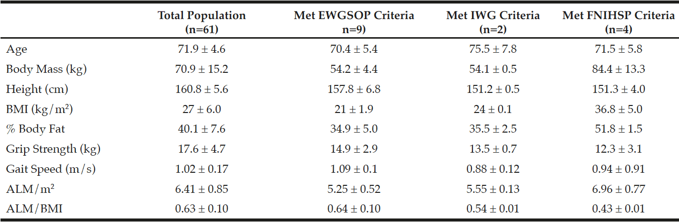
Table 1 Baseline characteristics of the population (n=61)

Table 1
Baseline characteristics of the population (n=61)

Data are presented as means ± standard deviations; Abbreviations: BMI = body mass index, ALM = sum of lean mass in both arms and both legs, m/s = meters per second, EWGSOP: European Working Group on Sarcopenia in Older People, IWG: International Working Group, FNIHSP: Foundation for the National Institutes of Health Sarcopenia Projet; Participants meeting EWGSOP criteria were sarcopenic (no pre-sarcopenia or severe sarcopenia); Participants meeting FNIHSP criteria had low lean mass and weakness (no low lean mass, weakness, and low physical function); Participants meeting IWG criteria (n=2) also met EWGSOP criteria and are included in that sample (n=9)


Discussion

These data indicate the large variation in sarcopenia prevalence depending on the classification criteria used. Within this sample, sarcopenia prevalence ranged from 3.3% to 14.8% with borderline significant differences in distribution frequency between EWGSOP and IWG criteria. This wide variation in prevalence is consistent with the findings of Cruz-Jentoft et al. (2014), who through systematic review found sarcopenia prevalence in community-dwelling women ranged from 1-30% when estimated using EWGSOP criteria (13). However, the authors expressed difficulty in comparing results of many studies due to inconsistent methodologies used in studies included in their review. In comparison, Patel et al. (2015) applied EWGSOP criteria to data from the Hertfordshire Cohort Study, which included 1,022 older women (23). While the baseline characteristics of that cohort closely resemble those of our sample, that study reported a 7.9% sarcopenia prevalence compared to our result of 14.8% using EWGSOP criteria. While those differences may be attributed to sample size, it may also be due to differences in grip strength. That study reported a mean grip strength of 26.3 kg while our results show a mean grip strength of only 17.6 kg, which is below the EWGSOP cut point for weakness in older women. This is consistent with the findings of Beaudart et al. (2014) who found grip strength criteria to largely influence sarcopenia prevalence (24). While there are considerably more data regarding sarcopenia prevalence using EWGSOP criteria, few studies have utilized IWG and/or FNIHSP criteria. However, Dam et al. in 2014 applied FNIHSP, IWG, and EWGSOP criteria to data collected from 2,950 older women through 9 different studies. That analysis found 2.3% of women to be weak and slow by FNIHSP criteria, 11.8% were sarcopenic by IWG criteria, and 13.3% were sarcopenic by EWGSOP criteria (18). Those researchers also noted that participants that had low lean mass by the ALM/BMI method were heavier with larger BMIs compared to those with low ALM/ht2. Our findings agree with those results, as every participant in our study who fell below the ALM/BMI cut-point had a BMI > 30 kg/m2. These results suggest that the FNIHSP criteria may be more effective at identifying sarcopenia in obese populations, while EWGSOP and IWG criteria may be more appropriate in non-obese populations. While our prevalence results vary with the findings of Dam et al. (18), possibly due to differences in sample size, it is evident that EWGSOP criteria consistently classify greater percentages of older women as sarcopenic when compared to FNIHSP and IWG criteria, and ALM adjusted for BMI may be the more effective method of identifying sarcopenia in obese, older women.
Reasons for variations in prevalence have recently been investigated by Masanés et al. (2016), who found that modification of EWGSOP lean mass cut points greatly varied sarcopenia prevalence, while modifying grip strength and gait speed cut points elicited little change in prevalence (25). However, those findings suggest that a large percentage of this population may have already been below the cut points for grip strength, as a combination of low ALM and weakness is required for a sarcopenia diagnosis by EWGSOP criteria.
Consequently our data show that the majority of participants considered sarcopenic by EWGSOP criteria had low ALM and weakness (n = 9), while no participants had low ALM accompanied with low gait speed. This also explains our low prevalence reported when using IWG criteria, which omits grip strength, and has a more liberal gait speed cut point. This suggests that inclusion of grip strength in sarcopenia diagnostic criteria may result in relatively higher prevalence estimates, and further screening for hand ailments (i.e. arthritis) may be necessary for accurate sarcopenia classification.
While the EWGSOP criteria are most prevalent within the literature, it does not take fat or body mass into consideration and may fail to classify those with sarcopenic obesity, as shown in our results (2). Moreover, the FNIHSP criteria may be ideal for the older female population as following menopause women typically experience increases in fat mass, which could prevent diagnosis by EWGSOP or IWG criteria (26). Our results underscore the discrepancies between different sets of sarcopenia classification criteria and therefore, inclusion of multiple sets of criteria may simplify the comparison of results and aid in determining population appropriate diagnostic criteria.
A small sample size, and a low number of participants who met classification criteria limited this study. A further limitation was the use of SMF-BIA to assess ALM rather than dual-energy x-ray absorptiometry (DXA). However, SMF-BIA has been found to be agreeable with DXA for measuring ALM in women, and BIA specific ALM/ht2 cut points presented by the EWGSOP were developed using prediction equations not applicable to the InBody 570 device (27, 28). Despite limitations, this study is novel in that EWGSOP, IWG, and FNIHSP criteria were all applied to the same sample of older, sedentary women from the same community. This allowed for the comparison of criteria without the need to adjust for sex, ethnicity, or activity levels. This study demonstrates the variability and limitations of current sarcopenia classification criteria, especially in obese individuals, and indicates the need for future research to develop current, criteria-appropriate cut-points for the measurement of ALM by SMF-BIA in this population to complement these findings.


Funding: This study was funded by the University of Rhode Island College of Human Sciences and Services.

Conflict of Interest: Matthew Delmonico has received research grants from the University of Rhode Island. All other authors report no conflicts of interest.

Ethical Approval: All procedures performed in studies involving human participants were in accordance with the ethical standards of the institutional and/or national research committee and with the 1964 Helsinki declaration and its later amendments or comparable ethical standards.

Informed consent: Informed consent was obtained from all individual participants included in the study.


References

1. Morley JE, Baumgartner RN, Roubenoff R, Mayer J, Nair KS. Sarcopenia. J Lab Clin Med 2001;137:231-243.
2. Newman AB, Kupelian V, Visser M, Simonsick E, Goodpaster B, Nevitt M, Kritchevsky SB, Tylavsky FA, Rubin SM, Harris TB et al. Sarcopenia: alternative definitions and associations with lower extremity function. J Am Geriatr Soc 2003;51:1602-1609.
3. Batsis JA, Mackenzie TA, Barre LK, Lopez-Jimenez F, Bartels SJ. Sarcopenia, sarcopenic obesity and mortality in older adults: results from the National Health and Nutrition Examination Survey III. Eur J Clin Nutr 2014;68:1001-1007.
4. Delmonico MJ, Beck DT. The Current Understanding of Sarcopenia: Emerging Tools and Interventional Possibilities. American Journal of Lifestyle Medicine, 2015.
5. Janssen I, Shepard DS, Katzmarzyk PT, Roubenoff R. The healthcare costs of sarcopenia in the United States. J Am Geriatr Soc 2004;52:80-85.
6. Antunes AC, Araújo DA, Veríssimo MT, Amaral TF. Sarcopenia and hospitalisation costs in older adults: a cross-sectional study. Nutrition & Dietetics, 2016.
7. Mijnarends D, Schols J, Halfens R, Meijers J, Luiking Y, Verlaan S, Evers S. Burden-of-illness of Dutch community-dwelling older adults with sarcopenia: Health related outcomes and costs. European Geriatric Medicine 2016;7:276-284.
8. Sousa A, Guerra R, Fonseca I, Pichel F, Ferreira S, Amaral T. Financial impact of sarcopenia on hospitalization costs. Eur J Clin Nutr 2016;70:1046-1051.
9. Administration on Aging, 2014. Projected Future Growth of the Older Population. U.S. Administration for Community Living. http://www.aoa.acl.gov/aging_statistics/future_growth/future_growth.aspx#age. Accessed 11/202016.
10. Borst SE. Interventions for sarcopenia and muscle weakness in older people. Age Ageing 2004;33:548-555.
11. Wen X, An P, Chen WC, Lv Y, Fu Q. Comparisons of sarcopenia prevalence based on different diagnostic criteria in Chinese older adults. J Nutr Health Aging 2015;19:342-347.
12. Tichet J, Vol S, Goxe D, Salle A, Berrut G, Ritz P. Prevalence of sarcopenia in the French senior population. J Nutr Health Aging 2008;12:202-206.
13. Cruz-Jentoft AJ, Landi F, Schneider SM, Zuniga C, Arai H, Boirie Y, Chen LK, Fielding RA, Martin FC, Michel JP et al. Prevalence of and interventions for sarcopenia in ageing adults: a systematic review. Report of the International Sarcopenia Initiative (EWGSOP and IWGS). Age Ageing 2014;43:748-759.
14. Cruz-Jentoft AJ, Baeyens JP, Bauer JM, Boirie Y, Cederholm T, Landi F, Martin FC, Michel JP, Rolland Y, Schneider SM et al. Sarcopenia: European consensus on definition and diagnosis: Report of the European Working Group on Sarcopenia in Older People. Age Ageing 2010;39:412-423.
15. Fielding RA, Vellas B, Evans WJ, Bhasin S, Morley JE, Newman AB, Abellan van Kan G, Andrieu S, Bauer J, Breuille D et al. Sarcopenia: an undiagnosed condition in older adults. Current consensus definition: prevalence, etiology, and consequences. International working group on sarcopenia. J Am Med Dir Assoc 2011;12:249-256.
16. Studenski SA, Peters KW, Alley DE, Cawthon PM, McLean RR, Harris TB, Ferrucci L, Guralnik JM, Fragala MS, Kenny AM et al. The FNIH sarcopenia project: rationale, study description, conference recommendations, and final estimates. J Gerontol A Biol Sci Med Sci 2014;69:547-558.
17. McLean RR, Shardell MD, Alley DE, Cawthon PM, Fragala MS, Harris TB, Kenny AM, Peters KW, Ferrucci L, Guralnik JM et al. Criteria for clinically relevant weakness and low lean mass and their longitudinal association with incident mobility impairment and mortality: the foundation for the National Institutes of Health (FNIH) sarcopenia project. J Gerontol A Biol Sci Med Sci 2014;69:576-583.
18. Dam TT, Peters KW, Fragala M, Cawthon PM, Harris TB, McLean R, Shardell M, Alley DE, Kenny A, Ferrucci L et al. An evidence-based comparison of operational criteria for the presence of sarcopenia. J Gerontol A Biol Sci Med Sci 2014;69:584-590.
19. Laukkanen P, Heikkinen E, Kauppinen M. Muscle strength and mobility as predictors of survival in 75-84-year-old people. Age Ageing 1995;24:468-473.
20. Rantanen T, Guralnik JM, Foley D, Masaki K, Leveille S, Curb JD, White L. Midlife hand grip strength as a predictor of old age disability. JAMA 1999;281:558-560.
21. Guralnik JM, Ferrucci L, Pieper CF, Leveille SG, Markides KS, Ostir GV, Studenski S, Berkman LF, Wallace RB. Lower extremity function and subsequent disability: consistency across studies, predictive models, and value of gait speed alone compared with the short physical performance battery. J Gerontol A Biol Sci Med Sci 2000;55:M221-31.
22. Guralnik JM, Simonsick EM, Ferrucci L, Glynn RJ, Berkman LF, Blazer DG, Scherr PA, Wallace RB. A short physical performance battery assessing lower extremity function: association with self-reported disability and prediction of mortality and nursing home admission. J Gerontol 1994;49:M85-94.
23. Patel HP, White MC, Westbury L, Syddall HE, Stephens PJ, Clough GF, Cooper C, Sayer AA. Skeletal muscle morphology in sarcopenia defined using the EWGSOP criteria: findings from the Hertfordshire Sarcopenia Study (HSS). BMC Geriatr 2015;15:171-015-0171-4.
24. Beaudart C, Reginster JY, Slomian J, Buckinx F, Locquet M, Bruyere O. Prevalence of sarcopenia: the impact of different diagnostic cut-off limits. J Musculoskelet Neuronal Interact 2014;14:425-431.
25. Masanés F, Rojano iL, Salvà A, Serra-Rexach J, Artaza I, Formiga F, Cuesta F, López Soto A, Ruiz D, Cruz-Jentoft A. Cut-off points for muscle mass — not grip strength or gait speed — determine variations in sarcopenia prevalence. J Nutr Health Aging: 2016;1-5.
26. Ley CJ, Lees B, Stevenson JC. Sex- and menopause-associated changes in body-fat distribution. Am J Clin Nutr 1992;55:950-954.
27. Anderson LJ, Erceg DN, Schroeder ET. Utility of multifrequency bioelectrical impedance compared with dual-energy x-ray absorptiometry for assessment of total and regional body composition varies between men and women. Nutr Res 2012;32:479-485.
28. Mahoney K, 2016. Validation of bioelectrical impedance analysis for the measurement of appendicular lean mass in older women. Open Access Master’s Theses. Paper 845:http://digitalcommons.uri.edu/theses/845.

EFFECTIVENESS OF PILATES AND CAMELLIA SINENSIS SUPPLEMENTATION ON CARDIOMETABOLIC RISK FACTORS AND REDOX MARKERS IN POSTMENOPAUSAL WOMEN: A PLACEBO-CONTROLLED, RANDOMIZED TRIAL


S. Junges1, R. Dias Molina2, C. Bittencourt Jacondino1, M. Lopes Da Poian3, E. Tatsch4, R. Noal Moresco4, M.G. Valle Gottlieb1


1. Graduate Program in Biomedical Gerontology of the Institute of Geriatrics and Gerontology, Pontifical Catholic University of Rio Grande do Sul (PUCRS), Porto Alegre, Brazil; 2. Graduate Program in Medicine and Health Sciences, Pontifical Catholic University of Rio Grande do Sul (PUCRS), Porto Alegre, Brazil; 3. Pharmacist, Graduate of the Lutheran University of Brazil, Porto Alegre, Brazil. 4. Department of Clinical and Toxicological Analysis, Health Sciences Center, Federal University of Santa Maria, Santa Maria, Brazil.

Corresponding Author: Maria Gabriela Valle Gottlieb, Avenida Ipiranga, 6681 – Prédio 81, 7º andar, sala 703, CEP: 90619-900, Porto Alegre, RS, Brasil, E-mail: maria.gottlieb@pucrs.br


J Aging Res Clin Practice 2017;6:80-87
Published online May 4, 2017, http://dx.doi.org/10.14283/jarcp.2017.7


Abstract

Objective: To verify the effectiveness of Pilates and Camellia sinensis extract (CSE) supplementation on cardiometabolic risk factors and redox markers in postmenopausal women. Design: A placebo-controlled, randomized trial. Setting: community-dwelling postmenopausal women without disability. Participants: fifty postmenopausal women volunteers with cardiometabolic risk factors (high waist circumference, triglycerides, HDL-c, glucose and blood pressure). The volunteers participants were randomized in four groups: Pilates+CSE (14); Pilates+Placebo (11); CSE (11); and Placebo (14). Intervention: the CSE and Pilates+CSE intervention groups consumed one 500mg CSE capsule with excipient per day for 24 weeks. The Placebo group consumed one capsule with a placebo excipient per day for 24 weeks. Pilates training was performed twice weekly for 60 minutes each time, over 24 weeks. Measurements: cardiometabolic risk factors (glucose, waist circumference (WC), systolic blood pressure (SBP), diastolic blood pressure (DBP), triglycerides, high-density lipoproteins-HDL-c) and oxidative metabolism markers (advanced oxidation protein products (AOPP), ferric-reducing ability of plasma (FRAP), nitrosative stress marker (NOx), ischemia modified albumin (IMA). Results: When baseline variables were adjusted, the WC of the Pilates + CSE was significantly lower than that of the CSE and Placebo groups after the intervention (p<0,001).The triglycerides levels of the Pilates + CSE and Pilates + Placebo groups were significantly lower than those of the Placebo group (p= 0,010) . The glucose levels of the Pilates + CSE group were significantly lower than those of the Placebo group (p= 0,041). Whitin-group pre and post intervention comparison showed that Pilates+CSE group presented the best effect in some cardiometabolic risk factors, with significant reductions in tree cardiometabolic risk factor: waist circumference, triglycerides, glucose and FRAP (P=0.003, P<0.001, P=0.021 and P=0.041, respectively). The Pilates+Placebo group was found to be effective in reducing triglycerides (P=0.002), while the CSE group presented increased post-intervention NOx levels (p= 0.009). Conclusion: Our results suggest that Pilates and Camellia sinensis extract intervention may help to reduce some cardiometabolic risk factors in postmenopausal women.


Key words: Cardiometabolic risk factor, Pilates method, camellia sinensis, oxidants, antioxidant, women.


Introduction

Cardiovascular diseases (CVD) are the main cause of morbidity and mortality in developed countries and development, as is the case in Brazil (1). There are several cardiovascular risk factor, however due to its high morbidity burden and prevalence in populations, obesity, diabetes, dyslipidemia and sedentary lifestyle stand out (1-3). Additionally, cardiometabolic risk factors are those involved in the development of type 2 DM and CVD due to a set of modifiable risk factors (1-3). In this sense, several studies have demonstrated that sedentarism is one of the main risk factors that can contribute to the onset of metabolic disorders and CVD (4-7). In parallel, studies involving lifestyle modifications, such as adherence to a balanced and healthy diet and the practice of regular physical exercise, have presented beneficial results for both weight reduction and MetS components (8-10). The evidence for this has been demonstrated in research indicating that both physical exercise and bioactive dietary compounds are able to maintain the oxidation process within physiological limits, controlling damage to macromolecules, which can lead to systemic damage that is often irreversible (8-9, 11). In this context, the Pilates exercise method (an exercise that combines strength and endurance) has emerged as a physical exercise with high numbers of participants in several countries, focusing on postural correction and acting on the back and abdominal muscles, thus helping prevent or mitigate the risk factors for cardiovascular and metabolic diseases (8). An important principle of this method is the respiratory maneuvers technique that demands a constant energy expenditure for all movements, preventing excessive cardiac stress, which is, in turn, reflected in the hemodynamic parameters (12, 13). Moreover, some studies involving Pilates have demonstrated its effectiveness in improving the body composition of postmenopausal women (12, 14). Yet physical exercise alone is not enough to prevent cardiometabolic risk factor and CVD, being essential the combination of a diet rich in bioactive components. The consumption of certain functional foods rich in bioactive and antioxidant substances, such as green tea that comes from the Camellia sinensis plant, can also be a strongally, contributing to the prevention of cardiometabolic risk factors and/or MetS, as well reducing oxidative processes (8, 11, 13). The Camellia sinensis plant presents a high content of flavonoids (catechins), which have a thermogenic effect and oxidize body fat. This can result in weight loss and decreased abdominal adiposity, and is therefore considered a powerful resource for the prevention of MetS components (15, 16). The antioxidant activity of green tea, other than it being rich in flavonoids, lies in its content of carotenoids, tocopherol, ascorbic acids and minerals (Zn, Ca, K, Mn), which enhance its antioxidant potential. Green tea polyphenols have in vitro antioxidant activity that neutralizes reactive oxygen and nitrogen species (17). Furthermore, they have also shown an ability to chelate metals, such as iron, preventing their participation in reactions that generate free radicals like Fenton and Haber-Weiss, which are extremely harmful to lipids, proteins and DNA (17). There is a gap in knowledge about the effects and benefits of the pilates method on cardiometabolic risk factors, as well as the combination of the method with functional foods such as camellia sinensis. Based on this evidence, this study aimed to verify the effectiveness of the Pilates training method and the use of Camellia sinensis extract (CSE) on cardiometabolic risk factors and redox markers in postmenopausal women.


Materials and methods

Study design

A placebo-controlled, randomized clinical trial

Population and sample: The population investigated were community-dwelling postmenopausal women without disability living in Porto Alegre, the capital city of Rio Grande do Sul state, Brazil. All women had a cardiometabolic risk factors and no history of cardiovascular event at the start of the study. Based on previous studies the initial sample size was 60 women, culminating in a final sample of 50 volunteers (9,12,18). The research was submitted to and approved by the Ethics Committee of the Pontifical Catholic University of Rio Grande do Sul (protocol n°474.390). Data collection began after approval and complied with requirements set out in Resolution 466/2012 of the Ministry of Health in relation to research involving humans. All participants signed the Consent Form. The project was sent to the Brazilian Clinical Trials Registry (ReBEC) and registered under number RBR-2sgtn2.

Recruitment

The recruitment campaign for community-dwelling post menopausal women with cardiometabolic risk factors was conducted via phone book, newspapers and were distributed of leaflets in pharmacies, health posts and supermarkets invited for the study. All women who presented clinical exams and a declared use of medications for diabetes, hypertension or dyslipidemia at the time of recruitment were considered to have cardiometabolic risk factors and were invited to participate in the study. After clarification of aspects related to the study, initial exams presented by the participants were analyzed in order to evaluate the cardiometabolic risk factors or MetS diagnosis, according to diagnostic criteria set by the National Cholesterol Education Program (NCEP), Expert Panel on detection, evaluation and treatment of high blood cholesterol in adults (Adult Treatment Panel) III NECP-ATPIII, of 2001 (2). After selection, volunteers were scheduled for blood collection and biochemical analysis to confirm the presence of the cardiometabolic risk factors or MetS components and to obtain anthropometric variables. The procedures for fasting blood collection, anthropometric measurements and blood pressure evaluation were explained to the participants and they were asked to sign the study Informed Consent Form. The inclusion criteria comprised: being in menopause for more than a year; and have cardiometabolic risk factors based on the NCEP-ATPIII (2001) criteria which include: altered WC: >102cm to man and >88cm (women), or dyslipidemia (TG ≥ 150mg/dl and HDL<40mg/dl (man), <50mg/dl (women);or systolic blood pressure ≥ 130 mmHg or diastolic blood pressure ≥ 85 mmHg (or the use of antihypertensive medication);and/or fasting glycemia ≥ 110mg / dl (2). Women were excluded from the study if they reported previous cardiovascular events (acute myocardial infarction, stroke, angina, heart failure), suffered from liver, gastric or thyroid diseases, presented memory problems, anxiety or sleep disorders, had any physical condition that might make Pilates training impossible, had undertaken Pilates or any other form of regular physical exercise, habitually or regularly drink green tea, or took vitamin supplements.

Randomization

All women with cardiometabolic risk factors were evaluated for exclusion criteria and those eligible were randomized, as shown in Figure 1. Randomization was performed through one-by-one drawing, using a box from which numbers corresponding to the women were randomly selected. In this way, the women were randomly divided into one of four groups: 1) Pilates+CSE (n=14); 2) Pilates+Placebo (n=11); 3) CSE (n=11); 4) Placebo (n=14).


Figure 1

Figure 1


Intervention

Camellia sinensis extract and Placebo. Both the placebo and Camellia sinensis extract capsules were of the same size, raw material quantity (500 mg) and color, and were prepared in the same batch acquired from the manipulation pharmacy, Dapelle Ltd. Each placebo capsule contained 1.0000 q.s corn starch, 0.5% magnesium stearate, 0.5% Aerosil (pharmaceutical talc, light magnesium carbonate, light magnesium oxide, talc, dibasic calcium phosphate, etc.) and 29% microcrystalline cellulose, and contained no detectable levels of polyphenol ingredients. Each CSE capsule had 500 mg dry extract of Camellia sinensis, with a total of 4.24% polyphenols, 3.60% tannin and 0.70% caffeine. The Pilates+CSE and Pilates+Placebo intervention group participants took one capsule containing 500 mg CSE or placebo per day, one hour after the main meal, for 24 consecutive weeks.

Training protocol

Pilates exercises were performed and completed by 25 randomized women in the following groups: Pilates+CSE (n=14) and Pilates+Palcebo (n=11). The intervention lasted 24 weeks, with two 60 min sessions per week. Exercises were conducted using specialized equipment: Reformer, Cadillac, Wall unit, Ladder Barrel and Chair; and floor exercises, which were classified as being of a basic level. All exercises were performed in a progressive and educational form, respecting the fundamental principles of the Pilates method. Training was divided into initial, main and final phases, consisting of four macrocycles. All training sessions began with a 10 min warm-up on an ergometric treadmill (Movement RT 150 pro), at a speed of 5.0 Km/h. The first four-week macrocycle was composed of educational exercises at a basic level aimed at promoting neural adaptation and an introduction of exercise for breathing techniques. The second macrocycle of eight weeks included specific strengthening exercises of the spinal column extensors, serratus, serratus anterior, abdominals, quadriceps (vastus medialis) and rotator cuff, and stretching of the pectoral (major and minor), rhomboid, trapezius, triceps, iliopsoas, hamstring and adductor muscles. The third macrocycle also lasted eight weeks, with performance of the same series of exercises but with increased spring resistance. The fourth macrocycle included variation of joint angles and distances with maintenance of load and exercise series, and ending with relaxation techniques. In addition to these, stretching exercises for the shortened muscle groups were carried out using a “tonic-ball” and “Franklin Ball”.

Blood collection

Blood sample (8-hour fasting) collections were performed on two different occasions (pre- and post-intervention). The samples were acquired via direct peripheral venipuncture using silicone-coated vacutainer blood collection tubes (Becton-Dickinson) containing 0.5 ml EDTA solution, with gel and a 19G (25 x 10) disposable needle, in the cubital fossa and under aseptic conditions. All sharps materials were disposed off in special containers and the biological waste discarded in white bags. All samples were sent to the Laboratory of Biochemistry, Molecular Genetics and Parasitology of the Institute of Geriatrics and Gerontology-PUCRS for due processing (separation of plasma, serum and buffy coat), and were then stored in a freezer at -20ºC.

Blinding

All researchers involved were blinded throughout this study. However, the pharmacist who distributed the CSE and placebo capsules, the Pilates group participants and instructors were not blinded for the Pilates training.

Evaluation of study adherence

The concept of adherence varies, but in general, a participant is understood to be adherent to a therapy using prescribed medications or other procedures when they observe the schedules, dosages and treatment durations for at least 80% of the total protocol. To evaluate the percentage of participant adherence in the Placebo, CSE and Pilates+CSE groups, participants were requested to present those capsules that were not consumed during the intervention period. Women who consumed less than 80% of capsules, together with those who failed to attend at least 80% of the Pilates training sessions, were excluded from the study (16,18). Four women (1 from the Pilates + CSE group and 4 from the Pilates + placebo group) were excluded from the study because they had a frequency of less than 80% in Pilates classes and/or in the use of the capsules. In addition, five women (4 CSE and 1 from placebo group) were excluded because they dropped out.

Evaluation of Adverse Effects

The study participants were asked to report adverse effects at any time in relation to the placebo, green tea or Pilates. None of the participants from any of the groups observed in this study complained of any adverse effects.

Variables and collection tools

The following variables were investigated for all study participants: age, marital status, education and income. Data were collected through use of a structured questionnaire. The cardiometabolic risk factors, such as fasting glucose and lipid profile, were measured using Labtest kits and analyzed by spectrophotometry, in accordance with manufacturer instructions. Weight was measured using a calibrated anthropometric balance scale with a 150kg capacity, graduated in 100g increments. Participants were weighed barefoot wearing a bathing suit only, with results recorded in kilograms (kg). Participant height was recorded using the measuring rod of the scales. Body Mass Index was then calculated through the equation, weight (kg) divided by height (m) squared. The minimum abdominal or waist circumference (WC) was recorded at the level of the iliac crest and values up to 80cm were considered normal for women. Above this value, individuals were considered to have central obesity (2). Evaluation of the levels of advanced oxidation protein products (AOPP) was performed according to Witko-Sarsat et al. (19), in a Cobas MIRA® analyzer (Roche Diagnostics, Basel, Switzerland). Nitrosative stress (NOx) were assessed based on the technique described by Tatsch et al. (20), in a Cobas MIRA® analyzer (Roche Diagnostics, Basel, Switzerland). The ferric reducing ability of plasma (FRAP) was performed following the technique described by Benzie et al. (21), in a Cobas MIRA® analyzer (Roche Diagnostics, Basel, Switzerland). Ischemia modified albumin (IMA) was measured by colorimetric assay with cobalt, described by Bar-Or et al. (22).
A nurse measured arterial blood pressure, recording systolic blood pressure (SBP) and diastolic blood pressure (DBP) using a mercury sphygmomanometer (Erka, Germany), equipped with an appropriately sized cuff for the right arm circumference.
Each participant remained at rest (sitting) for at least 5 min before the measurement was taken. Two measurements were recorded with an approximate interval of 30 min between them. The appearance of sounds was used for identification of SBP and their disappearance (Korotkoff phase V) for DBP. The Osler maneuver for measurement of systolic arterial blood pressure (considered positive when the radial artery is still palpable after inflating the cuff above the SBP level) was concomitantly used due to the possibility of pseudohypertension caused by stiffening of the brachial artery, secondary to atherosclerosis (can increase arterial blood pressure by 30 mmHg or more). These cases were excluded from the study. Blood pressure levels up to 130/85 mmHg were considered normal in accordance with recommendations from the National Cholesterol Education Program (NCEP) expert panel on detection, evaluation and treatment of high blood cholesterol in adults (Adult Treatment Panel III) (2). Diagnosis of MetS was made according to the NCEP-ATP III diagnostic criteria (2).

Statistical Analysis

Data were recorded using an Excel spreadsheet and analyzed through the IBM statistics package, SPSS, version 21.0. Continuous variables were expressed as mean and standard deviation/standard error or median and interquartile range. Categorical variables were described by absolute and relative frequencies. One-way analysis of variance (ANOVA) was applied followed by Tukey’s post hoc test to compare the mean values. The Kruskal-Wallis test was used in the case of asymmetry. The Analysis of Covariance (ANCOVA), complemented by the Bonferroni test, was applied to adjust for differences concerning baseline measurements. Intragroup comparisons were evaluated using Student’s t test for paired samples (symmetrical distribution) or the Wilcoxon test (asymmetric distribution). A significance level of 5% was adopted (p<0.05).


Results

The sample comprised of 50 volunteers with a cardiometabolic risk factors, with a mean age of 61.3±6.6 years (range 51-77 yr). The mean age did not differ between the groups, being respectively: Pilates + CSE group (60.5±6.2 yr), Pilates + Placebo group (62.6±6.7 yr), CSE group (59.1±4.7 yr) and Placebo group (62.9 ± 8.0 yr), with p= 0.462. No statistically significant differences between the investigated groups were observed for the sociodemographic variables of race, income and education (p> 0.05).
Investigation of the components of MetS for the 50 study participants revealed that 35 (70%) were verified as presenting three MetS diagnostic criteria components and 15 (30%) presented two (NCEP ATP III). All participants recorded a waist circumference >88 cm. A comparison was made between the initial measurement results for the four groups in all the recorded variables (Table 1). The triglyceride (TG) levels for those patients from the Camellia sinensis group were observed to be significantly lower than the other groups. No statistically significant differences between the groups were seen for the remaining variables before the intervention began.

Table 1 Comparison of cardiometabolic risk factors (CRF) and redox markers between the groups in the pre-intervention period

Table 1
Comparison of cardiometabolic risk factors (CRF) and redox markers between the groups in the pre-intervention period

CRF: cardiometabolic risk factors; CSE: Camellia sinensis extract; WC: waist circumference; SBP: systolic blood pressure; DBP: diastolic blood pressure; TG: triglycerides; HDL: high-density lipoproteins; AOPP: advanced oxidation protein products; FRAP: ferric-reducing ability of plasma; NOx: nitrosative stress marker; IMA: ischemia modified albumin; * Described by median (Percentis 25-75)


The post-intervention variables between the four groups are compared in Table 2. After adjustment for the baseline measurements, the post-intervention WC variable for the Pilates+CSE group was significantly lower than the CSE and Placebo groups.

Table 2 Comparison between groups of cardiometabolic risk factors (CRF) and redox markers in the post-intervention period

Table 2
Comparison between groups of cardiometabolic risk factors (CRF) and redox markers in the post-intervention period

*adjusted for baseline measurement; a,b, Equal letters are not significantly different using the Tukey test with a significance level of 5%; **variables underwent log-transformation for conducting ANOVA.CRF: cardiometabolic risk factors; CSE: Camellia sinensis extract; WC: waist circumference; SBP: systolic blood pressure; DBP: diastolic blood pressure; TG: triglycerides; HDL: high-density lipoproteins; AOPP: advanced oxidation protein products; FRAP: ferric-reducing ability of plasma; NOx: nitrosative stress marker; IMA: ischemia modified albumin.


The TG levels for the Pilates+CSE and Pilates+Placebo groups were significantly lower than the Placebo group. Blood glucose for the Pilates+CSE group were significantly lower than the Placebo group. No statistically significant differences were observed in the redox markers.
Table 3 compares intragroup variables. A significant reduction in WC, TG, blood glucose levels and FRAP was found in the Pilates+CSE group. In the Pilates+Placebo group, a significant reduction was seen only in the TG variable. A significant increase in NOx levels was verified in the CSE group, while in the Placebo group a significant increase in WC and FRAP and significant reduction of triglycerides was observed.

Table 3 Intragroup comparison of cardiometabolic risk factors (CRF) and redox markers, pre- and post-intervention

Table 3
Intragroup comparison of cardiometabolic risk factors (CRF) and redox markers, pre- and post-intervention

* Described by median (Percentile 25-75); CRF: cardiometabolic risk factors; CSE: Camellia sinensis extract; WC: waist circumference; SBP: systolic blood pressure; DBP: diastolic blood pressure; TG: triglycerides; HDL: high-density lipoproteins; AOPP: advanced oxidation protein products; FRAP: ferric-reducing ability of plasma; NOx: nitrosative stress marker; IMA: ischemia modified albumin


Discussion

This study verified the effectiveness of Pilates training and the use of Camellia sinensis extract on the cardiometabolic risk factors of postmenopausal women. The results suggested that the combination of Pilates+CSE can be the most effective method for reducing three important cardiometabolic risk factor, being waist circumference, triglycerides and blood glucose, and for increasing levels of FRAP. In addition, supplementation with CSE alone suggested increase NOx levels. Furthermore, the Pilates+Placebo combination also seemed to be effective in reducing triglyceride levels. Researches on this topic are relatively recent and yet inconclusive, especially in relation to the use of Pilates training. Most studies focused on this method have investigated its effectiveness in chronic low back pain, postural correction and improvement of dynamic balance, flexibility, strength and endurance, increasing muscle mass and reducing fat mass, rehabilitation, and improvements in quality of life (12, 14, 18, 23). The effects of Pilates on cardiometabolic risk factors has been investigated more recently, however, the literature is still scarce (8, 9, 24). Studies are more abundant in relation to the consumption of Camellia sinensis extract, with reports on its beneficial effects on adipogenesis, mitigation of MetS, particularly with regard to hyperglycemia, hypercholesterolemia, and waist circumference, as well as in lessening oxidative stress (11, 15, 16). These effects have been observed in human and nonhuman experimental models, and for its consumption both in the form of tea (infusion) and capsules (extract).
Pilates combined with CSE supplementation presented better efficacy in mitigating three components of MetS that have a high level of morbidity and mortality (waist circumference, blood levels of triglyceride and glucose) in postmenopausal women. Double-blind randomized clinical trials, such as the Heart Outcomes Prevention Evaluation (HOPE), have already shown that obesity, especially abdominal, worsens the prognosis of patients with cardiovascular diseases. This study demonstrated that abdominal obesity alone is associated with a 20-23% increase in relative risk for myocardial infarction, 38% for heart failure, and 17% for total mortality (25). Abdominal obesity, which is metabolically active, produces chronic low-grade inflammation, increased flow of free fatty acids and an increase in LDL-c plasma levels (26). The union of Pilates and CSE can promote a better distribution of adipose tissue through increased mobilization of fat deposited in the abdominal region (12, 14, 15, 27). In addition, studies have demonstrated that catechins (EGCG) of Camellia sinensis promote weight loss, affect fat distribution, inhibit adipogenesis and cause apoptosis in mature adipocytes (12, 14, 15, 27). The findings of the present study reinforce the hypothesis that Pilates exercises involve a considerable energy expenditure through the performance of multidimensional and resistance exercises, which are of low and moderate intensity, contributing to the burning of lipids and preventing or decreasing their accumulation in adipose tissue (28). This in turn may contribute to the prevention of cardiovascular diseases (8,9,28). One study analyzed the effect of Pilates on a diabetic group but found no statistical difference in the mean glycated hemoglobin and daily doses of subcutaneous insulin between the intervention and control groups (24). A hypothesis proposed by the authors suggested that participants increased their carbohydrate intake prior to the physical activity due to the risk of hypoglycemia while performing the exercise. However, their caloric intake was not evaluated (24). In relation to lipid profile, no statistically significant differences were found in the mean levels of triglycerides, HDL-c, LDL-c and total cholesterol (24) in the Pilates intervention group. Nonetheless, the findings of the present study demonstrated that both Pilates+CSE (reduction of three components of MetS) and Pilates+Placebo were able to reduce at least one of the diagnostic criterion of MetS.
With regard to effects of intervention on the redox markers, the present study noted an increase in FRAP levels in the Pilates+CSE group and in NOx levels in the CSE group. According to Benzie et al., individuals that present high levels of FRAP have a higher antioxidant capacity in reducing iron (21). An in vitro study with CSE conducted by Sohrab et al., exhibited the ability of CSE polyphenols to chelate metals, such as iron, preventing their participation in reactions that generate free radicals like Fenton and Haber-weiss (29). Besides the antioxidant properties of CSE, according to Steinbacher and Eckl, oxidative stress induced by exercise serves as an important signal to stimulate muscle adaptation to antioxidant systems through activation of redox-sensitive signaling pathways (30). While an intense contraction is sufficient to activate these pathways, the upregulation of protein synthesis requires cumulative effects of repetitive exercise movements, which Pilates provides. This formation and adaptation also allows the cell to incorporate high levels of exogenous antioxidants in the form of dietary supplementation. This corroborates the results of the present study, in which only the Pilates+CSE group showed increases in FRAP levels. In relation to NOx findings, the bioactive components and antioxidant and anti-inflammatory activity of CSE may contribute to raise NO levels, thus increasing its metabolites.
Despite the beneficial results presented with respect to the cardiometabolic risk factors, it is important to consider some limitations of the present research. The first concerns is to reduced sample sizes. The second relates to the non-assessment of the macro- and micronutrient intake of the sample, which could have affected the obtained results, as well as causing unexpected results in the Placebo group, especially in relation to triglycerides and FRAP. Nevertheless, despite the limitations of the present trial, our results suggest that Pilates and supplementation with Camellia sinensis decrease cardiometabolic risk factors and increase FRAP levels in postmenopausal women, which may positively influence the global health of this sample. Additionally, the study reinforces the recommendation of Pilates method exercises for this age group and a diet rich in bioactive compounds, such as the flavonoids of Camellia sinensis, not only as an adjuvant treatment, but also for the prevention cardiometabolic risk factors.


Conclusion

The presented results suggest the effectiveness of the Pilates method with CSE supplementation on cardiometabolic risk factors, such as triglycerides, blood glucose and waist circumference, as well as on FRAP. However, the Pilates+Placebo group also appear to be effective in reducing triglycerides. In addition, ingestion of CSE also appear to be effective in increasing NOx levels. Our results suggest that this type of intervention, combining Pilates and Camellia sinensis may help to reduce some cardiometabolic risk factors in postmenopausal women.


Acknowledgements: Our thanks to the National Postdoctoral Program of the Coordination for the Improvement of Higher Education Personnel (PNPD/CAPES) and the Dapelle manipulation pharmacy for providing the extracts, and to the women who participated in the study. Trial registration: RBR-2sgtn2 (Brazilian Clinical Trials Registry- ReBEC)

Conflict of interest: The authors declare no conflict of interest involving this study.

Ethical standards: The study followed all the ethical recommendations of Resolution 466/12 of the Brazilian Ministry of Health on Research involving human beings. The investigators undertook to respect the protocol in all respects.


References

1. Brasil. Ministério da Saúde. Cadernos de Atenção Básica, no. 38, 2014. [cited 2015 March] Available at: http://bvsms.saude.gov.br/bvs/publicacoes/estrategias_cuidado_doenca_cronica_obesidade_cab38.pdf
2. National Cholesterol Education Program – NCEP. Executive Summary of the Third Report of the National Cholesterol Education Program.Expert Panel on Detection, Evaluation, and Treatment of High Blood Cholesterol in Adults – ATPIII. JAMA 2001;285:2486–2497. http://dx.doi.org/10.1001/jama.285.19.2486
3. Diretrizes da Sociedade Brasileira de Cardiologia. Pocket Book.5ª edição 2011-2013. p.4-446.
4. González-Gross M, Meléndez A. Sedentarism, active lifestyle and sport: Impact on health and obesity prevention. Nutr Hosp. 2013;28(Suppl 5):89-98.
5. Nauman J, Nilsen TI, Wisloff U, Vatten LJ. Combined effect of resting heart rate and physical activity on ischaemic heart disease: mortality follow-up in a population study (the HUNT study, Norway). J Epidemiol Community Health. 2010;64:175-81.
6. Myers J, Prakash M, Froelicher V, Do D, Partington S, Atwood JE. Exercise capacity and mortality among men referred for exercise testing. N Engl J Med. 2002;346:793-801.
7. Aravindalochanan V1, Kumpatla S, Rengarajan M, Rajan R, Viswanathan V. Risk of diabetes in subjects with sedentary profession and the synergistic effect of positive family history of diabetes. Diabetes Technol Ther. 2014;16(1):26-32.
8. Junges S, Jacondino CB, Gottlieb MGV. Pilates effect on risk factors for cardiometabolic disease: a systematic review. Scientia Medica. 2015;25(1): 1-8.
9. Fourie M, Gildenhuys M, Shaw I, Shaw B, Toriola A, Goon DT. Effects of a MatPilates program on cardiometabolic parameters in elderly women. Pak J MedSci. 2013; 29(2):500-4.
10. Finelli C, Padula MC, Martelli G, Tarantino G. Could the improvement of obesity-related co-morbidities depend on modified gut hormones secretion? World J Gastroenterol. 2014;20(44):16649-64.
11. Yousaf S, Butt MS, Suleria HAR, Iqbala MJ. The role of green tea extract and powder in mitigating metabolic syndromes with special reference to hyperglycemia and hypercholesterolemia. Food Funct. 2014;5(3):545-56.
12. Bergamin M, Gobbo S, Bullo V, et al. Effects of a Pilates exercise program on muscles trength, postural controland body composition: results from a pilot study in a group of post-menopausal women. Age (Dordr). 2015;37(6):118.
13. Qian G, Xue K, Tang L, et al. Mitigation of oxidative damage by green tea polyphenols and Tai Chi exercise in postmenopausal women with osteopenia. PLoS One. 2012;7(10).
14. Vaquero-Cristóbal R, Alacid F, Esparza-Ros F, López-Plaza D, Muyor JM, López-Miñarro PA. The effects of a reformer Pilates program on body composition and morphological characteristics in active women after a detraining period. Women Health. 2015;19:1-23.
15. Lin J, Della-Fera MA, Baile CA. Green tea polyphenol epigallocatequin Gallate inhibits adipogenesis and induces apoptosis in 3T3-L1 adipocytes. Obes Res. 2005;13(6):982-990.
16. Vieira Senger AE, Schwanke CH, Gomes I, Valle Gottlieb MG. Effect of green tea (Camellia sinensis) consumption on the components of metabolic syndromein elderly. J. Nutr Health Aging. 2012;16(9):738-42.
17. Higdon JV, Frei B. Tea catechins and polyphenols: health effects, metabolism, and antioxidant functions. Crit Rev Food Sci Nutr. 2003;43:89-143.
18. Junges S, Gottlieb MG, Babtista RR, Quadros CB, Resende TL, Gomes I. Pilates method effect for posture and flexibility in women with kyphosis. Brazilian journal of Science and Movement. 2012;20(1):21-25.
19. Witko-sarsat V, Friedlander M, Nguyen Khoa T, et al. Advanced Oxidation Protein Products as Novel Mediators of Inflammation and Monocyte Activation in Chronic Renal Failure. Journal Of Immunology. 1998; 161 (5): 2524-2532.
20. Tatsch E, Bochi, GV, Pereira RS. A simple and inexpensive automated technique for measurement of serum nitrite/nitrate. Clin Biochem. 2011; 44: 348-350.
21. Benzie IF, Strain JJ. The ferric reducing ability of plasma (FRAP) as a measure of “antioxidant power”: the FRAP assay. Anal Biochem. 1996; 239 (1):70-76.
22. Bar-or D, Lau E, Winkler JV. Novel assay for cobalt-albumin binding and its potential as a marker for myocardial ischemia–a preliminary report. J Emerg Med. 2000; 19: 311-315.
23. Natour J, Cazotti L de A, Ribeiro LH, Baptista AS, Jones A. Pilates improves pain, function and quality of life in patients with chronic low back pain: a randomized controlled trial. Clin. Rehabil. 2015;29(1):59-68.
24. Tunar M, Ozen S, Gaksen D, Asar G, Bedizds S, Darcan S. The effects of Pilates on metabolic control and physical performance in adolescents with type 1 diabetes mellitus. J. Diabetes Complications. 2012;26(4):348-51.
25. Dagenais GR, Yi Q, Mann JF, Bosch J, Pogue J, Yusuf S. Prognostic impact of body weight and abdominal obesity in women and men with cardiovascular disease. Am Heart J. 2005;149(1):54-60.
26. Boden G. Obesity and Free Fatty Acids (FFA). Endocrinol Metab Clin North Am. 2008; 37(3): 635–ix.
27. Lin JK, Lin-Shiau SY. Mechanismis of hypolipidemic and anti-obesity effects of tea and tea polyphenols. Mol Nutr Food Res. 2006;50:211-7.
28. Torabian M, Taghadosi M, Ajorpaz NM, Khorasanifar L. The effect of Pilates exercises on general in women with type 2 diabetes. Life Science Journal .2013;10: 283-288.
29. Sohrab G; Hosseinpour-Niazi S, Hejazi J, Yuzbashian E, Mirmiran P, Azizi F. Dietary polyphenols and metabolic syndrome among Iranian adults. Int J Food Sci Nutr. 2013; 64 (6): 661-667.
30. Steinbacher P, Eckl P. Impact of oxidative stress on exercising skeletal muscle. Biomolecules. 2015;5(2):356-77.