jarlife journal
Sample text

AND option

OR option

EFFECTS OF VITAMIN D SUPPLEMENTATION ON WELL-BEING, POSTURAL CONTROL, MUSCLE STRENGTH, BONE AND CALCITROPIC HORMONES – A RANDOMIZED DOUBLEBLIND PLACEBO CONTROLLED TRIAL


S.A. Eriksen1, J. Starup-Linde2, R.P. Hirata3, K.K. Petersen4, T. Graven-Nielsen4, P. Vestergaard1,5


1. Department of Clinical Medicine, Aalborg University Hospital, Aalborg, Denmark; 2. Department of Endocrinology and Internal Medicine, Aarhus University Hospital, Aarhus, Denmark; 3. SMI, Department of Health Science and Technology, Aalborg University, Aalborg, Denmark; 4. Center for Neuroplasticity and Pain (CNAP), SMI, Department of Health Science and Technology, Aalborg University, Aalborg, Denmark; 5. Department of Endocrinology, Aalborg University Hospital, Aalborg, Denmark

Corresponding Author: Stine Aistrup Eriksen, PhD cand.scient.med, Department of Clinical Medicine, Aalborg University Hospital, Aalborg, Denmark, stineaistrup@hotmail.com,  Phone: +45 23 98 60 72

J Aging Res Clin Practice 2019;8:49-56
Published online June  5, 2019, http://dx.doi.org/10.14283/jarcp.2019.9


Abstract

Background: Antidepressants may increase the risk of fractures through negative effects on the musculoskeletal system that could be hindered by vitamin D supplements. Objectives: To study the pleiotropic effects of daily vitamin D supplementation in depressed patients treated with citalopram (patients) and healthy controls. Design: Randomised double blind placebo controlled trial. Setting: A study of Danish women in the age 50 to 90 years. Participants: A total of 21 patients and 50 controls. Intervention: Participants received daily vitamin D supplementation (50 micrograms) or placebo in one year. Measurements: Bone Densitometry by dual-energy x-ray absorptiometry. Serum 25-hydroxyvitamin D, intact-Parathyroid Hormone, type 1 procollagen N terminal peptide, tartrate resistant acid phosphatase type 5b. Pain sensitivity measures based on pain detection thresholds by cuff algometry, temporal summation of pain, conditioned pain modulation, and cutaneous pain sensitivity by pinprick test. Degree of depression by the Major Depression Inventory. Physical performance was assessed by Timed up and go, isomeric handgrip exercise, and postural control by force plate. Results: Serum 25(OH)D levels increased in the vitamin D treated patients compared with controls at the 12 months visit (P<0.05). Conversely, intact- Parathyroid Hormone decreased among the patients and controls receiving vitamin D compared with placebo (P<0.05). Vitamin D improved Major Depression Inventory scores in patients and controls compared with placebo (P<0.05). In patients receiving vitamin D, handgrip strength improved (P<0.05). Conclusions: Vitamin D may improve depressive symptoms, and improve handgrip strength among patients compared to controls.

Key words: Vitamin D, citalopram, BMD, pain sensitivity, physical performance.


Introduction

Several studies have shown that vitamin D supplementation have a positive influence on Bone Mineral Density (BMD). Higher doses (700 to 800 IU a day) of vitamin D supplementation seemed to be more effective in fracture prevention and prevention of falls than smaller doses (400 to 600 IU a day) among middle-aged and older adults (1).
Vitamin D may also affect skeletal muscle cell contractility, differentiation, and proliferation (2), although the effects remain controversial. The synthesis of vitamin D in the skin decreases with age, as well as absorption in the intestines as well as activation in the kidneys and peripheral tissues is lower among elderly people. Further, the level and capacity of the vitamin D receptor is lower with advancing age, with a resultant higher prevalence of vitamin D deficiency within this population. Especially among elderly people, vitamin D deficiency has been associated with an important determinant of disability, comprising poor physical performance, muscle fatigue, falls and fractures (3). Falls and fractures are likewise strongly associated with muscle weakness, gait and balance deficits (4), as well as presence of musculoskeletal pain (5).
Major depressive disorder (MDD) is prevalent especially in the geriatric population. Medical treatment with antidepressants have significantly advanced the outcome of depression although a number of remitted depressed persons still experience residual symptoms like fatigue, myalgia, and bone pain (6). These symptoms resemble those seen in patients deficient in vitamin D and recent meta-analyses have reported evidence for an association between hypovitaminosis D and MDD (7). Low exposure to sunlight because of isolation at home and poor dietary habits may be consequences of the depressive condition, affecting vitamin D levels negatively.
Chronic pain is common in MDD where approximately 70% of patients with MDD may experience chronic pain (8). The association is most likely bidirectional since depression predict persistent pain and pain predict depressive relapse and persistence, as prognosis and treatment outcome of depression are affected negatively by the co-existence of pain (9). Antidepressants are known to have analgesic properties and have, in addition to treatment of depressive symptoms, been prescribed in the treatment for pain. Specifically patients with impaired pain inhibitory pathways have been shown to achieve greater pain efficacy to duloxetine, a serotonin-noradrenalin reuptake inhibitory, compared to patients with less impaired pain inhibitory pathways (10), suggesting common pathways between depression and chronic pain.
A recent meta-analysis showed that vitamin D, among other vitamins, comprises a potential for reducing depressive symptoms when supplemented in adjunction to antidepressant treatment (11). Another meta-analysis (7) found evidence for an association between 25(OH)D levels and depressive symptoms. Such effects are likely mediated through the recently discovered involvement of vitamin D in regulation of neurotransmitters such as serotonin and noradrenaline (12), which are seen as central components of MDD pathophysiology, and are the main targets of antidepressants. Vitamin D deficiency has also been found to be associated with increased pressure pain sensitivity (13), but the results of treatment with vitamin D on pain varies (14, 15).
Selective serotonin reuptake inhibitors (SSRIs) are often prescribed as first line treatment for MDD in elderly people due to its favourable side-effect profile compared to other types. Recently, SSRIs have however been associated with decreased bone mineral density (BMD) (16) and an increased risk of bone fractures (17).
The aim of this study was in a randomized placebo controlled setting to evaluate the effects of 50 micrograms of daily vitamin D in patients treated with citalopram for major depressive disorder as well as healthy controls on: 1) General well-being expressed as major depression inventory (MDI) score and EQ-5D as a measure of quality of life. 2) Symptoms associated with depression and lack of well-being such as pain and pain sensitivity. 3) Muscle function and strength expressed as postural control measured by force plate and handgrip strength. 4) The more classical vitamin D on bone and bone turnover as well as calcitropic hormones.


Methods

The study consisted of a randomised double blind controlled trial of 50 micrograms (µg) (2000 IU) cholecalciferol (vitamin D3) per day versus matching placebo for 12 months. The study was approved by the local ethics committee (N-20130052) and registered on clinical trials.gov (NCT01932931). The study followed the Helsinki declaration and all participants gave informed written consent.

Subjects

Subjects were recruited between March 2014 and February 2015 through local newspaper advertisement, local hospitals, and pharmacies as well as advertisement on social media.
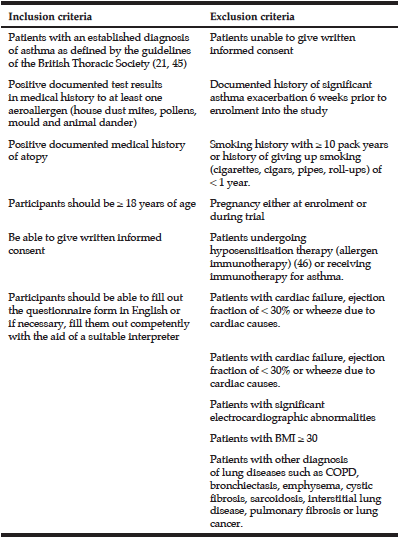
Inclusion criteria were women aged 50 to 90 years, who were current citalopram or mirtazapine users, or individuals who were going to initiate treatment of either drug within the following two months due to a diagnosis of major depressive disorder; or healthy controls, i.e. not depressed or receiving antidepressants. Due to few participants (n=3) in the Mirtazapine group as originally planned, results from this group will not be presented. The exclusion criteria were: 1) Current or use within the past 6 months of drugs affecting bone turnover such as corticosteroids, hormone replacement therapy in postmenopausal women, drugs against osteoporosis or other bone diseases (Paget’s disease of bone), vitamin D supplementation >35 micrograms daily, depot Medroxyprogesterone Acetate (DMPA), Cyclosporine (CsA), Antiretroviral Therapy (ART). 2) Impaired renal function (serum creatinine > 150 micromolar/l). 3) Pregnant women. 4) Individuals diagnosed with cancer or a metabolic disorder such as diabetes. 5) Individuals with prosthetic material in hip or spine. 6) Individuals diagnosed with a disease that affects bone such as Paget’s disease of the bone, or fibrous dysplasia. 7) Individuals which is not considered eligible for the clinical trial e.g. individuals diagnosed with dementia, severely psychotic or depressed individuals. 8) Individuals that have been taking other antidepressants than citalopram or mirtazapine for more than 6 months prior to the inclusion and if this treatment persisted for a period of minimum 12 months. 9) Individuals who cannot stand up and stand still without support or a helping device due to physically impairment. 10) The presence of other pain problems (e.g. osteoarthritis) or sensory dysfunction (e.g. fibromyalgia, neuropathic pain).

Bone mineral density

Bone Densitometry (BMD) of total hip, femoral neck, and lumbar spine (L1-L4) was assessed for all subjects at baseline and after 12 months using by dual-energy x-ray absorptiometry (DXA) scans on a GE Lunar Prodigy Scanner (GE Healthcare Lunar prodigy, USA). Quality control procedures were run every day including calibration by use of QA Phantom (Lunar DPX Series QC Phantom and a Block Phantom). The coefficient of variation (CV) between the two scanners was 1%.

Biochemical measures

Samples were immediately frozen at -80 °C. Serum vitamin D [total serum 25-hydroxyvitamin D (25(OH)D)] was measured using an electrochemiluminescence-binding assay ECLIA (Elecsys® Vitamin D total, modular analytics E170). CV was 1.7-7.8 % for intra-assay and CV 2.2-10.7 % for inter-assay variation.
Procollagen type 1 N-terminal peptide (P1NP), Tartrate-resistant phosphatase type 5b (TRAP5b – iSYS), and intact PTH were determined using an iSys machine and kits from IDS plc. Intact PTH was measured using the IS3200 and control kit (detection limit 5 pg/ml, CV 4.1-8.2%). P1NP was measured using the IS4000 and control kit (detection limit 2 ng/ml, CV4,4-5.3%). TRAP5b was measured using the IS4100 and control kit (detection limit 0.9 IU/l, CV 5.0-13.6%).

Self-rated questionnaires

All subjects filled out questionnaires at each visit at the clinic including information regarding mental and physical health status: Major Depression Inventory (MDI) (18), and EQ-5D (19).

Postural control

Subjects were tested during quiet bipedal stance on a force platform (Plux Biosignals S.A, Arruda dos Vinhos, Portugal) in four different sensory conditions: i) eyes open, standing on a firm surface, ii) eyes closed on firm surface, iii) eyes open on soft surface, iv) eyes closed on soft surface, each lasting for 35 seconds. One trial consisted of the four sensory conditions i) to iv) recorded in the same order and in one sequence, repeated three times separated by short breaks (30-60 seconds) in between. For the soft surface condition, a soft foam pillow (O’live Balance pad, Denmark) was placed on top of the force platform. Average parameters from the three trials, for each of the four sensory conditions, were used for analysis. The center of pressure (CoP) position was estimated from the vertical force data extracted from the force plate. Coefficient of variation (CV) for the four different conditions was determined in a pilot study of seven healthy adults (aged 24-31years) and was <8.1% for both CoP range and velocity in the anterior-posterior and medial-lateral direction.

Timed up and go test (TUG)

The subject started from a sitting position in an arm chair (seat height approximately 43-47 cm) and were asked to rise from the chair on an “three, two, one, go” start-signal and walk three meters (marked on the floor), and walk back to the chair and sit down again. Time was recorded manually from the “go” signal and stopped when the subject again was sitting in the chair. The test was repeated three times and the best (fastest) selected for further analysis. CV was calculated in a pilot study on healthy adults (n=8), aged 24-53 years, and was 3.7%.

Handgrip strength

Isometric handgrip strength was measured using a hand dynamometer (NC70144, Procare.dk, Denmark). Each subject was instructed to perform maximal contraction force with their dominant hand (defined as their “writing hand”), in a seated upright position and the test hand pointing downwards, parallel with the trunk and unsupported. The test was repeated three times. The maximal strength of the three trials was used for further analysis. CV for handgrip strength was 2.9% in a pilot study of healthy adults (n=6) aged 17-55 years.

Cutaneous pinprick sensitivity

The pinprick test is conducted by use of eight metallic weight calibrated pins, flat contact area, diameter of 0.6 mm, with fixed stimulus intensities (weights: 0.8, 1.6, 3.2, 6.4, 12.8, 25.6 and 60.0 g, Aalborg University, Denmark). The test was conducted in a single point on the skin above the left upper trapezius muscle. Pinprick score was defined as the lightest weighted needle, which consistently elicits a sharp or pricking sensation. The average of the three pinprick scores was used in further analyses.

Computer controlled cuff pressure algometry

Pressure-induced pain cuff stimulation was induced using computerised cuff algometry (NociTech, Denmark, and Aalborg University, Denmark). Zero cm on the VAS was defined as “no pain” and 10 cm as “maximal pain”. Three different experimental setups were conducted in the following order: Pressure pain detection and tolerance thresholds, temporal summation of pain (TSP), and conditioning pain modulation (CPM).
One cuff was placed around the left lower leg at the belly of the m. gastrocnemius and inflated to a maximum pressure limit of 100 kPa. During cuff inflation, the subjects were instructed to continuously rate their pressure-induced pain intensity on the VAS, from the time at which the pressure was perceived as painful, and pressing the handheld button when the pain became intolerable, defined as the cuff Pressure Pain Tolerance Threshold (cuff PTT). If the subject did not reach their pain tolerance level before the maximum stimulation intensity, 100 kPa was used as an estimate for the cuff PTT value. The pressure pain detection threshold (cPDT) was defined as the pressure at which the VAS score exceeded 1 cm [20, s.]. Cuff stimulation was repeated three times, separated by a two-minute resting interval and the mean cuff PDT and PTT were used in further calculation.
TSP was evaluated by 10 sequential stimuli delivered at 0.5 Hz at the intensity of 75% of PTT. Pain intensity was rated continuously on the VAS during the sequential stimuli, and a total of 10 VAS scores were extracted. The test was repeated twice separated by a minimum one-minute break, and the 10 VAS score means were used in the analysis. For analysis of TSP, the mean VAS score was calculated from the first to the 4th stimulus (VAS-I) and from the 7th to the 10th stimulus (VAS-II). The TSP-effect was defined as the difference between VAS-I and VAS-II (i.e. VAS-II minus VAS-I) [21].
CPM was assessed by applying a conditioned stimulus followed by a test stimulus. The conditioning stimulation was applied through a computer-controlled cuff stimulation, where a constant pressure 30 kPa with one tourniquet cuff placed around the belly of the m. gastrocnemius on the contralateral lower leg (right side) acted as a conditioning stimulus. During the cuff test stimulus, cuff PDT and cuff PTT were reassessed. PDT was extracted and the CPM effect was calculated as the difference between the unconditioned and the conditioned test stimulus (i.e. PDTconditioned minus PDTunconditioned).

Power calculation

With a one-year change in BMD of 2% and a SD of 2%, 21 patients were desirable in each group (42 in total) with a risk of 5% for type 1 error and 10% for type 2 error. Due to potential drop outs, the number was set at 50 (25 in the active and 25 in the placebo group).
Primary outcome measures were plasma vitamin D, BMD by DXA of the lumbar spine, femoral neck, and total hip.

Statistical analyses

Results were reported as means ± standard error of the mean (SEM). Differences in baseline characteristics comparing citalopram users and controls and vitamin D treatment versus placebo treatment were tested using t-tests for independent samples, Fisher’s Exact test and Chi-Square Test for Independence as appropriate. To test for significant changes in major outcome variables (BMD and biochemistry) among groups (patients/controls) and according to treatment (vitamin D/placebo) during the 12 months follow-up period, a three-way mixed repeated-measures analysis of variance (ANOVA) (time x group x treatment) was used, controlling for both age and BMI. All post-hoc tests were adjusted for multiple comparisons (Bonferroni). Pearson’s coefficients was used to examine correlations between the 12 months difference in 25(OH)D and the major covariates (e.g. citalopram dose or MDI score). All analyses were performed using SPSS Statistics for Windows, version 24.0 (Armonk, NY: IBM Corp).


Results

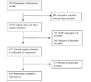
A total of 21 patients treated with citalopram and 50 controls met the inclusion criteria and completed all baseline measures (Fig. 1). No serious adverse events were recorded during the trial in neither the vitamin D, nor the placebo group.
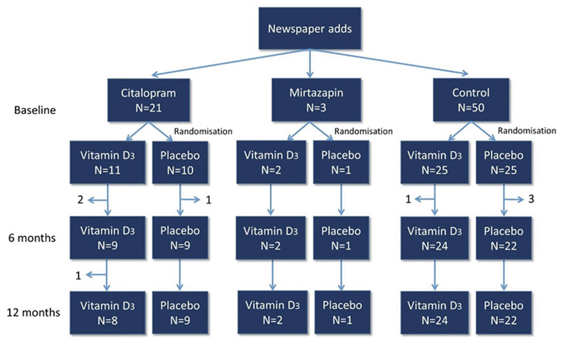
Figure 1 Flow-chart of inclusion

Figure 1
Flow-chart of inclusion

Baseline characteristics

Table 1 shows baseline characteristics. Average age and systolic blood pressure was higher among patients compared to controls (62.6±1.6 versus 57.1±0.8 years, P=0.001) and (139.6±4.2 versus 126.4±2.0 mmHg, P=0.009), respectively. Moreover, alcohol consumption per month was borderline higher among patients compared to controls (43±7 versus 28±3 units/month, P=0.054). There was no difference in the amount of vitamin D or calcium supplement use at inclusion (normal daily use) at baseline within or between the groups.
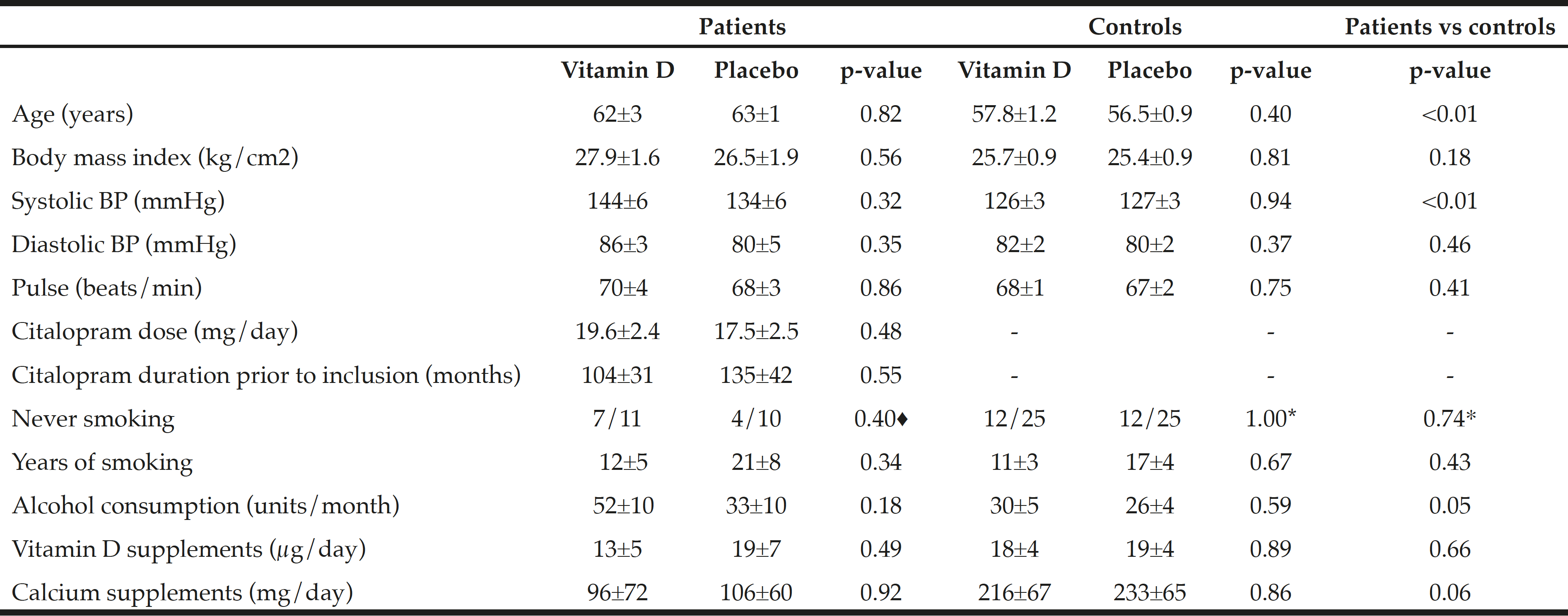
Table 1 Baseline characteristics. Mean and SEM or proportion

Table 1
Baseline characteristics. Mean and SEM or proportion

♦ Fisher’s exact test, * Chi square test


Serum Vitamin D

Figure 2 shows the calcitropic hormones. Serum 25(OH)D levels increased more among the vitamin D treated than among the placebo treated for both the citalopram group and the control group. The increase in serum 25(OH)D levels was higher in the vitamin D treated citalopram users (+57.3±14.5 nmol/l) than among the controls (+36.0±4.3 nmol/l, p=0.03), Fig. 2.
A three-way mixed repeated-measures ANOVA with time (baseline, 6 and 12 months follow-up) as within-subject factors and group (patient and control) and treatment (vitamin D and placebo) as between-subject factors, adjusting for age and BMI, showed a significant main effect of treatment on serum 25(OH)D levels after 12 months: F (2, 114) 44.86, p<0.001, ηp2=0.44.
iPTH decreased more with vitamin D treatment after 12 month among the citalopram treated (change during 12 months -8.4±6.1 pg/ml with vitamin D vs. +6.6±5.5 pg/ml with placebo) than among the controls (-4.2±2.3 vs. +0.8±2.3 pg/ml, p<0.01), Fig. 2.
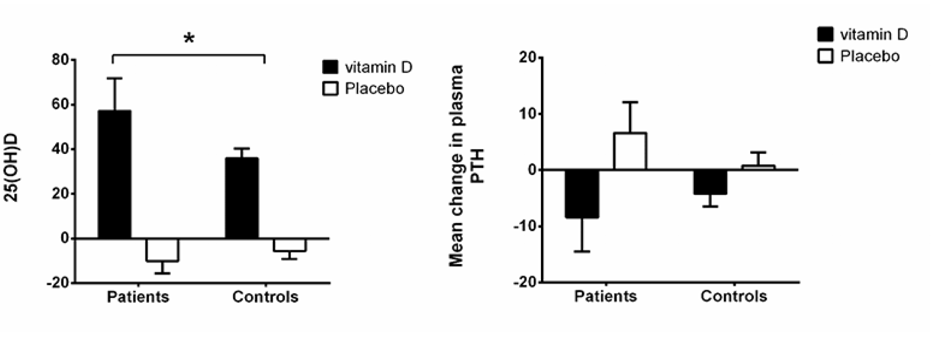
Figure 2 Absolute changes in calcitropic hormones (25-OH-vitamin D and PTH) from baseline to 12 months among citalopram treated and controls stratified by treatment group (vitamin D 50 µg or placebo)

Figure 2
Absolute changes in calcitropic hormones (25-OH-vitamin D and PTH) from baseline to 12 months among citalopram treated and controls stratified by treatment group (vitamin D 50 µg or placebo)

* p<0.05 by one-tail t-test


Measures of degree of depression

Baseline MDI total score was higher among the patients compared to the healthy controls (P<0.01, and remained different at 6 and 12 months follow-up, by ANOVA (P<0.01, Fig. 3A). Among the controls, a significant lower MDI score was rated among the vitamin D treated compared to placebo treated at six months follow-up (p=0.01 by t-test for two samples), which became non-significant again at 12 months (p=0.15 by t-test for two samples).
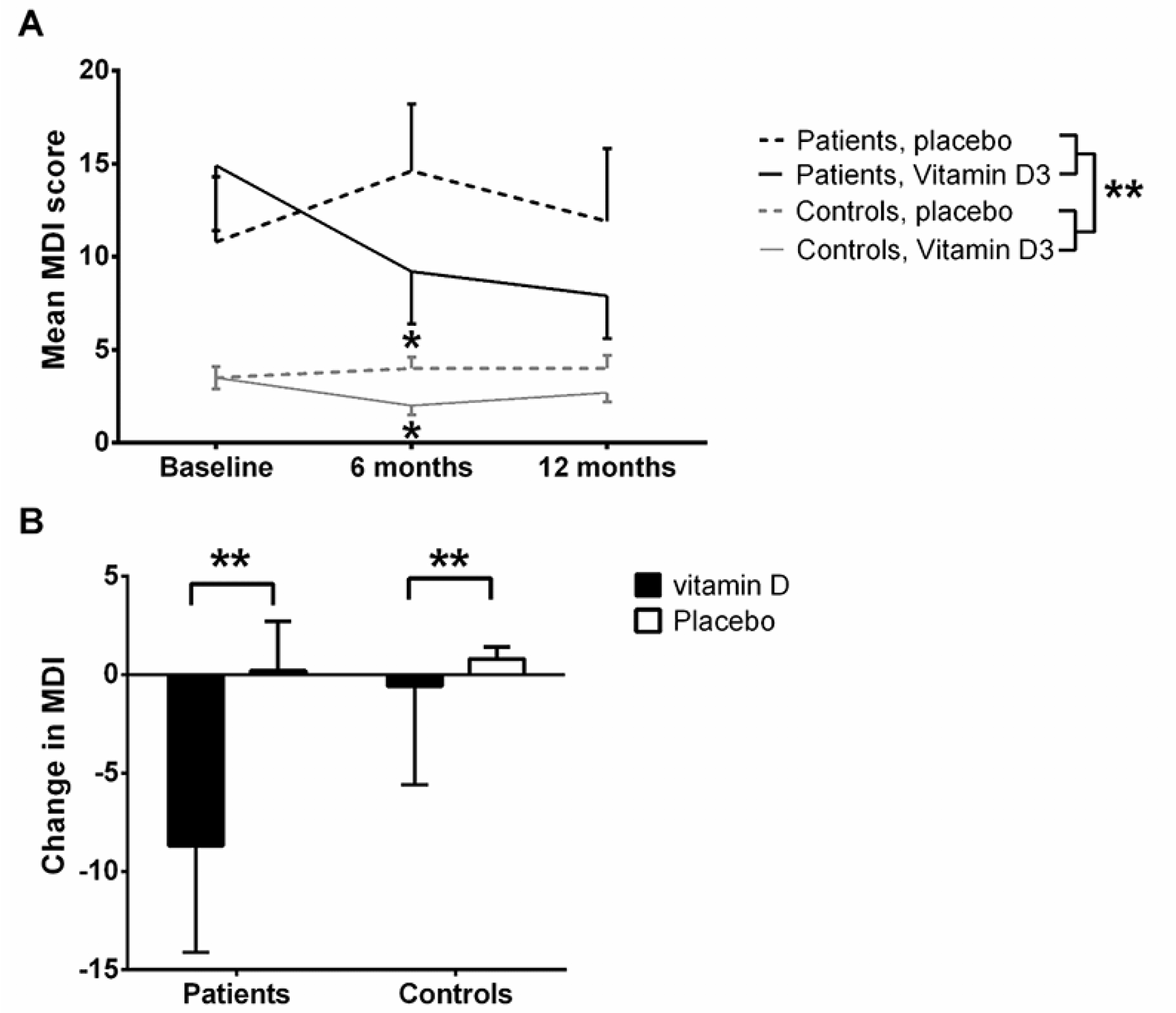
Figure 3 MDI score over time in patients and controls by treatment (vitamin D vs. placebo)

Figure 3
MDI score over time in patients and controls by treatment (vitamin D vs. placebo)

*P<0.05, **P<0.01; A: Absolute MDI total score over time (lower MDI total score is better self-perceived mental health). By ANOVA, no difference between vitamin D and placebo treated (p=0.39) was present, whereas a significant difference was present between patients and controls (p<0.01); B: Change in MDI depression score (delta MDI total score) from baseline to one year stratified by citalopram treated and controls randomised to vitamin D or placebo. Mean and SEM for changes.


An analysis of the changes in total MDI score over 12 month (delta MDI total score) revealed an improvement among vitamin D treated patients after 12 months as compared to placebo treated patients (mean improvement in MDI score when treated with vitamin D was -8.7±5.4 vs. a worsening with placebo of 0.2±2.5 equalling a mean difference of 8.9±2.7, p<0.01 by standard normal distribution) (Fig. 3B). A difference was also present for the controls (difference in MDI score: -0.6±0.5 vs. 0.8±0.6 equalling a mean difference of 1.4±0.3, p<0.01 by standard normal distribution – Fig. 3B). The symptoms that tended to improve were primarily the more severe, including all three core-symptoms of depression: feeling sad and sorry, lack of interest in daily activities, lack of energy, and the associated symptoms: decreased self-confidence, bad conscience, feeling restless, and to some degree that life was not worth living, difficulties concentrating, and decreased appetite (data not shown).
Overall, for patients and controls a positive correlation was present between changes in plasma 25(OH)D and changes in MDI total scores after 12 months of vitamin D treatment, with higher plasma 25(OH)D correlated to lower MDI scores (i.e. fewer depressive symptoms). This was significant among controls (Pearson’s r=0.-43, p<0.01), but not among the citalopram treated.

Handgrip strength

Fig. 4 shows that after 12 month handgrip strength was significantly higher among the patients receiving vitamin D supplement compared to the patients receiving placebo (31.1 vs. 25.2 kg, P=0.03). Among the controls, no differences were present after 6 and 12 months. There were no correlations between the improvements in MDI and handgrip strength.
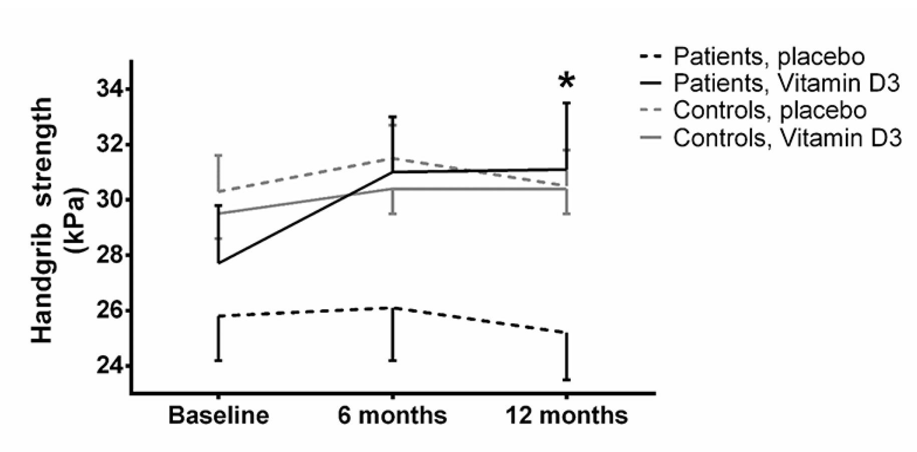
Figure 4 Handgrip strength. Values are mean and SEM. Dashed line is placebo, continuous line is vitamin D. Black line is controls, grey line are patients

Figure 4
Handgrip strength. Values are mean and SEM. Dashed line is placebo, continuous line is vitamin D. Black line is controls, grey line are patients

*: p<0.05, when comparing vitamin D with placebo treatment at 12 months


Timed Up and Go

At baseline the TUG performance was significantly inferior (i.e. longer time needed to complete the test) among the patients when compared to controls (P=0.01). After 6 and 12 months this difference became less pronounced (P=0.03 and P=0.08).

Postural control and clinical measures

The changes in parameters of postural control as well as blood pressure and pulse did not differ between vitamin D and placebo treated neither among patients with major depressive disorder on citalopram nor among the healthy controls (data not shown). Body weight and height also remained unchanged between all groups.

Pain measures

At baseline patients experienced a higher level of self-perceived pain in daily life (higher EQ-5D pain rating) compared to controls (1.75 ± 0.14 and 1.24 ± 0.06, respectively, P<0.01) with no effect over time.
No significant effect of vitamin D3 treatment was present for cuff PDT and cuff PTT (t-tests, p>0.05 and ANOVA, p>0.05 – data not shown). Baseline TSP and CPM did not differ significantly when comparing patients and controls. No significant effect from 12-months vitamin D3 vs. placebo treatment could be detected on TSP and CPM (t-tests, p>0.05 and ANOVA, p>0.05).

BMD and bone turnover markers

The changes in BMD and bone turnover markers (TRAB5b and P1NP) did not differ between vitamin D and placebo treated neither among citalopram treated patients nor among the healthy controls.


Discussion

In this study, it was demonstrated shown that vitamin D may improve depressive symptoms in patients treated with citalopram, and improve handgrip strength.

Vitamin D and depression

The improvement in delta MDI total score among the patients treated with vitamin D after 12 months, suggest a potential beneficial effect that may be used as a simple and effective supplementary treatment for depression. This is supported by results from recent studies (11). The correlation between improvements in the MDI total score and increased plasma 25(OH)D for all subjects further indicates vitamin D as a likely contributor to improvement in mental health. This is in line with a large prospective cohort study of non-depressed women aged 55 to 69 years, who observed a significantly lower mental health-related quality of life when consuming <400 IU/day [<10 µg/day] of vitamin D compared to those who consumed ≥400 IU/day (22).
The absolute decrease of around nine of 50 possible points in the individual depressive symptoms is a rather large absolute gain in well-being among the patients, and it may thus have a rather large clinical importance if confirmed in further studies. Furthermore, it is interesting, that the symptoms that tended to improve were primarily the more severe, including all three core-symptoms of depression: feeling sad and sorry, lack of interest in daily activities, lack of energy, and the associated symptoms: decreased self-confidence, bad conscience, feeling restless, and to some degree that life was not worth living, difficulties concentrating, and decreased appetite. Also among the controls, a trend of fewer depressive symptoms was seen among vitamin D treated compared to placebo.

Potential cytochrome p450 (CYP)-enzyme interaction for citalopram and vitamin D

The higher increase in serum 25(OH)D among citalopram treated patients compared to controls may be caused by an interaction between citalopram and the CYP system. Hence citalopram may occupy the CYP system preventing this from turning 25-OH Vitamin D into 24,25-OH-vitamin D, instead facilitating the formation of 1,25-OH2-Vitamin D and thus a higher biological effect mirrored in the trend towards a larger decrease in PTH. Citalopram is metabolised by the CYP2C19 system, the inactive 25,25-dihydroxy vitamin D is metabolised by the CYP24A1 enzyme, and 1,25-dihydroxy vitamin D is metabolised by the CYP27B1, while 25-hydroxy-vitamin D is metabolised from cholecalciferol by CYP27A1 [23]. Cholecalciferol may interact with CYP2C19 [24], and citalopram may thus potentially interact with cholecalciferol metabolism. Both CYP27B1 and CYP24A1 are expressed in the brain tissue suggesting that there may exist both synthesis and elimination, respectively, of vitamin D3 in the brain (25).
Voshaar et al. (26) investigated serum vitamin D levels among depressed elderly patients treated with antidepressants (n=355) compared to non-depressed control subjects (n=124). In contrast to the present study, they reported that TCA usage correlated with low 1,25-(OH)2 vitamin D3 levels, but not 25(OH)D, and neither SSRI nor newer types of antidepressants did effect any of those two compounds (26). This may be explained by possible different interactions between CYP enzymes and antidepressants during low basal 25(OH)D levels as compared to higher 25(OH)D levels induced by cholecalciferol supplements. It should be noted that baseline 25(OH)D levels was relatively higher among citalopram treated patients of the present study, than that reported in the study by Voshaar and colleagues (26).

Grip strength, effect from vitamin D supplementation

Despite the fact that patients of the present study were vitamin D replete (mean 25(OH)D ≥86 nmol/L) at baseline, an improvement in handgrip strength was found among patients receiving 2000 IU vitamin D supplement during the one year follow-up period compared to the placebo group. Actually – despite being older and thus having a priori lower hand grip strength – the patients on citalopram improved their hand grip strength to that of the controls. Currently, the effect of Vitamin D on muscle strength is controversial. A recent study report a decrease in hand grip strength in elderly vitamin D-sufficient women following vitamin D supplementation (27). However, this study (27) reports a relatively short follow up of three months whereas the present study has a longer follow up, which may suggest a dynamic in the effect of Vitamin D; vitamin D at steady state may improve muscle strength. However, short-term supplementation with no steady state may decrease muscle strength. Improved well-being from diminished depressive symptoms may potentially lead to increased physical activity and thus increased strength. However, no correlations were present in our study between improvements in MDI and handgrip strength.

Vitamin D, pain and pain sensitivity

The patients had higher levels of self-perceived pain at baseline compared to controls. Despite this, no differences were found in pain sensitivity measures between patients and controls and no changes were observed following treatment.
High clinical pain ratings and long painful durations (years) have been found associated with increased pain sensitivity and this has been documented in multiple painful conditions (28). Pain sensitivity is influenced by multiple factors and cognitive factors and pain sensitivity have been found associated in recent studies (28). Duloxetine (an antidepressant) has demonstrated analgesic effects in chronic pain patients (28) and has been suggested to target the pain inhibitory pathways (10) suggesting a link between depression and pain sensitivity. Despite this, the clinical pain ratings in the current study were low, which could explain why patients treated with anti-depressants and controls were not found different in regards to the pain sensitivity measures. The vitamin D-induced analgesic effect of antidepressants, may be an involvement of vitamin D in neuronal functioning through production and/or regulation of various monoamines including serotonin and noradrenaline (29) and neurotrophic factors with the potential to alter pain sensitivity. Serotonin and noradrenaline are mainly described to be involved in pain inhibitory pathways (30) but the current study was unable to demonstrate a significant change in pain inhibitory pathways after treatment, which might be due to the lack of impairment in the pain inhibitory pathways at baseline.

Strengths and limitations

The main advantage of the present randomized double-blinded placebo controlled study was the homogeneous group of subjects using only one type of antidepressant (citalopram), i.e. confounding from multiple drugs was avoided. Furthermore, a standardized dose of vitamin D that induced a significant increase in serum vitamin D was used, which was raised from within the normal level to the upper range of the normal level, i.e. it was possible to study the effects of high levels of vitamin D, and also at these levels no effects were seen.
The main limitation of our study was the low number of patients in the patient group and that no males were studied.


Conclusion

In conclusion, vitamin D may improve depressive symptoms in patients treated with citalopram, and potentially improve handgrip strength. However, further research on larger cohorts is needed to clarify the clinical implications.


Funding: Peter Vestergaard reports grants from The Obel Family Foundation (#25243), grants from The AP Møller and Chastine Maersk Mc. Kinny Møller Foundation (#01) during the conduct of the study; Thomas Graven-Nielsen and Kristian Kjær Petersen reports grants from The Danish National Research Foundation (#DNRF121), during the conduct of the study. The sponsors had no role in the design and conduct of the study; in the collection, analysis, and interpretation of data; in the preparation of the manuscript; or in the review or approval of the manuscript. Stine Aistrup Eriksen, Jakob Starup-Linde and Rogerio Pessoto Hirata has nothing to disclose. Therefore all authors: Stine Aistrup Eriksen, Jakob Starup-Linde, Rogerio Pessoto Hirata, Kristian Kjær Petersen, Thomas Graven-Nielsen and Peter Vestergaard report no conflicts of interests.

Acknowledgements: The Obel Family Foundation and the AP Møller and Chastine Maersk Mc. Kinny Møller Foundation are acknowledged for providing financial support. Center for Neuroplasticity and Pain (CNAP) is supported by the Danish National Research Foundation. The management and staff at Center for Clinical and Basic Research (CCBR) Aalborg, are acknowledged for providing optimal clinical facilities and professional execution of clinical measures (DXA and blood sampling), as well as continuous support and guidance in good clinical practice.

Conflict of interest: No conflicts of interests.

Ethical standard: The study followed the Helsinki declaration and all participants gave informed written consent.


References

1.    H. A. Bischoff-Ferrari m.fl., “Effect of Vitamin D on falls: a meta-analysis”, JAMA, bd. 291, nr. 16, s. 1999–2006, apr. 2004.
2.    L. Ceglia og S. S. Harris, “Vitamin D and its role in skeletal muscle”, Calcif. Tissue Int., bd. 92, nr. 2, s. 151–162, feb. 2013.
3.    C. Annweiler, A. M. Schott, G. Berrut, B. Fantino, og O. Beauchet, “Vitamin D-related changes in physical performance: a systematic review”, J Nutr Health Aging, bd. 13, nr. 10, s. 893–898, dec. 2009.
4.    S. W. Muir og M. Montero-Odasso, “Effect of vitamin D supplementation on muscle strength, gait and balance in older adults: a systematic review and meta-analysis”, J Am Geriatr Soc, bd. 59, nr. 12, s. 2291–2300, dec. 2011.
5.    B. Stubbs, L. Eggermont, S. Patchay, og P. Schofield, “Older adults with chronic musculoskeletal pain are at increased risk of recurrent falls and the brief pain inventory could help identify those most at risk”, Geriatr Gerontol Int, bd. 15, nr. 7, s. 881–888, jul. 2015.
6.    A. Fekadu, L. J. Rane, S. C. Wooderson, K. Markopoulou, L. Poon, og A. J. Cleare, “Prediction of longer-term outcome of treatment-resistant depression in tertiary care.”, The British journal of psychiatry : the journal of mental science, bd. 201, s. 369–75, nov. 2012.
7.    R. E. S. Anglin, Z. Samaan, S. D. Walter, og S. D. McDonald, “Vitamin D deficiency and depression in adults: systematic review and meta-analysis.”, The British journal of psychiatry : the journal of mental science, bd. 202, nr. 2, s. 100–7, feb. 2013.
8.    E. W. de Heer m.fl., “The association of depression and anxiety with pain: a study from NESDA”, PLoS ONE, bd. 9, nr. 10, s. e106907, 2014.
9.    J. F. Karp, J. Scott, P. Houck, C. F. Reynolds, D. J. Kupfer, og E. Frank, “Pain predicts longer time to remission during treatment of recurrent depression”, J Clin Psychiatry, bd. 66, nr. 5, s. 591–597, maj 2005.
10.    D. Yarnitsky, M. Granot, H. Nahman-Averbuch, M. Khamaisi, og Y. Granovsky, “Conditioned pain modulation predicts duloxetine efficacy in painful diabetic neuropathy”, Pain, bd. 153, nr. 6, s. 1193–1198, jun. 2012.
11.    J. Sarris m.fl., “Adjunctive Nutraceuticals for Depression: A Systematic Review and Meta-Analyses”, American Journal of Psychiatry, bd. 173, nr. 6, s. 575–587, jun. 2016.
12.    E. Garcion, N. Wion-Barbot, C. N. Montero-Menei, F. Berger, og D. Wion, “New clues about vitamin D functions in the nervous system.”, Trends in endocrinology and metabolism: TEM, bd. 13, nr. 3, s. 100–5, apr. 2002.
13.    R. von Känel, V. Müller-Hartmannsgruber, G. Kokinogenis, og N. Egloff, “Vitamin D and central hypersensitivity in patients with chronic pain”, Pain Med, bd. 15, nr. 9, s. 1609–1618, sep. 2014.
14.    M. Gaikwad, S. Vanlint, M. Mittinity, G. L. Moseley, og N. Stocks, “Does vitamin D supplementation alleviate chronic nonspecific musculoskeletal pain? A systematic review and meta-analysis”, Clin. Rheumatol., bd. 36, nr. 5, s. 1201–1208, maj 2017.
15.    Z. Wu, Z. Malihi, A. W. Stewart, C. M. Lawes, og R. Scragg, “Effect of Vitamin D Supplementation on Pain: A Systematic Review and Meta-analysis”, Pain Physician, bd. 19, nr. 7, s. 415–427, okt. 2016.
16.    S. Schwan og P. Hallberg, “SSRIs, bone mineral density, and risk of fractures–a review”, Eur Neuropsychopharmacol, bd. 19, nr. 10, s. 683–692, okt. 2009.
17.    P. Vestergaard, D. Prieto-Alhambra, M. K. Javaid, og C. Cooper, “Fractures in users of antidepressants and anxiolytics and sedatives: effects of age and dose”, Osteoporos Int, bd. 24, nr. 2, s. 671–680, feb. 2013.
18.    L. R. Olsen, D. V. Jensen, V. Noerholm, K. Martiny, og P. Bech, “The internal and external validity of the Major Depression Inventory in measuring severity of depressive states”, Psychol Med, bd. 33, nr. 2, s. 351–356, feb. 2003.
19.    S. J. Coons, S. Rao, D. L. Keininger, og R. D. Hays, “A comparative review of generic quality-of-life instruments”, Pharmacoeconomics, bd. 17, nr. 1, s. 13–35, jan. 2000.
20.    T. Graven-Nielsen, H. B. Vaegter, S. Finocchietti, G. Handberg, og L. Arendt-Nielsen, “Assessment of musculoskeletal pain sensitivity and temporal summation by cuff pressure algometry: a reliability study”, Pain, bd. 156, nr. 11, s. 2193–2202, nov. 2015.
21.    K. K. Petersen, T. Graven-Nielsen, O. Simonsen, M. B. Laursen, og L. Arendt-Nielsen, “Preoperative pain mechanisms assessed by cuff algometry are associated with chronic postoperative pain relief after total knee replacement”, Pain, bd. 157, nr. 7, s. 1400–1406, jul. 2016.
22.    S. Motsinger, D. Lazovich, R. F. MacLehose, C. J. Torkelson, og K. Robien, “Vitamin D intake and mental health-related quality of life in older women: the Iowa Women’s Health Study”, Maturitas, bd. 71, nr. 3, s. 267–273, mar. 2012.
23.    D. E. Prosser og G. Jones, “Enzymes involved in the activation and inactivation of vitamin D”, Trends Biochem. Sci., bd. 29, nr. 12, s. 664–673, dec. 2004.
24.    H. Yamazaki og T. Shimada, “Effects of arachidonic acid, prostaglandins, retinol, retinoic acid and cholecalciferol on xenobiotic oxidations catalysed by human cytochrome P450 enzymes”, Xenobiotica, bd. 29, nr. 3, s. 231–241, mar. 1999.
25.    L. R. Harms, T. H. J. Burne, D. W. Eyles, og J. J. McGrath, “Vitamin D and the brain”, Best Pract. Res. Clin. Endocrinol. Metab., bd. 25, nr. 4, s. 657–669, aug. 2011.
26.    R. C. Oude Voshaar, W. J. Derks, H. C. Comijs, R. A. Schoevers, M. H. de Borst, og R. M. Marijnissen, “Antidepressants differentially related to 1,25-(OH)₂ vitamin D₃ and 25-(OH) vitamin D₃ in late-life depression”, Transl Psychiatry, bd. 4, s. e383, apr. 2014.
27.    L. S. Bislev, L. Langagergaard Rødbro, L. Rolighed, T. Sikjaer, og L. Rejnmark, “Effects of Vitamin D3 Supplementation on Muscle Strength, Mass, and Physical Performance in Women with Vitamin D Insufficiency: A Randomized Placebo-Controlled Trial”, Calcif. Tissue Int., jun. 2018.
28.    L. Arendt-Nielsen m.fl., “Assessment and manifestation of central sensitisation across different chronic pain conditions”, Eur J Pain, bd. 22, nr. 2, s. 216–241, 2018.
29.    E. A. Shipton og E. E. Shipton, “Vitamin D and Pain: Vitamin D and Its Role in the Aetiology and Maintenance of Chronic Pain States and Associated Comorbidities”, Pain Res Treat, bd. 2015, s. 904967, 2015.
30.    K. Bannister og A. H. Dickenson, “What the brain tells the spinal cord”, Pain, bd. 157, nr. 10, s. 2148–2151, 2016.

SERUM 25-HYDROXY VITAMIN D, PHYSICAL ACTIVITY AND COGNITIVE FUNCTION AMONG OLDER ADULTS


H. Eymundsdottir1,2, M. Chang2,3, O.G. Geirsdottir1,2, P.V. Jonsson2,4,5, V. Gudnason4,6, L. Launer7, M.K. Jonsdottir8,9, A. Ramel1,2


1. Faculty of Food Science and Nutrition, University of Iceland, Reykjavik, Iceland. 2. The Icelandic Gerontological Research Center, The National University Hospital of Iceland, Reykjavik, Iceland; 3. Sports Science, School of Science and Engineering, Physical Activity, Physical Education, Health and Sport (PAPESH) Research Centre, Reykjavik University; 4. Faculty of Medicine, University of Iceland; 5. Department of Geriatrics, The National University Hospital of Iceland, Reykjavik, Iceland; 6 Icelandic Heart Association, Kopavogur, Iceland; 7 Laboratory of Epidemiology and Population Sciences, National Institute on Aging, National Institutes of Health (NIH), Bethesda, Maryland, USA; 8 Department of Psychology, Reykjavik University, Reykjavik; 9 Mental Health Services (Memory Clinic) Landspitali – The National University Hospital of Iceland.

Corresponding Author: Hrafnhildur Eymundsdottir, MPH. The Icelandic Gerontological Research Center, The National University Hospital of Iceland, Tungata 26, 101 Reykjavik, Iceland, (+354 8457415), E-mail hre6@hi.is

J Aging Res Clin Practice 2018;7:143-148
Published online October 15, 2018, http://dx.doi.org/10.14283/jarcp.2018.24


Abstract

Objective: To investigate the association between 25-hydroxy vitamin D (25OHD) and cognitive function with particular consideration of physical activity (PA) in Icelandic older adults. Design: Cross-sectional study. Setting: Iceland. Participants: Old adults aged 65-96. The final analytical sample included 4304 non-demented participants. Measurements: Serum 25OHD was categorized into deficient (≤ 30 nmol/L, 8%), insufficient (31-49 nmol/L, 25%) and normal-high levels (>50 nmol/L, 67%). Cognitive function assessments included measurements of memory function (MF), speed of processing (SP) and executive function (EF) all categorized as low and high (divided by 50th percentile). Multivariate logistic regression analysis was used to calculate the odds ratio (OR) for having high cognitive function. Results: Serum 25OHD was positively associated with cognitive function. Adjustment for PA and other potential confounders diminished this association only partially. Compared to participants with normal-high levels of 25OHD, those with deficient levels had decreased odds for high SP (OR: 0.74, CI: 0.57-0.97), high MF (OR: 0.55; CI: 0.43-0.71) and high EF (OR: 0.76, CI: 0.57-1.0). Conclusion: Serum 25OHD below ≤30 nmol/L was associated with decreased odds for high cognitive function among community dwelling old adults as compared to those with 25OHD above > 50 nmol/L. Neither PA nor other potential confounders explained the associations between 25OHD and cognitive function. Future studies should explore mechanisms and the potential clinical relevance of this relationship.

Key words: Vitamin D, memory function, speed of processing, executive function.


Introduction

Serum 25-hydroxy vitamin D (25OHD) has been recognized as crucial for maintaining calcium and phosphate homeostasis which is important for bone health (1). Further, epidemiological studies have indicated associations between low 25OHD levels and a variety of chronic illnesses including type 1 and type 2 diabetes (2), autoimmune diseases and liver disease (3, 4) In recent years there has been a growing interest in the potential role of vitamin D in cognitive function (5-9). Older adults are at a higher risk for 25OHD deficiency than younger people (10) and several studies have reported a positive correlation between 25OHD and cognitive function in this vulnerable group (6, 8, 9).
In general, aging is associated with reduction in brain tissue volume with concomitant declines in cognitive function (11). Impairment in cognitive function has also been linked to lifestyle factors, such as physical inactivity in older adults (12, 13). Evidence from physical activity (PA) intervention studies also indicates that PA might have a role in preserving cognitive function among older individuals (14).
Interestingly, low PA has also been associated with low levels of serum 25OHD (15), possibly related to the lack of sunlight exposure. Since several previous studies, that have investigated serum 25OHD and cognitive function, have not used statistical correction for PA (16, 17), it remains unclear whether the association between 25OHD and cognitive function among older adults is direct or whether it is mediated by PA.
The aim of the current study was thus to investigate the cross-sectional associations between 25OHD and cognitive function with particular consideration of the potentially mediating effects of PA using data from the Age Gene/Environment Susceptibility-Reykjavik Study (AGES-Reykjavik), a large population based cohort of older adults living at a northern latitude.


Subjects and methods

Study population

The AGES-Reykjavik Study (AGES-RS) examined risk factors for diseases in old age, including environmental factors and genetic susceptibility, and their interactions. Briefly, the AGES–RS is a continuation of the Reykjavik Study in Iceland. The Reykjavik Study was initiated in 1967 by the Icelandic Heart Associations and included men and women born in 1907–1935 living in the Reykjavik area [18]. During 2002 – 2006, 5764 persons randomly chosen from survivors of the Reykjavik Study cohort were re-examined for the AGES–RS. Participants completed a questionnaire, underwent a clinical examination, and completed a cognitive test battery. Details on the study design and the baseline AGES-RS assessments have been given elsewhere (19). The study was approved by the National Bioethics Committee in Iceland (approval VSN-00-063), The Data Protection Authority, and by the National Institute on Aging Intramural Institutional Review Board. Written informed consent was obtained from all participants.

Serum 25-hydroxy vitamin D measurement

The accredited IHA laboratory performed 25OHD measurements in batch using unfrozen serum samples and the Liaison chemiluminescence immunoassay (DiaSorin Inc, Stillwater, Minnesota). The inter-assay coefficient of variation was < 6.5 % when calculated data are from measurements using a frozen serum pool as the control sample and < 12.7 % when calculated data is from measurements using Liaison quality controls. Existing serum 25OHD levels were then standardized according to the international Vitamin D Standardization Program (VDSP) as previously described (20). Standardized serum 25OHD was used as categorized variable in statistical analyses based on Guidelines for Health Professionals from the National Institutes of Health (2014) (21). The cut points of serum 25OHD used in this study were as follows: deficient (≤ 30 nmol/L), insufficient (31-49 nmol/L), normal-high levels (≥ 50 nmol/L).

Cognitive function assessment and dementia

Assessment of cognitive function included nine tests, or test components, focusing on three cognitive domains, i.e., memory, processing speed and executive function. For each of the domains, a composite score was constructed based on a theoretical grouping of the tests and by converting raw scores into standardized z scores reflecting the distribution within the study sample. The inter-rater reliability for all tests was excellent (Spearman correlation coefficients range 0.96–0.99) (22).
The memory composite measure included the immediate and delayed-recall portions of a modified version of the California Verbal Learning Test (23) . The processing speed composite measure included the Digit Symbol Substitution Test (24), the Figure Comparison Test (25) and the Stroop Test (26) Part I (reading) and Part II (color naming). The executive function composite measure included the Digits Backward Test (24), a shortened version of the CANTAB Spatial Working Memory test (27)and the Stroop Test, Part III (word-color interference). The three domains of memory, processing speed and executive function composite measures were each categorized into low (lower 50%) and high (higher 50%) making each domain a binary variable.
A consensus diagnosis of dementia made by a team composed of a geriatrician, neurologist, neuropsychologist, and a neuroradiologist was made according to international guidelines from the Diagnostic and Statistical Manual of Mental Disorders, Fourth Edition (28).

Covariates

A number of covariates were included in the present study including demographic, anthropometric, lifestyle, laboratory and disease-related variables:
Education was categorized into four levels (elementary school, high school, undergraduate, more than undergraduate education).
Body mass index (BMI) was calculated as kg/m2. Smoking status was evaluated as ever vs. never smoker. Alcohol consumption was evaluated as either currently consuming vs. not consuming. Cod liver oil consumption (never, less than once a week, 1-6 times a week, daily) and multivitamins (yes/no) were assessed via questionnaire.
Leisure time physical activity was assessed by a self-reported questionnaire. Participants were asked, how many hours per week they participated in moderate/vigorous intensity physical activity in the past 12 months. Predefined answer categories were never, rarely, weekly but <1 hour per week, 1–3 hours per week, 4-7 hours per week and more than 7 hours per week. In final analysis physical activity categories were combined into 1. none, 2. ≤ 3 hours/week or 3. > 3 hours/week.
Participants were instructed in advance to bring all medication they had used during the preceding two weeks before the clinic visit and were categorized into ≤ 4 medication vs. ≥ 5 medication. Diabetes mellitus was defined by a physician’s diagnosis of diabetes, use of diabetes medication and/or fasting blood glucose of >7.0 mmol/L. Hypertension was defined at baseline by a physician’s diagnosis of hypertension, use of hypertension medications and/or blood pressure above 140/90 mm Hg.
A high level of depressive symptoms was classified as a score of ≥ 6 on the 15-item Geriatric Depression Scale (29). APOE alleles were genotyped on a subsample of 2113 people using standard methods (30). APOE genotypes were grouped as APOE ε4 carrier (ε3/4, and ε4/4 genotype) and APOE ε4 non-carrier (ε2/2, ε2/3and ε3/3).

Analytical Sample

Of the total cohort (N=5764), 5519 had measurement of serum 25OHD, and 4699 of those had complete data on cognitive function. All subjects with dementia diagnosis (n=180, 3.1%) and APOE genotypes ε2/4 (n=115, 2.0%) were excluded from analysis. Participants with APOE genotypes 2/4 were excluded since the allele ε 2 and ε 4 have opposite effects on the risk for cognitive impairment and dementia[30]. The final sample having complete data, included 4304 participants (Figure 1). Compared with those who were not included in the analytical sample, those included were significantly younger (76.31±5.4 vs. 80.42±6.7 years, p < 0.001) and less likely to have diabetes (11.9% vs. 18.4 %, p < 0.001), but the two groups did not differ significantly by gender, blood pressure or medication use.

Statistical analysis

Statistical analyses were carried out using IBM SPSS version 22.0 (SPSS, Chicago, IL, USA). Differences between participants in the 25OHD- or PA-categories were calculated using chi-square test for categorical variables and analysis of variance (ANOVA) for continuous variables (normally distributed).
The associations between 25OHD and each of the three cognitive domains were examined using logistic regression models with a different degree of statistical correction. Therefore, the z scores of the three domains were converted into binary variables (low and high) using the 50th percentile as a cut point. Model 1 included 25OHD categories, age, sex and education; model 2 added PA; and model 3 additionally included BMI, medication use, diabetes, hypertension, depressive symptoms, alcohol consumption and smoking status as covariates. The level of statistical significance was set at p < 0.05.


Results

Deficient and insufficient levels of 25OHD were observed in 8% and 25% of the participants, respectively, whereas 67% had normal-high levels (Table 1). All of the lifestyle-, and disease-related variables were different between the three 25OHD groups. Cod liver oil and use of multivitamin supplements were associated with higher 25OHD.
Figure 2 shows the associations between cognitive domain scores and PA stratified by 25OHD. PA was associated with higher cognitive function in the normal-high and in the insufficient 25OHD category, however, these associations were less clear in the deficient category.

Figure 1 Flow chart

Figure 1
Flow chart

Figure 2 Standardized composite scores of each cognitive domain according to three categories of leisure time physical activity(PA); categorized as none, ≤ 3 hours/week or > 3 hours/week. The figures are stratified by three categories of serum 25OHD levels

Figure 2
Standardized composite scores of each cognitive domain according to three categories of leisure time physical activity(PA); categorized as none, ≤ 3 hours/week or > 3 hours/week. The figures are stratified by three categories of serum 25OHD levels

*Significantly different from the PA never group.

Table 1 Demographic and health characteristics according to serum 25-hydroxy vitamin D concentrations among AGES/Reykjavik participants

Table 1
Demographic and health characteristics according to serum 25-hydroxy vitamin D concentrations among AGES/Reykjavik participants

Data are shown as mean ± SD or as %; * Significant differences between the three 25OHD categories according to chi-square test for categorical variables and analysis of variance for continuous variables; 1. Hypertensive: systolic BP > 140 mmHg, diastolic BP > 90 mmHg or medication for hypertension; 2.. Diabetes mellitus was defined by physician’s diagnosis of diabetes or use of diabetes medication; 3 Standardized composite score


Results from the logistic regression analyses are shown in Table 2. We found that participants with deficient 25OHD levels were less likely (odds ratios between 0.52 – 0.76 depending on the statistical model) to have high cognitive function as compared to participants with normal-high 25OHD levels. Differences between participants with insufficient 25OHD and participants with normal-high 25OHD were not significant in the fully corrected models.
The differences in odds gradually diminished with additional covariate adjustment and thus the greatest differences between the three 25OHD categories were observed in the least corrected model 1. After additional adjustment for PA in model 2, the odds ratios for high function changed only marginally in the deficient group, e.g., from 0.58 to 0.61 for speed of processing. The changes were similar for the other domains.
In the fully corrected models the odds ratio for high function of the deficient group remained significantly lower for speed of processing (OR: 0.74, CI: 0.57-0.97) and memory function (OR:0.55, CI:0.43-0.71), but not in executive function (OR: 0.76, CI: 0.58-1.00).

Table 2 Odds Ratio* for high cognitive function dependent on three categories of 25-hydroxy vitamin D among AGES-Reykjavik participants

Table 2
Odds Ratio* for high cognitive function dependent on three categories of 25-hydroxy vitamin D among AGES-Reykjavik participants

* Odds ratio based on multivariate logistic regression analysis; CI = confidence interval; OR = odds ratio; Model 1: Adjusted for age, gender, education; Model 2: Adjusted for age, gender, education and physical activity; Model 3: Adjusted for age, gender, education, physical activity, body mass index, depression symptoms, medication, hypertension, diabetes, current smoking, and alcohol consumption


Discussion

In this large cross-sectional study we investigated the associations between 25OHD and cognitive function among older individuals living in Iceland with attention to the potentially mediating effect of PA. We found that participants with deficient levels of 25OHD were significantly less likely to have high cognitive functioning as compared to participants with normal-high levels. PA itself was significantly associated with cognitive function, which has been reported previously. It has been suggested that PA sustains cerebral blood flow by decreasing blood pressure, lowering lipid levels and inhibiting platelet aggregation (31). However, in our study PA did not seem to mediate the associations between 25OHD and cognitive function, as inclusion of PA as covariate in statistical models barely changed the outcomes.
In general, our participants had mean levels of serum 25OHD comparable to those reported in other northern latitude countries (32-35). When looking at the characteristics of our study sample, we found that participants with deficient 25OHD levels had unhealthier lifestyles and poorer health compared to participants with normal-high 25OHD. They smoked more frequently, had lower engagement in PA and a higher BMI. They also had a higher incidence of depressive symptoms, diabetes, but lower educational levels (Table 1).
For the interpretation of the results it is important to consider that the above mentioned lifestyle and health-related variables can act as confounders. As seen in our calculations, statistical correction for these variables attenuated the relation between 25OHD and cognitive function to a certain degree. However, the association remained significant for memory function and speed of processing and borderline significant for executive function.
There is a biological basis for the role of vitamin D in cognitive function (5). Vitamin D receptors are found in the brain area most vulnerable to aging. They are localized in both neurons and glial cells of the brain (36). In animals, vitamin D deficiency can affect concentrations of neurotransmitters necessary for the normal function of the brain (37) . Further, animal studies have reported that vitamin D supplementation improves learning and memory impairment related to disease (38) or inflammation (39), which are important factors in the ageing process. Finally, vitamin D supplementation prevented cognitive decline in aging rats (37) in an animal model that tried to mimic the range of human 25OHD, i.e., from deficient to normal.
In the past few years several epidemiological studies have been published on vitamin D and cognitive function which were recently summarized in an extensive systematic review by van der Schaft et al. (40). This review included 25 cross-sectional studies on vitamin D and cognitive function. In agreement with our study, the main result of the review was a statistically significant worse outcome on one or more cognitive function tests or a higher frequency of dementia with lower vitamin D levels or -intake in 18 out of 25 (72%) studies. Importantly, van der Schaft et al. (40)discussed and analyzed adjustment for potential confounders to assess the relationship between vitamin D and cognition. There are many potential confounders, including general health, exercise and socio-economic class, of which age, level of education, BMI and gender are the most important ones (40). They found that e.g., around half of the included studies did not adjust for education or BMI. Here is where the present study adds to prior knowledge, because we could show that associations between cognitive function and 25OHD remained significant despite extensive adjustment for potential confounders. However, considering the differences in the majority of health and lifestyle variables between participants in the three 25OHD categories, we cannot exclude the possibility of residual confounding, although the inclusion of a wide range of covariates covering anthropometric, social, psychological, medical and lifestyle variables reduces such a risk.
Being cross-sectional, our study cannot determine whether low 25OHD is a cause or a consequence of low cognitive function. Longitudinal study designs can address this question. The above mentioned review (39) included five prospective cohort studies showing that participants with low 25OHD had faster cognitive decline than those with high 25OHD. This was also shown in a more recently published prospective cohort study by Karakis et al. (41). However, as prospective cohort studies are still sensitive to the effects of confounding, and thus placebo-controlled randomized clinical trials are needed to confirm results obtained from both cross-sectional and prospective cohort studies

Strengths and limitations

It is a strength of the current study that we used detailed cognitive assessment that allowed us to examine specific cognitive domains in relation to 25OHD. Also, several health-related, socioeconomic, and lifestyle variables were available for our sample, so we could adjust for number of important confounders in the statistical analysis. Finally, the final sample size was large, comprising 4304 participants. However, it is an inherent limitation of this study like all other cross-sectional studies that one cannot disentangle cause and effect.


Conclusion

In our sample of community dwelling old adults, participants with deficient 25OHD were less likely to have high memory function or high speed of processing. PA was associated with high cognitive function, however it did not explain the associations between 25OHD and cognitive function.


Acknowledgments: Funding: This work was supported by The Foundation of St. Josef´s Hospital in cooperation with The Icelandic Gerontological Research Center, National University Hospital of Iceland. The AGES-RS study was supported by the National Institutes of Health (Intramural Research Programs of the National Institute of Aging and the National Eye Institute, ZIAEY00401), National Institutes of Health contract number N01-AG-1-2100, the Icelandic Heart Association, and the Icelandic Parliament.

Conflict of Interest: The authors declare that they have no conflict of interest.

Ethical approval: AGES-Reykjavik was approved by the National Bioethics Committee in Iceland that acts as the institutional review board for the IHA (approval number VSN-00-063) and the National Institute on Aging Intramural Institutional Review Board.  A multistage consent is obtained for AGES–Reykjavik to cover participation and access to administrative records.  Release of data for analysis is governed by rules created by these bodies to protect the privacy of Icelandic participants.

Informed consent: Informed consent was obtained from all individual participants included in study.


References

1.    Dusso, A.S., A.J. Brown, and E. Slatopolsky, Vitamin D. Am J Physiol Renal Physiol, 2005. 289(1): p. F8-28.
2.    Afzal, S., S.E. Bojesen, and B.G. Nordestgaard, Low 25-hydroxyvitamin D and risk of type 2 diabetes: a prospective cohort study and metaanalysis. Clin Chem, 2013. 59(2): p. 381-91.
3.    Wang, J., et al., Meta-analysis of the association between vitamin D and autoimmune thyroid disease. Nutrients, 2015. 7(4): p. 2485-98.
4.    Iruzubieta, P., et al., Vitamin D deficiency in chronic liver disease. World J Hepatol, 2014. 6(12): p. 901-15.
5.    Buell, J.S. and B. Dawson-Hughes, Vitamin D and neurocognitive dysfunction: preventing “D”ecline? Mol Aspects Med, 2008. 29(6): p. 415-22.
6.    Wilkins, C.H., et al., Vitamin D deficiency is associated with low mood and worse cognitive performance in older adults. Am J Geriatr Psychiatry, 2006. 14(12): p. 1032-40.
7.    Przybelski, R.J. and N.C. Binkley, Is vitamin D important for preserving cognition? A positive correlation of serum 25-hydroxyvitamin D concentration with cognitive function. Arch Biochem Biophys, 2007. 460(2): p. 202-5.
8.    Slinin, Y., et al., Association between serum 25(OH) vitamin D and the risk of cognitive decline in older women. J Gerontol A Biol Sci Med Sci, 2012. 67(10): p. 1092-8.
9.    Miller, J.W., et al., Vitamin D Status and Rates of Cognitive Decline in a Multiethnic Cohort of Older Adults. JAMA Neurol, 2015. 72(11): p. 1295-303.
10.    Ramel, A., et al., Vitamin D deficiency and nutritional status in elderly hospitalized subjects in Iceland. Public Health Nutrition, 2009. 12(7): p. 1001-5.
11.    Ikram, M.A., et al., Brain tissue volumes in relation to cognitive function and risk of dementia. Neurobiol Aging, 2010. 31(3): p. 378-86.
12.    Chang, M., et al., The effect of midlife physical activity on cognitive function among older adults: AGES–Reykjavik Study. J Gerontol A Biol Sci Med Sci, 2010. 65(12): p. 1369-74.
13.    Colcombe, S.J., et al., Aerobic fitness reduces brain tissue loss in aging humans. J Gerontol A Biological Science and Medical Science, 2003. 58(2): p. 176-80.
14.    Kirk-Sanchez, N.J. and E.L. McGough, Physical exercise and cognitive performance in the elderly: current perspectives. Clin Interv Aging, 2014. 9: p. 51-62.
15.    Brock, K., et al., Low vitamin D status is associated with physical inactivity, obesity and low vitamin D intake in a large US sample of healthy middle-aged men and women. J Steroid Biochem Mol Biol, 2010. 121(1-2): p. 462-6.
16.    Etgen, T., et al., Vitamin D deficiency, cognitive impairment and dementia: a systematic review and meta-analysis. Dement Geriatr Cogn Disord, 2012. 33(5): p. 297-305.
17.    Annweiler, C., et al., Vitamin D and cognitive performance in adults: a systematic review. Eur J Neurol, 2009. 16(10): p. 1083-9.
18.    Bjornsson, H.B., OJ. Davidson, D et al. , Health survey in the Reykjavik area-women (No. abc 24 (XXIV)):The Icelandic Heart Association. 1982.
19.    Harris, T.B., et al., Age, Gene/Environment Susceptibility-Reykjavik Study: multidisciplinary applied phenomics. Am J Epidemiol, 2007. 165(9): p. 1076-87.
20.    Cashman, K.D., et al., Vitamin D deficiency in Europe: pandemic? Am J Clin Nutr, 2016. 103(4): p. 1033-44.
21.    health, N.i.o. Vitamin D. 2016, February 11; Available from: https://ods.od.nih.gov/factsheets/VitaminD-HealthProfessional/.
22.    Qiu, C., et al., Diabetes, markers of brain pathology and cognitive function: the Age, Gene/Environment Susceptibility-Reykjavik Study. Ann Neurol, 2014. 75(1): p. 138-46.
23.    Delis DC, K.J., Kaplan E, Ober BA., California Verbal Learning Test Manual—Adult Version (Research Edition). 1987, New York Psychological Corporation.
24.    Wechsler, D., Adult Intelligence Scale 1955, New York: Psycological Corporation.
25.    Salthouse, T.A. and A.W. Kersten, Decomposing adult age differences in symbol arithmetic. Mem Cognit, 1993. 21(5): p. 699-710.
26.    Stroop, J., Studies of interference in serial verbal reactions. Journal of Experimental Psyocology 1933. 121(1): p. 15-21.
27.    Robbins, T.W., et al., Cambridge Neuropsychological Test Automated Battery (CANTAB): a factor analytic study of a large sample of normal elderly volunteers. Dementia, 1994. 5(5): p. 266-81.
28.    Sheehan, D.V., et al., The Mini-International Neuropsychiatric Interview (M.I.N.I.): the development and validation of a structured diagnostic psychiatric interview for DSM-IV and ICD-10. J Clin Psychiatry, 1998. 59 Suppl 20: p. 22-33;quiz 34-57.
29.    Yesavage, J.A., et al., Development and validation of a geriatric depression screening scale: a preliminary report. J Psychiatr Res, 1982. 17(1): p. 37-49.
30.    Corder, E.H., et al., Gene dose of apolipoprotein E type 4 allele and the risk of Alzheimer’s disease in late onset families. Science, 1993. 261(5123): p. 921-3.
31.    Rogers, R.L., J.S. Meyer, and K.F. Mortel, After reaching retirement age physical activity sustains cerebral perfusion and cognition. J Am Geriatr Soc, 1990. 38(2): p. 123-8.
32.    Bates, B., Lennox, A., Prentice, A., Bates, C., and P. Page, Nicholson, S. and Swan, G., National Diet and Nutrition Survey: Results from Years 1, 2, 3 and 4 (combined) of the Rolling Programme (2008/2009 – 2011/2012). 2014, Public Health England.
33.    Snijder, M.B., et al., Adiposity in relation to vitamin D status and parathyroid hormone levels: a population-based study in older men and women. J Clin Endocrinol Metab, 2005. 90(7): p. 4119-23.
34.    Andersen, R., et al., Teenage girls and elderly women living in northern Europe have low winter vitamin D status. Eur J Clin Nutr, 2005. 59(4): p. 533-41.
35.    Holick, M.F., High prevalence of vitamin D inadequacy and implications for health. Mayo Clin Proc, 2006. 81(3): p. 353-73.
36.    Eyles, D.W., et al., Distribution of the vitamin D receptor and 1 alpha-hydroxylase in human brain. J Chem Neuroanat, 2005. 29(1): p. 21-30.
37.    Latimer, C.S., et al., Vitamin D prevents cognitive decline and enhances hippocampal synaptic function in aging rats. Proc Natl Acad Sci U S A, 2014. 111(41): p. E4359-66.
38.    Moghadamnia, A.A., et al., Vitamin D Improves Learning and Memory Impairment in Streptozotocin-Induced Diabetic Mice. Arch Iran Med, 2015. 18(6): p. 362-6.
39.    Tian, A., et al., Vitamin D improves cognitive function and modulates Th17/T reg cell balance after hepatectomy in mice. Inflammation, 2015. 38(2): p. 500-9.
40.    van der Schaft, J., et al., The association between vitamin D and cognition: a systematic review. Ageing Res Rev, 2013. 12(4): p. 1013-23.
41.    Karakis, I., et al., Association of Serum Vitamin D with the Risk of Incident Dementia and Subclinical Indices of Brain Aging: The Framingham Heart Study. J Alzheimers Dis, 2016. 51(2): p. 451-61.

LONG SUN-EXPOSURES INFLUENCING HIGH SUB-CUTANEOUS SYNTHESIS OF VITAMIN-D3 MAY BE ASSOCIATED WITH EXACERBATION OF SYMPTOMS IN ALLERGIC-ASTHMA


L.G. D’Cruz1,2,3,4, S.A. Husain3, T. Wells2, C. Morgan3,5, P.J. Stanczyk2, A. Satgunarajah3, J. Kashir2, B.L. Calver2, L.M. Blayney2, F.A. Lai2,6


1. Northern Ireland Centre for Stratified Medicine, University of Ulster, Northern Ireland; 2. College of Biomedical & Life Sciences, School of Biosciences, Cardiff University, Cardiff, UK; 3. Respiratory Medicine Department, Maidstone & Tunbridge Wells NHS Trust, Maidstone, Kent, UK; 4. Department of Cardiology and Cardiothoracic Surgery, University Hospital of Wales, Cardiff & Vale NHS Trust, Cardiff University, Cardiff, UK; 5. Imperial College Respiratory Research Unit, St. Mary’s Hospital, London, UK; 6. College of Medicine, Member of QU Health, Qatar University, Doha, Qatar

Corresponding Author: Dr. Leon Gerard D’Cruz, Northern Ireland Centre for Stratified Medicine, Altnagelvin Hospital, University of Ulster, Londonderry, Northern Ireland – BT47 6SB. Email: l.dcruz@ulster.ac.uk; DCruzLG2@cardiff.ac.uk, Tel: +44-(0) 28716 75867  

J Aging Res Clin Practice 2018;7:47-54
Published online March 26, 2018, http://dx.doi.org/10.14283/jarcp.2018.10


Abstract

Objectives: Does excessive sun-exposure, non-use of sunscreen and/or high doses of vitamin-D3 supplements provoke exacerbation of asthma? Design: Clinical examinations, retrospective records-access and questionnaire surveys were distributed to a convenience sample of allergic-asthma patient (n=183). Setting: Patients (19-89 years) attending the outpatient respiratory clinics at Maidstone Hospital were enrolled. Results: 90.3% of patients (total IgE levels ≥75 kU/L ; n=103) exposed to direct sunlight of ≥ 15 minutes per day continuously for 6-7 days presented with wheeze (χ2(1) = 7.46; p< 0.05) compared to only 9.7% patients of similar atopy-status, presenting with wheeze if exposed to sunlight of < 15 minutes per day for 6-7 days.  68.9% patients (with IgE levels ≥ 75 kU/L ; n=103), non-users of sunscreen (SPF 30 and above), exposed to direct sunlight of ≥ 15 minutes per day continuously for 6-7 days developed a wheeze, compared to fewer users of sunscreen (9.7%, n=103), exposed to the same duration of sunlight who developed asthma symptoms (p< 0.05).  Vitamin-D3 supplementation in asthma-patients with clinical signs of hypovitaminosis-D (n=21), produced symptoms of morning chest-tightness (76.2%), allergic rhinitis (61.9%) and wheeze (100%), 2 weeks after initiation of treatment. Conclusions: Our results advocate direct sunlight exposure < 15 minutes per day and use of sunscreen as a novel approach to preventing atopic-asthma symptoms in allergic-asthma patients.. Activated vitamin-D3 is well-recognised to shift the immune-balance towards Th2 predominance, favouring allergic asthma. These results suggest that limiting subcutaneous synthesis of vitamin-D3 in asthma patients and re-addressing dosage of vitamin-D3 supplementation is necessary may contribute to prevent exacerbation of symptoms.

Key words: Vitamin D, asthma, atopy, sunlight, Th1/Th2.


Introduction

With an increasingly elderly population, prone to falls, fractures and coupled with increased general bone fragility and/or fracture risk poses a significant challenge for health service provision.  The biomechanical strength of bone declines when bone demineralization (or “bone-resorption”) exceeds bone deposition or accrual.  Conventional treatment to prevent the bone demineralization that occurs during osteoporosis or osteopenia is with calcium and vitamin-D supplementations, promoting consumption of a diet rich in calcium and vitamin-D while encouraging sunlight-exposure to elevate subcutaneous synthesis of endogenous vitamin-D3.
Subcutaneous synthesis of previtamin-D3 is processed by the liver resulting in 25-hydroxyvitamin D (25D3), the main circulating “inactive” form of vitamin-D with a half-life of 15 days.  25D3 is converted to activated vitamin-D3; 1,25(OH)2-vitaminD3 (1,25D3) by 25-hydroxy-vitamin D-1-α-hydroxylase (CYP27B1) in the kidneys (1, 2). The exposure levels necessary for optimal subcutaneous vitamin-D3 synthesis is dependent to a certain extent on skin pigmentation, darker skin colouring require longer exposures to fair skin since melanin absorbs UV-B radiation in the 290–320 nm range, melanin also serves as a light filter limiting the incident UV radiation available for the subcutaneous production of previtamin D3 (3).  Clinical estimates of response to UV-light is determined using a phototyping scale by grading shades of skin colour with the Fitzpatrick scale (4)
Optimal levels of serum 1,25D3 are necessary to facilitate the absorption of calcium in the intestine via a transporter molecule called calcium-binding protein (CaBP) (also known as: calbindin-D) (5-8). Thus, vitamin-D3 is essential for normal duodenal absorption of dietary calcium, particularly in the elderly, although additional vitamin-D3-independent mechanisms for calcium absorption clearly exist (9).
In the clinical context, vitamin-D3 insufficiency is noted when serum levels of 25D3 is < 25 nmol/L whereas levels of > 50 nmol/L generally indicates sufficiency (10 , 11).  Vitamin-D3 supplementation is initiated when serum levels indicate hypovitaminosis-D or clinical judgement warrants it.
However, vitamin-D3 has another important role as it regulates the balance in T-helper cells in the immune system; in which T-helper 1 (Th1) cells are pro-inflammatory whereas Th2 cells exert a pro-surveillance/anti-inflammatory influence (12-14).
1,25D3 favours the proliferation of Th2 type cells in culture (15), preferentially switching the immune-balance to Th2 predominance (16).  This is potentially significant in atopic or allergic asthma in which the cytokines interleukin-4 (IL4) and IL13 drive a Th2- mediated quiescent yet surveillant immune response.
Asthma has traditionally been considered a disease of the young, but these preconceptions are changing, with emerging evidence that asthma may be significant in the elderly.  In this age group asthma has often been underdiagnosed, leading to sub-optimal treatment (17-19), and an increased risk of progression to critical stages, associated with elevated mortality -risk (20, 21).
Here lies the conundrum:, adequate levels of vitamin-D3 are necessary for optimal absorption of calcium (for healthy bone-maintenance) from diet, whereas high-levels of vitamin-D3 could potentially shift the immune balance to one of Th2-predominance, predisposing patients, especially the elderly, to an increased risk of an exacerbation of atopic asthma.
Thus, this study was conducted to determine the optimal levels of exposure to direct sunlight that would facilitate subcutaneous synthesis of vitamin-D3 without causing exacerbation of asthma symptoms that would be detrimental to lung function especially in elderly asthmatic patients.


Methods

Selection of patients with asthma, clinical criteria

Patients either referred for management of asthma from primary care or from previous admission to our respiratory specialist unit for asthma-exacerbations were approached for initial screening and possible inclusion into the study.  Criteria for the diagnosis of asthma were as recommended by the 2016 British Thoracic Society (BTS)/ Scottish Intercollegiate Guidelines Network (SIGN) guidelines.  Briefly, this included a documented history of recurrent coughing, wheeze, dyspnoea on exertion or at rest, improvement of symptoms with use of β2 agonists and detailed clinical assessment (22).
Diagnosis of asthma was confirmed by previous demonstration of one of the following (i) reversibility of airflow obstruction (defined as improvement in FEV1 by 12%) in 20 minutes following inhalation of a short-acting bronchodilator (eg. two puffs of 100 micrograms of Salbutamol) or (ii)  ≥20% increase in diurnal variability in peak expiratory flow rate (PEFR) for 3 days in a week or over several weeks or (iii) ≥ 20% PEFR increase on standard asthma treatment (22).

Recruitment and enrolment of participants to the study

Patients (>18 years of age) with a previously confirmed asthma diagnosis (by bronchodilator reversibility testing and/or by diurnal PEFR variability), clinical assessment and a confirmed history of atopy with raised allergen-specific immunoglobulin E (IgE) or positive radio-allergosorbent test (RAST) to at least one aero-allergen (from documented history in case-notes) (23, 24) were approached for recruitment into the VIDAS (Vitamin D in atopy syndromes) study. Inclusion and exclusion criteria are summarised in table 1. Written informed consent was obtained from each patient and documented in the allocated section at end of the questionnaire, following the explanation of the aims of the study by the members of the research team.

Design of the questionnaire and study

This was a cohort study on a convenience sample size (n=183), recruited from a patient population demonstrating a history of atopy and asthma.  The questionnaire (designed following extensive feedback from colleagues and patients) was distributed to all consenting inpatients and those attending follow-up outpatient respiratory clinics at Maidstone General Hospital (Maidstone, UK) between November 2015 and March 2016.  The content of the questionnaire is akin to a standard clinical history-taking session, including an assessment of the vitamin-D status and their personal assessment of their nutritional and life-style choices.
The VIDAS questionnaire was designed to address the lack of suitable validated questionnaires to assess the effect that consumption of nutrition containing vitamin-D, vitamin-D supplementation, life-style choices and subcutaneous synthesis of vitamin-D can have on various respiratory symptoms in asthma.

Evaluation of amount of sunlight exposure in patients

Patients were asked via the VIDAS questionnaire, how much sunlight exposure they were exposed to, for the purpose of statistical analysis, the responses were collected in three groups, (i) less than 15 minutes of exposure, (ii) 15-30 minutes exposure or (iii) > 30 mins exposure.


Table 1 Inclusion and Exclusion criteria of the VIDAS study

Table 1
Inclusion and Exclusion criteria of the VIDAS study


Data of sunlight exposure and pollen counts

Duration of sunlight between November 2015 and March 2016 for the geographical area surrounding Maidstone, Kent, UK was obtained from the webservice “Timeanddate.com” (Stavanger, Norway) using the “sunrise and sunset calculator” option (25).  Data for the UK pollen and spore counts are maintained by the Midlands Asthma and Allergy Research Association, MAARA  (26, 27).

Ethics and institutional approvals

The study was approved by the Maidstone and Tunbridge Wells (MTW) NHS Trust Research & Development Department, the MTW-Trust Audit department and the Institutional Review Board of MTW NHS Trust.  The study was carried out in accordance with GCP guidelines (28).  All patients were treated appropriately with due care as per the requirements in the Declaration of Helsinki (29).

Statistics

Results were analysed using descriptive statistics. Chi square and the Fisher’s exact test was used to test for independence and associations.  All statistical analysis was carried out using SPSS ver 23 (IBM Corp), with P<0.05 considered as statistically significant.


Results

250 patients were initially identified as being suitable for enrolment into the study; however a total of 183 patients finally consented to participating in the study over a staggered period of 4 months.  Non-participants cited time as a factor, anxiousness whilst waiting for their outpatient appointments while others declined participation without stating reasons.


Figure 1 Distribution of gender and age of participants in the study

Figure 1
Distribution of gender and age of participants in the study

The study enrolled patients from a diverse age distribution, 47.6% of males and 79% of female participants were above the age of 50


Of the patients recruited, 40 % were males and, 60% females, between the ages of 19 and 89 years, with the distribution of age ranges shown in Figure 1. It is important to note that, 47.6% of males and 79% of female participants were aged ≥ 50. There was only one participant in the study from a non-Caucasian background, and although that patient (male) was of mixed Asian origin, he had fairly fair skin pigmentation.
All patients recruited to this study lived locally, within the catchment area of the hospital covering the south of West-Kent and the north of East-Sussex.  Questionnaires were delivered by hand, to patients at their outpatients’ appointment, forms were returned and checked by a member of the research staff, and thus the response rate was 100% for this study.
The average duration of daily sunlight hours, in the area of Kent, during the months of November 2015 and March 2016 (the period of the study) was among the lowest in a year (winter to early spring in the United Kingdom), averaging between 8 and 11 hours per day (Figure 2).


Figure 2 Duration of daily sunlight observed in Kent, England Aug 2015-July-2016

Figure 2
Duration of daily sunlight observed in Kent, England Aug 2015-July-2016

Results compiled for the town of Maidstone from the webservice – “Timeanddate.com”, Stavanger, Norway (24).


Patients in this study with a clinically documented rise in specific IgE while demonstrating symptoms of wheeze and other symptoms of atopy were classified as having a “wheeze due to atopic causes”.  A total of 103 patients fell into this category, presenting with symptoms of atopic wheeze.
Exposure to direct sunlight of ≥15 minutes per day for a continuous period of 6-7 days produced symptoms of atopic-wheeze in 93 patients (90.3%, n=103) compared to only 10 (9.7%, n=103) patients who developed atopic wheeze when exposed to direct sunlight of <15 minutes per day for a continuous period of 6-7 days (Figure 3a).
The Chi-square test was used to check if the development of atopic wheeze in asthma patients was independent of (not associated with) the duration of direct sunlight they were exposed to.  However, the results indicated that a significant correlation was found, (χ2  (1) = 7.46; P < 0.05), the duration of direct sunlight that asthma patients were exposed to, in excess of 15 minutes per day continuously for 6-7 days, did influence the development of wheeze among asthma sufferers (Figure 3b).
There were fewer total number of patients who presented a wheeze from non-atopic causes in relation to the influence of the duration of exposure to direct sunlight (n=14, Figure 3).  The Fisher’s exact test was used to explore the association between non-atopic wheeze and the duration of direct sunlight exposure.  A significant association between the duration of daily sunlight and the development of wheeze was noted, (Figure 3a; P = 0.018), indicating that these asthma patients, despite not having a rise in their total IgE levels, were significantly affected by sunlight exposure.


Figure 3 Development of wheeze after exposure to direct sunlight

Figure 3
Development of wheeze after exposure to direct sunlight

Serum total IgE levels were determined following presentation of wheeze.  Wheeze associated with levels of total IgE ≥ 75 kU/L was interpreted clinically as a positive atopic reaction to aeroallergens (house dust mites, pollens, mould or animal dander), common in allergic asthma whereas wheeze manifesting with low IgE levels (IgE< 75kU/L) is commonly associated with an infectious aetiology such as an upper respiratory infection.  The number of asthma patients in each category is shown above the bars, there were a total of 103 patients who presented with wheeze due to atopic aetiology and 14 who presented with wheeze while also experiencing an upper respiratory tract infection with low total IgE levels (non-atopic wheeze).


The use of sunscreen among patients with asthma was investigated in this study, since subcutaneous vitamin-D3 synthesis would be reduced when the transmission of ultra-violet light to the skin is impeded.  The majority of patients (68.9%, n=103) who presented with a wheeze “associated with atopic causes” (specific IgE levels ≥ 75 kU/L), were not habitual sun-screen users, even when they ventured outdoors and were exposed to direct sunlight for approximately ≥15 mins per day (Figure 4a).
We used the Chi-square test to explore if unimpeded subcutaneous synthesis of Vitamin-D3, by non-use of sunscreen, when exposed to sunlight ≥ 15 mins per day, continuously for 6-7 days, has any effect on the development on wheeze.
The results showed a significant association between the non-use of sunscreen and the development of wheeze (χ2 (1) = 10.098; P < 0.05).  This strongly suggested that unimpeded synthesis of subcutaneous vitamin-D3 from not using sunscreen, contributes to the development of wheeze in patients with serum levels of IgE ≥ 75 kU/L.
Figure 4b shows the majority of patients that presented with wheeze without any “associated atopic causes” (specific IgE levels < 75 kU/L and no concomitant symptoms of atopy), were habitual users of sun-screen (SPF 30 and above) whenever they were exposed to direct sunlight (73.33%, n=15).  The Fisher’s exact test showed that a significant association exists, correlating the use of sun-screen and the development of wheeze (P=0.002) in patients with serum levels of IgE < 75 kU/L.  These results are opposite to findings in figure 4a, where the majority of atopic wheeze is seen among the non-users of sunscreen.


Table 2 Manifestation of asthma symptoms in patients treated with cholecalciferol (vitamin-D3) for low serum vitamin-D levels

Table 2
Manifestation of asthma symptoms in patients treated with cholecalciferol (vitamin-D3) for low serum vitamin-D levels

Patients treated for low vitamin-D levels reported the manifestation of mild symptoms of asthma presenting approximately two weeks after taking vitamin-D3 supplements.


Figure 4 The effect of using sun-screen (impeding the subcutaneous synthesis of vitamin-D3) on the development of wheeze when patients are exposed to direct sunlight for ≥ 15 mins per day

Figure 4
The effect of using sun-screen (impeding the subcutaneous synthesis of vitamin-D3) on the development of wheeze when patients are exposed to direct sunlight for ≥ 15 mins per day

(a) 68.9% (n=103) of patients who did not use sun-screen whenever being exposed to direct sunlight developed an atopic-wheeze (IgE ≥ 75 kU/L),  compared to 31.1% of patients who were habitual users of sunscreen. (b) 73.33% of patients (n=15) who were habitual users of sunscreen and with IgE levels < 75 kU/L developed wheeze compared to 26.67% who did not use sun-screen.


In addition we observed the gradual clinical manifestation of mild symptoms commonly noted in an exacerbation of asthma (wheeze, chest tightness, dyspnoea) in patients who were being treated for low serum vitamin-D3  levels (25D3 concentration < 25 nmol/L) or clinical signs of hypovitaminosis-D (high risk of fragile bones, osteoporosis or displaying clinical symptoms of osteomalacia, hypocalcaemia or Paget’s disease) with 800 units of cholecalciferol (D3), once daily as recommended by the standard UK medical guidelines (30).
Two weeks after initiation of treatment, patients had reported mild symptoms of wheeze (100 %, n=21), dyspnoea and chest tightening on waking in the morning (76.2%) and mild symptoms of allergic rhinitis; 61.9% (Table 2).  As there was no clinical need for determination of serum total IgE levels for these patients, we do not know the serum-atopy status of these patients.


Discussion

Given the emerging evidence linking vitamin D3 with the progression of asthma, this study presents evidence in support of the hypothesis that elevated sunlight exposure exacerbates atopic wheeze, particularly in more mature patients.
The demographics of this study showed a preponderance of females to males 3:2 (P=0.001) asthma patients.  This is probably reflective of the population demographics in Kent, where 51% of the population was female (total population in Kent = 1.46 million (31)).  Our data also showed a shifting trend in the demographics of asthma, once regarded to be a disease of the young, where 79% of female asthma patients and just under half of all male asthma patients were over the age of 50.  Previously, inadequate knowledge about the disease, impairment of cognitive function, and non-optimal inhaler techniques all contributed to high mortality figures in the elderly asthma patient, however with better public educational resources, this trend is beginning to reverse.
We conducted our study during the winter-months, when sunlight-dependent vitamin-D synthesis in subcutaneous tissue is at its synthesis, would be lowest point in the calendar.  Vitamin-D is usually synthesised as a result of the conversion of a precursor found under the skin following activation by ultra-violet radiation, UVB (wavelengths 270-300nm) from the sun (32).  As a result, normal plasma calcium levels can be maintained if patients have sufficient sunlight exposure. It is widely recognised that approximately 15 minutes of exposure per day is sufficient to allow optimal subcutaneous synthesis of vitamin-D (33 , 34).  However, during the winter-months in countries situated north of the tropic of cancer, patients often record low-vitamin D levels due to a combination of unpleasant cold temperatures limiting their time outdoors and the reduced daylight hours with overcast skies (35).  This study evaluated the responses of patients in three categories of sun-exposure duration-times and correlated them with adverse respiratory symptoms.  We hope to extend the study in future to examine other molecular markers that might be upregulated on subcutaneous light exposure such as calcidol (a Vitamin-D metabolite), p53 and c-Jun (transcription factors that are upregulated on UV exposure) and Toll-like receptor-9 (CD 289 ; an activator of the innate immune system) (36).
Epidemiological studies carried out previously have shown that Vitamin-D deficiency is significantly associated with the development of asthma and is a useful predictor for the risk of developing asthma in future (37).  Epidemiological studies and experimental research demonstrating the association of Vitamin-D with asthma have been reviewed extensively elsewhere (38 , 39), however few studies have addressed the duration of exposure of direct sunlight or the amount of vitamin-D either by prescription supplementation or diet and its impact on the exacerbation of symptoms in asthma.  Our study explores the link between the duration of exposure to sunlight and the development of symptoms in asthma, particularly wheeze.
Wheeze can arise from various causes including “non-atopic causes” that can manifest during upper-respiratory-tract infections (40 , 41)    without the concomitant symptoms of atopy (eg. conjunctivitis, atopic dermatitis, immune-urticaria, allergic rhinitis) or without a rise in specific IgE or high total IgE (>75kU/L).  IgE is the antibody class that is produced in response to allergic reactions, usually raised in exacerbations of symptoms in asthma which is why, omalizumab, the humanised antibody against the Fc receptor of IgE, is such an effective treatment for patients with severe allergic asthma (42).
Our data indicate that exposure to direct sunlight for more than 15 minutes per day, continuously for nearly a week produced wheeze in a significant number of patients with the concomitant rise in IgE levels above 75 kU/L (Figure 3a).  However, we also noted a similar sunlight-induced elevation in wheeze in patients who did not show a concomitant rise in IgE levels (Figure 3a).  Thus, the development of wheeze was unlikely to be due to aeroallergens which would have triggered the increase in total serum IgE levels.  As the duration of sunlight was the key variable in this observation, and since significantly less numbers of patients developed a wheeze if their daily exposure to direct sunlight was limited to less than 15 minutes per day. This supports the hypothesis that promoting the subcutaneous synthesis of vitamin-D3 may have been responsible.
If this is the case, then the use of sun-screen (SPF 30 and above) should theoretically improve the symptoms in asthma patients with atopic wheeze. We observed significant numbers of patients not using sun-screen developed wheeze with a rise in total IgE, whereas lower numbers of patients presented with wheeze among those that used sunscreen and had high IgE levels.  Thus, in patients with high total IgE and wheeze, the use of sunscreen was beneficial, presumably limiting the amount of subcutaneous Vitamin-D synthesis.  This is also a positive message to asthma sufferers since the use of adequate UV protection is preventative against the development of cutaneous squamous cell carcinoma (43).
However, it was confounding to note that significant numbers of patients with lower IgE levels (< 75kU/L) who habitually used sun-screen developed a wheeze.  One possible interpretation for these results is that patients who had low circulating IgE levels and yet exhibiting symptoms of wheeze predominantly from inflammatory causes, were “Th1-switched” (Figure 4b).  Patients fighting off an infection (with an immune balance switched to Th1) often present with acute wheezing through the production of pro-inflammatory mediators such as interleukin-1 (IL-1), IL-12, and IL-18, tumour-necrosis factor (TNF), interferon gamma (IFN-γ ), and granulocyte-macrophage colony stimulating factor), triggering a systemic neutrophilic response.  This leads to pulmonary neutrophilia (44), with subsequent airway injury, remodelling, and small airway constriction contributing to wheeze
Throughout this study, the subcutaneous synthesis of vitamin-D3 from exposure to sunlight was indirectly implied since we were unable to directly measure the levels of vitamin-D3 in a cohort that were actively being exposed to sunlight.  A rational approach to test the contribution of vitamin-D3 levels to the development of asthma symptoms would be to administer vitamin-D to patients, then observing the development of wheeze, however such a study would be ethically unjustifiable.
However, we observed the development of mild asthma symptoms in patients who had low serum vitamin-D levels (< 25 nmol/L) and/or had clinical signs of hypovitaminosis-D, thus having a clinical need for the administration of cholecalciferol (a vitamin-D3 supplement).  21 patients in this study were on treatment for hypovitaminosis-D and were treated with cholecalciferol.  These patients had their asthma symptoms under good control at the outset (no wheeze, no chest-tightening or dyspnoea).  We accept that the sample size for this observation was small, and therefore statistically under-powered, further observational trials probably involving multiple hospital sites is warranted.
As the asthma symptoms were mild, treatment for hypovitaminosis under senior clinician-care continued.  The symptoms recorded for each of these patients were managed clinically with success by titrating their inhaler medication.  These observations strongly suggest the role of vitamin-D3 in shifting the balance of the immune system towards a Th2 predominant balance, where symptoms of atopic wheeze predominate.
The threshold levels for hypovitaminosis-D are still under debate. However, the UK National Osteoporosis Society has applied threshold levels recommended by the US Institute of Medicine (IOM); serum levels of 25D3 > 50 nmol/L should be recognised as vitamin-D “sufficient” (45, 46).  The treatment of patients, presenting with clinical symptoms of hypovitaminosis-D, with supplements is a necessary measure. However, we propose that the dosage of vitamin-D supplementation needs to be readdressed for asthmatic patients.

Limitations of the study

Unfortunately, selection bias cannot be ruled out in this study, despite the fact that home visits were offered to those who were unable to come to the research centre. Older and more infirm subjects did not participate in the study to the same extent as younger and healthier individuals.  Consequently, the group of non-participants were comprised of relatively older, and probably frailer, subjects than those in the study population.
The study was conducted between the months of November and March, due to cold ambient conditions during this period, we expect that limited amount of skin would be exposed and therefore the application of sunscreen would be to less exposed areas than if the study were conducted in the summer-months.  It was difficult to determine the amount of exposed areas, we hope to carry out a longer study incorporating assessment of all the months during the year, to address this limitation in this study.
This study exposes a very difficult and important conundrum; high vitamin-D is important for healthy-bones in a mid-elderly population prone to falls and fractures, but conversely high levels can worsen some lung-conditions. Recommending high-levels of vitamin-D supplementation or life-style choices that encourage high natural synthesis of vitamin-D in those at risk from falls and fractures can predispose an elderly asthmatic patient to frequent asthma exacerbations which can sometimes be fatal.  Our study proposes that 15 minutes of direct sunlight exposure is sufficient to facilitate optimal subcutaneous synthesis of Vitamin-D3 that maintains bone-integrity among the vulnerable elderly population while keeping asthma symptoms at bay.
This study explores a number of areas in asthma, the importance of Vitamin-D and raises a number of questions that could be designed as separate studies for future research, particularly the appropriate dosage of vitamin-D supplementation in asthmatics.  We aim to recruit larger numbers of asthma patients, in multiple hospital sites over a wider geographical area in order to refine and further elaborate the various findings noted in this study.


Acknowledgements: We acknowledge the contribution of the R&D department at Maidstone Hospital, particularly Hazel Everest, Julie Knowles, Denise Day, research nurses Stephanie McKinley, Tracey Nolan, the Patient Research Ambassador– Frances Mossie and Nathan Boakes.  We thank the Kent Lung Awareness Charity for assistance, support and critically assessing the questionnaires from a lay-perspective.  We also would like to thank Prof. Zaheer Yousef, the HCRW All Wales clinical lead for Cardiovascular Research in Wales, for critical review of the manuscript.

Funding: LG D’Cruz was funded by the NIHR at Maidstone Hospital, The Sir Geraint Evans Wales Heart Research Institute, Cardiff, and currently funded by InvestNI in Northern Ireland. FAL was supported by the UK Science & Innovation Network and The British Council.

Author Contributions: LG D’Cruz: Designed the study, recruited, clinically examined & assessed the patients, carried out the statistical analysis and drafted the manuscript. SA Husain: clinically examined patients, involved in recruitment to study, reviewed manuscript. FA Lai: advised on design of study, reviewed manuscript. T Wells: reviewed manuscript and data. C Morgan & A Satgunarajah : Assisted in Clinical examination of patients and assisted in recruitment, reviewed manuscript; PJ Stanczyk, J Kashir, BL Calver, LM Blayney: Reviewed statistics, manuscript and data.

Disclosure: All authors in this manuscript declare that they have no conflicting interest


References

1.    Lips P, Hosking D, Lippuner K, Norquist JM, Wehren L, Maalouf G, et al. The prevalence of vitamin D inadequacy amongst women with osteoporosis: an international epidemiological investigation. J Intern Med2006 Sep;260(3):245-54.
2.    Nair R, Maseeh A. Vitamin D: The “sunshine” vitamin. J Pharmacol Pharmacother2012;3(2):118–26.
3.    Norman AW. Sunlight, season, skin pigmentation, vitamin D, and 25-hydroxyvitamin D: integral components of the vitamin D endocrine system. Am J Clin Nutr1998 Jun;67(6):1108-10.
4.    Fitzpatrick TB. The validity and practicality of sun-reactive skin types I through VI. Arch Dermatol1988 Jun;124(6):869-71.
5.    Bronner F. Mechanisms and functional aspects of intestinal calcium absorption. J Exp Zool A Comp Exp Biol2003 Nov 1;300(1):47-52.
6.    Buckley M, Bronner F. Calcium-binding protein biosynthesis in the rat: regulation by calcium and 1,25-dihydroxyvitamin D3. Arch Biochem Biophys1980 Jun;202(1):235-41.
7.    Pansu D, Bellaton C, Roche C. [Intestinal absorption of calcium and its regulation. Tissue, membrane and molecular events]. Diabete Metab. 1984 May;10(2):106-20.
8.    Christakos S, Gill R, Lee S, Li H. Molecular aspects of the calbindins. J Nutr1992 Mar;122(3 Suppl):678-82.
9.    Wongdee K, Charoenphandhu N. Vitamin D-enhanced duodenal calcium transport. Vitam Horm 2015;98:407-40.
10.    Spiro A, Buttriss JL. Vitamin D: An overview of vitamin D status and intake in Europe. Nutr Bull2014 Dec;39(4):322-50.
11.    Lowe NM, Bhojani I. Special considerations for vitamin D in the south Asian population in the UK. Ther Adv Musculoskelet Dis2017 Jun;9(6):137-44.
12.    Lemire JM. Immunomodulatory actions of 1,25-dihydroxyvitamin D3. J Steroid Biochem Mol Biol. 1995 Jun;53(1-6):599-602.
13.    D’Ambrosio D, Cippitelli M, Cocciolo MG, Mazzeo D, Di Lucia P, Lang R, et al. Inhibition of IL-12 production by 1,25-dihydroxyvitamin D3. Involvement of NF-kappaB downregulation in transcriptional repression of the p40 gene. J Clin Invest1998 Jan 01;101(1):252-62.
14.    Cantorna MT, Humpal-Winter J, DeLuca HF. In vivo upregulation of interleukin-4 is one mechanism underlying the immunoregulatory effects of 1,25-dihydroxyvitamin D(3). Arch Biochem Biophys2000 May 01;377(1):135-8.
15.    Sloka S, Silva C, Wang J, Yong VW. Predominance of Th2 polarization by vitamin D through a STAT6-dependent mechanism. J Neuroinflammation2011 May 24;8:56.
16.    Bansal AS, Henriquez F, Sumar N, Patel S. T helper cell subsets in arthritis and the benefits of immunomodulation by 1,25(OH)(2) vitamin D. Rheumatol Int2012 Apr;32(4):845-52.
17.    Scichilone N, Pedone C, Battaglia S, Sorino C, Bellia V. Diagnosis and management of asthma in the elderly. Eur J Intern Med2014 Apr;25(4):336-42.
18.    Battaglia S, Benfante A, Scichilone N. Asthma in the older adult: presentation, considerations and clinical management. Expert Rev Clin Immunol2015;11(12):1297-308.
19.    Al-Alawi M, Hassan T, Chotirmall SH. Advances in the diagnosis and management of asthma in older adults. Am J Med2014 May;127(5):370-8.
20.    Arjona N. Near-fatal asthma in the elderly. Dimens Crit Care Nurs2015 Jan-Feb;34(1):26-32.
21.    Bom AT, Pinto AM. Allergic respiratory diseases in the elderly. Respir Med2009 Nov;103(11):1614-22.
22.    BTS. 2016 British Thoracic Society (BTS)/ Scottish Intercollegiate Guidelines Network (SIGN) guideline no.153 – British guideline on the management of asthma- A national clinical guideline: https://www.brit-thoracic.org.uk/document-library/clinical-information/asthma/btssign-asthma-guideline-2016. Last accessed 7th Aug, 2017
23.    National Institute for Health and Care Excellence (NICE). Asthma: diagnosis and monitoring of asthma in adults, children and young people, London: NICE; 2015. https://www.nice.org.uk/guidance/gid-cgwave0640/documents/draft-guideline Last accessed 7th Aug, 2017
24.    Eysink PE, ter Riet G, Aalberse RC, van Aalderen WM, Roos CM, van der Zee JS, et al. Accuracy of specific IgE in the prediction of asthma: development of a scoring formula for general practice. Br J Gen Pract2005 Feb;55(511):125-31.
25.    Time and Date server, 2016: https://www.timeanddate.com/sun/uk/maidstone. Last accessed 7th Aug 2017
26.    Midlands Asthma and Allergy Research Association, (MAARA2).  2016; Available from: http://www.maara.org/2013-08-19-06-21-06/spore-calendar  Last accessed 7th Aug 2017
27.    Midlands Asthma and Allergy Research Association, (MAARA). http://wwwmaaraorg/2013-08-19-06-21-06/pollen-calendar http://www.maara.org/2013-08-19-06-21-06/pollen-calendar  Last accessed 7th Aug 2017
28.    ICH Harmonised Tripartite Guideline: Guideline for Good Clinical Practice E6(R1). ICH working group; 1996 http://www.ich.org/fileadmin/Public_Web_Site/ICH_Products/Guidelines/Efficacy/E6/E6_R1_Guideline.pdf Last accessed 7th Aug 2017
29.    Declaration of Helsinki – Ethical Principles for Medical Research Involving Human Subjects.  Helsinki, Finland: World Medical Association; 1964 [updated 2013];  https://www.wma.net/policies-post/wma-declaration-of-helsinki-ethical-principles-for-medical-research-involving-human-subjects/ Last accessed 7th Aug 2017
30.    British National Formulary. British Medical Association and Royal Pharmaceutical Society of Great Britain, 2017.
31.    Kent Census- 2011 Census population: Age and gender profile (unrounded).  Maidstone: Kent County Council; 2011 https://www.kent.gov.uk/__data/assets/pdf_file/0005/12479/2011-Census-population-age-and-gender-profile.pdf Last accessed 7th Aug 2017
32.    Sahay M, Sahay R. Rickets vitamin D deficiency and dependency. Indian Journal of Endocrinology and Metabolism 2012 Mar-Apr;16(2):164-76.
33.    Tsiaras WG, Weinstock MA. Ultraviolet irradiation and oral ingestion as sources of optimal vitamin D. J Am Acad Dermatol 2010 Jun;62(6):935-6.
34.    Diffey BL. Modelling the seasonal variation of vitamin D due to sun exposure. Br J Dermatol2010 Jun;162(6):1342-8.
35.    Lawson DE, Paul AA, Black AE, Cole TJ, Mandal AR, Davie M. Relative contributions of diet and sunlight to vitamin D state in the elderly. Br Med J1979 Aug 4;2(6185):303-5.
36.    Pacini L, Ceraolo MG, Venuti A, Melita G, Hasan UA, Accardi R, et al. UV Radiation Activates Toll-Like Receptor 9 Expression in Primary Human Keratinocytes, an Event Inhibited by Human Papillomavirus 38 E6 and E7 Oncoproteins. J Virol2017 Oct 01;91(19).
37.    Bener A, Ehlayel MS, Tulic MK, Hamid Q. Vitamin D deficiency as a strong predictor of asthma in children. Int Arch Allergy Immunol2012;157(2):168-75.
38.    Ali NS, Nanji K. A Review on the Role of Vitamin D in Asthma. Cureus2017 May 29;9(5):e1288.
39.    Jat KR, Khairwa A. Vitamin D and asthma in children: A systematic review and meta-analysis of observational studies. Lung India2017 Jul-Aug;34(4):355-63.
40.    McKean MC, Leech M, Lambert PC, Hewitt C, Myint S, Silverman M. A model of viral wheeze in nonasthmatic adults: symptoms and physiology. Eur Respir J2001 Jul;18(1):23-32.
41.    Strina A, Barreto ML, Cooper PJ, Rodrigues LC. Risk factors for non-atopic asthma/wheeze in children and adolescents: a systematic review. Emerg Themes Epidemiol2014;11:5.
42.    Walker S, Monteil M, Phelan K, Lasserson TJ, Walters EH. Anti-IgE for chronic asthma in adults and children. Cochrane Database Syst Rev2006 Apr 19(2):CD003559.
43.    Green AC, Olsen CM. Cutaneous squamous cell carcinoma: an epidemiological review. Br J Dermatol2017 Feb 16. doi: 10.1111/bjd.15324. [Epub ahead of print]
44.    Liu J, Pang Z, Wang G, Guan X, Fang K, Wang Z, et al. Advanced Role of Neutrophils in Common Respiratory Diseases. J Immunol Res 2017;:6710278. doi: 10.1155/2017/6710278. Epub 2017 May 15
45.    Institute of Medicine (US) Committee to Review Dietary Reference Intakes for Vitamin D and Calcium; Ross AC, Taylor CL, Yaktine AL, et al., editors. Dietary Reference Intakes for Calcium and Vitamin D. Washington (DC): National Academies Press (US); 2011. Available from: https://www.ncbi.nlm.nih.gov/books/NBK56070/  Last accessed 7th Aug 2017
46.    National Osteoporosis Society, 2013. Vitamin D and bone health: a practical clinical guideline for patient management. Available at: https://nos.org.uk/media/2073/vitamin-d-and-bone-health-adults.pdf   last accessed 7th Aug 2017.

DEPRESSIVE SYMPTOMS AND LEVEL OF 25-HYDROXYVITAMIN D IN FREE-LIVING OLDEST OLD


M.T. da Rocha Lima1, O. Custódio2, P. Ferreira do Prado Moreira3, L.M. Quirino Araujo2, C. de Mello Almada Filho2, M. Seabra Cendoroglo4


1. Geriatrics Division, Paulista School of Medicine, Federal University of Sao Paulo, Brazil; 2. Geriatrics Division, Paulista School of Medicine, Federal University of Sao Paulo, Brazil; 3. Nutritionist at the Geriatrics Division, Paulista School of Medicine, Federal University of Sao Paulo, Brazil; 4. Professor at the Geriatrics Division, Paulista School of Medicine, Federal University of Sao Paulo, Brazil

Corresponding Author: Márcio Tomita da Rocha Lima, Rua Professor Francisco de Castro, 105, Vila Clementino, CEP 04020-050, São Paulo – São Paulo, Brazil, Telephone number: +551155764848 – extension line 2298, marciotrl@yahoo.com.br

J Aging Res Clin Practice 2016;5(3):142-146
Published online June 16, 2016, http://dx.doi.org/10.14283/jarcp.2016.101


Abstract

Background: Nowadays, the relation between hypovitaminosis D and depression has been reported and it is estimated that 1 billion people worldwide have vitamin D deficiency or insufficiency. However, the oldest old people are not included or are under-represented in most of the studies. Objective: To examine the association between depressive symptoms and 25-hydroxyvitamin D level (25(OH)Vit D) in elderly aged 80 and over who are physically more active and independent. Design: Cross-sectional study. Setting and Participants: Data collected from 182 oldest old people, aged 80 and over in the Geriatric Division from Federal University of São Paulo. Measurements: The functionality was evaluated by the Instrumental activities of their daily living (IADL). The approach of the depressive symptoms was done by the Geriatric Depression Scale (GDS) in its reduced 15 item version. 25-hydroxyvitamin D (25(OH)Vit D) analyses was done in serum sample refrigerated and protected from solar exposition. We considered deficiency serum level of 25(OH)Vit D <10ng/mL, insufficiency between 10 and 30ng/mL and sufficiency >30ng/mL. Results: According to blood level of 25(OH)Vit D we found difference between GDS score comparing the groups: “deficiency” (U=144,50; z=-3,126; p=0,002) and “insufficiency” groups (U=975,50; z=-2,793; p=0.005) are different than “sufficiency” group. Conclusion: In free-living independent oldest old people the goal of 25(OH)Vit D levels can be higher to avoid depressive symptoms, levels under 30ng/mL can be inadequate. Considering that the costs are low and side effects are not common, 25(OH)Vit D supplementation can be an important public health action.

Key words: Oldest old, aged, 80 and over, vitamin D, depression.


Introduction

The accelerated aging of Brazil has one of its epidemiologic consequences: the increased number of elderly with chronic diseases and incapacities that generate dependency (1). Late-life depression (LLD) affects from 10% to 22% of the growing geriatric population living in the community (2,3), it´s a risk factor for all-cause mortality in the elderly (4) and adults 85 and older appear to be more vulnerable to depression than other age groups (5). Wu et al. (6) demonstrates that the age-related growth of depressive symptoms occurs wholly in the context of medical comorbidity and does not have an independent effect. Weyerer et al. (7) found that the incidence of depression symptoms, measured using the GDS-15 Geriatric Depression Scale, increases significantly with age in non-demented primary care attenders aged 75 years and older. The presence of depressive symptoms as a risk factor for disability occurs in both genders (8) and it is associated with development of cognitive decline in older patients (9).
Nowadays, the relation between hypovitaminosis D and depression has been reported and it is estimated that 1 billion people worldwide have vitamin D deficiency or insufficiency (10). Hoogendijk et al. (11), in a cohort study, found that the lower levels of vitamin D were associated with higher intensity of depression. Milaneschi et al. (12), in 2010, also in a cohort study (InCHIANTI study), evaluated elderly of ages 65 and up and observed that hypovitaminosis D was a risk factor for the development of depressive symptoms in elderly. On the other hand, Toffanello et al. (13), in a prospectively studied population (Pro.V.A. study), showed that there was no direct effect between vitamin D deficiency and the onset of late-life depressive symptoms.
The oldest old people are not included or are under-represented in most of the studies. Because of that, we want to know if there is association between depressive symptoms and vitamin D in elderly aged 80 and over who are physically more active and independent.


Methods

Studied population

The analyzed data is part of a cohort study about free-living independent elderly aged 80 and over. The elderly have been following in the Geriatric Division from Federal University of São Paulo. We didn´t include oldest old people with dementia, cancer, acute disease, dialytic therapy, chemotherapy or radiotherapy.
The studied population included 182 oldest old people evaluated from the period of January 2010 to January 2012. The experimental protocols were approved by the appropriate institutional review committee and meet the guidelines of their responsible governmental agency. Informed consent was obtained from all individual participants included in the study (Federal University of São Paulo Ethical Committee approval number 1532/09).

Clinical assessment

The collected data were sex, age, ethnicity, precedence, smoking history (current, previous or more than one year without smoking, never smoked), alcohol history (drinking any amount of alcohol in the last 10 years), health perception (excellent, good, regular or bad), chronic pain (presence of pain for more than 3 months), and any exposition to sunlight. The neuropsychological evaluation was made by the Mini–mental state examination (MMSE) developed by Folstein and validated in Brazil by Brucki et al. (14). The functionality was evaluated by the Instrumental activities of their daily living (IADL) (15). The nutritional evaluation was made by the means of the Body mass index (BMI) (16), abdominal circumference (AC – we considered as a high AC value in elderly ≥ 102cm in men and ≥ 88cm in women), hip circumference (HC) and waist-to-hip ratio (WHR=CA/HC; WHR > 0,99cm2 in men or > 0,97 in women is associated with an increased cardiovascular risk) (17).
The approach of the depressive symptoms was done by the Geriatric Depression Scale (GDS) in its reduced 15 item version. Paradela et al. (18) validated the Portuguese version of the GDS to track depressive symptoms in ambulatory elderly, with a cut mark at 5/6 showing sensibility of 81% and specificity of 71%.

Biochemical analysis

The biochemical analysis of creatinine, fasting glycemia and serum hemoglobin was measured on a fasting blood specimens (collected after 10-hour fast). 25(OH)VitD analyses was done in serum sample refrigerated and protected from solar exposition. We used the DiaSorin LIAISON® 25(OH)VitD, which one is based on chemiluminescence technology (CLIA). We considered deficiency serum level of 25(OH)VitD <10ng/mL, insufficiency between 10 and 30ng/mL and sufficiency >30ng/mL (10).

Statistical analysis

For data processing we used “Statistical Package for the Social Sciences (SPSS) for Windows” (version 13). A measure of central tendency was represented by median and interquartile amplitude (IA) when appropriate. We used the bootstrapping method for assigning confidence intervals from the proportion and median. Levene’s test was used to assess the equality of variances for a variable calculated for two or more groups. We also used t Student´s test to determine if two sets of data were significantly different from each other, and the non-parametric tests Mann-Whitney (U) and Kruskal-Wallis (KW). When the Kruskal-Wallis (KW) test leads to significant results, Mann-Whitney (U)´s test was used with Bonferroni-corrected significance level. Chi-square test (X2) was used considering the recommendations of Cochran and the Fisher’s exact test when these recommendations were violated.


Results

We studied independent oldest old people, with a IADL median 26,0 (IA 5,0) for men and 24,0 (IA 5,0) for women (p=0,187). Most of them were women and 82% of oldest old women never smoked (Table 1). The women had more depressive symptoms than men. On the other hand, oldest old men had a better performance on MMSE, with a schooling median 4,0 (IA= 5,5) for men and 3,0 (IA 3,3) for women (p=0,09). 83,7% of men and 88,7% of women had insufficiency or deficiency blood levels of 25(OH)Vit D although 52,2% declared sunlight exposition.
We also observed that 66,7% of men and 49,2% of women had excellent or good health perception (p=0,154). 77,6% of men and 62,4% of women did not have chronic pain (p=0,076) and 36,2% of men and 64,6% of women had abdominal circumference increased (X2=11,280; gl=1, p=0,001). There were no differences between serum levels of fasting glycemia of men compared to women (median 87,0 +/- 16,0 and 88,0 +/- 17,0 respectively). However, the men hemoglobin (average 13,7 +/- 1,6) was greater than for women (average 13,2 +/- 1,5; p=0.005).
According to blood level of 25(OH)Vit D (Table 2) we found difference between GDS score comparing the groups: “deficiency” (U=144,50; z=-3,126; p=0,002) and “insufficiency” groups (U=975,50; z=-2,793; p=0.005) are different than “sufficiency” group; but there was no difference between “deficiency” and “insufficiency” groups (U=1460,00; z=-1.263; p=0,206).

Table 1 Characterization of elderly aged 80 years and over according to gender

Table 1
Characterization of elderly aged 80 years and over according to gender

IA: interquartile amplitude; CI: confidence interval; SD: standard deviation; MMSE: Mini–mental state examination; GDS: Geriatric depression scale; BMI: Body mass index; Missing values a=9; *U=3116,50, z=-0,452; **U=2399,00, z=-2,678; ***U=2444, z=-2,605; ****U=2835,50, z=-0,428; #X2=1,505, gl=2; &Fisher’s exact test.


Table 2 Characterization of elderly aged 80 years and over according to levels of 25(OH)Vit D

Table 2
Characterization of elderly aged 80 years and over according to levels of 25(OH)Vit D

IA: interquartile amplitude;  CI: confidence interval; SD: standard deviation; MMSE: Mini–mental state examination; GDS: Geriatric depression scale; BMI: Body mass index; WHR= waist-to-hip ratio ; Clearance of creatinine: estimated clearance of creatinine. Missing values a= 9; b= 8; c=8; d=13; e=2. *KW=5,503, gl=2; **KW=1,539, gl=2; ***KW=10,743, gl=2; ****KW=1,954, gl=2; *****KW=9,103, gl=2; ******KW=2,485, gl=2; #X2=1,505, gl=2; ##X2=4,48, gl=2; ###X2=4,249, gl=2; & Fisher’s exact test.


Discussion

In our cross-sectional study, we observed that there was association between worst GDS scores with < 30ng/mL of 25(OH)Vit D in oldest old people. It´s already known that depressive symptoms are associated with clinical 25(OH)Vit D deficiency (levels <10ng/mL) (19) in elderly 65 years of age. This was confirmed in a systematic review and meta-analysis of epidemiological studies: depression risk was found to be inversely associated with serum 25(OH)Vit D in both cross-sectional and cohort studies (20). But it seems that in free-living independent oldest old people the goal of 25(OH)Vit D levels can be higher to avoid depressive symptoms, levels under 30ng/mL can be inadequate. We have to consider that there is a decline of 25(OH)Vit D levels with age and also a gender difference (21) that is going to increase the risk of depression in oldest old age and can compromise functionality.
Low blood levels of 25(OH)Vit D can be related with the inflammatory status observed in depressed patients, because in these conditions, auto-reactive T cells against tissues and synthesis of the interleukins and the pro-inflammatory cytokines (IL-12, interferon gama) are stimulated by the immunologic system (22). Synthesis and metabolism of serotonin (5-hydroxytryptamine) is influenced by cytokine signaling pathways (23). In physiologic conditions, indoleamine 2,3-dioxigenase (IDO) compete by tryptophan hydroxylase (TH) in tryptophan metabolism. The activation of IDO metabolizes the tryptophan in kynurenine and in the end quinolinic acid. It decreases brain tryptophan and the serotonin levels.
The functional reserve decline with age and also the capability to the oldest old to maintain a health life style and independency. It´s interesting to note that these oldest old people are independent, free-living individuals and even so had 25(OH)Vit D levels under 30ng/mL. This was found for others researchers in elderly above 60 years of age (24, 25) despite their high sun exposure during the summer months and regarding the nutritional status (26).
It´s suggested that 25(OH)Vit D supplementation is indicated as a complement of depression treatment (27). Zanetidou et al. (28) demonstrated that administering 25(OH)Vit D to patients 65 years or older as an adjunct to antidepressant therapy was associated with a significant improvement in the depressive symptomatology. Considering that the costs are low and side effects are not common, 25(OH)Vit D supplementation is very cost-effective and can be a good choice to prevent depressive symptoms. This can be an important public health action to avoid depressive humor in oldest old people (29). We already know that to prevent fractures the goal is > 30ng/mL of 25(OH)Vit D (30) and it seems that, in oldest old people these levels are also recommended to avoid depressive symptoms. It´s important to establish if to avoid depressive symptoms in oldest old the goal is also > 30ng/mL of 25(OH)Vit D.
Our study has limitations: selection was by convenience and the GDS is a screening instrument and detect depression symptoms and not the diagnosis of depression.  It´s also important to note that 56,9% of the sample had an increased abdominal circumference that can be related with low levels of 25(OH)Vit D and also with a more inflammatory condition.
We conclude that the goal of 25(OH)Vit D levels can be higher to avoid depressive symptoms in free-living independent oldest old people and levels under 30ng/mL can be inadequate. Considering that the costs are low and side effects are not common, 25(OH)Vit D supplementation can be an important public health action.


Funding: This study was funded by FAPESP (Fundação de Amparo à Pesquisa do Estado de São Paulo – São Paulo Research Funding Foundation) – grant number 2011/12753-8. The sponsors had no role in the design and conduct of the study; in the collection, analysis, and interpretation of data; in the preparation of the manuscript; or in the review or approval of the manuscript.

Acknowledgements: We acknowledge and thank Ana Beatriz Galhardi Di Tommaso, Renato Laks, Paulo Mateus Costa Affonso, and all doctors who contributed in data collection.

Conflict of interest: Márcio Tomita da Rocha Lima, Osvladir Custódio, Patricia Ferreira do Prado Moreira, Lara Miguel Quirino Araujo, Clineu de Mello Almada Filho and Maysa Seabra Cendoroglo have no conflicts of interest to declare.

Ethical standards: This experiment complies with the current laws of the country in which they were performed.


References

1.     World Bank. Growing old in an older Brazil: implications of population aging on growth, poverty, public finance, and service delivery. 2011. https://openknowledge.worldbank.org/bitstream/handle/10986/2351/644410PUB00Gro00ID0188020BOX361537B.pdf?sequence=1. Accessed 09 Dec 2015.
2.       Snowdon J. How high is the prevalence of of depression in old age? Rev Bras Psiquiatr 2002;24(Suppl. I):42-47. http://dx.doi.org/10.1590/S1516-44462002000500009. Accessed 11 August 2015.
3.      Blay SL, Andreoli SB, Fillenbaum GG, Gastal FL. Depression morbidity in later life: prevalence and correlates in a developing country. Am J Geriatric Psychiatry 2007;15(9):790–799.
4.        Diniz BS, Reynolds CF 3rd, Butters MA, et al. The effect of gender, age, and symptom severity in late-life depression on the risk of all-cause mortality: The Bamuí Cohort Study of Aging. Depress Anxiety 2014;31(9):787-795.
5.       Jeon HS, Dunkle RE. Stress and depression among the oldest-old: A longitudinal analysis. Res Aging 2009;31(6):661-687.
6.         Wu Z, Schimmele CM, Chappell NL. Aging and late-life depression. J Aging Health 2012;24(1):3-28.
7.       Weyerer S, Eifflaender-Gorfer S, Wiese B, et al. Incidence and predictors of depression in non-demented primary care attenders aged 75 years and older: results from a 3-year follow-up study. Age and Ageing 2013;42(2):173-180.
8.       Alexandre Tda S, Corona LP, Nunes DP, Santos JL, Duarte YA, Lebrão ML. Gender differences in incidence and determinants of disability in activities of daily living among elderly individuals: SABE study. Arch Gerontol Geriatr 2012;55(2):431-437.
9.       Boyle LL, Porsteinsson AP, Cui X, King DA, Lyness JM. Depression predicts cognitive disorders in older primary care patients. J Clin Psychiatry 2010;71(1):74–79.
10.        Holick MF. Vitamin D deficiency. N Engl J Med 2007;357:266-281.
11.      Hoogendijk WJ, Lips P, Dik MG, Deeg DJ, Beekman AT, Penninx BW. Depression is associated with decreased 25-hydroxyvitamin D and increased parathyroid hormone levels in older adults. Arch Gen Psychiatry 2008;65(5):508-512.
12.      Milaneschi Y, Shardell M, Corsi AM, et al. Serum 25-hydroxyvitamin D and depressive symptoms in older women and men. J Clin Endocrinol Metab 2010;95(7):3225-3233.
13.      Toffanello ED, Sergi G, Veronese N. Serum 25-hydroxyvitamin d and the onset of late-life depressive mood in older men and women: the Pro.V.A. study. J Gerontol A Biol Sci Med Sci 2014;69(12):1554-1561.
14.     Brucki SM, Nitrini R, Caramelli P, Bertolucci PH, Okamoto IH. Suggestions for utilization of the mini-mental state examination in Brazil. Arq Neuropsiquiatr 2003;61(3B):777-781.
15.      Lawton MP, Brody EM. Assessment of older people: self-maintaining and instrumental activities of daily living. Gerontologist 1969;9:179-186.
16.       Lipschitz DA. Screening for nutritional status in the elderly. Prim Care 1994;21:55-67.
17.     Gravina CF, Franken R, Wenger N, et al. II Guidelines of Brazilian Society of Cardiology in geriatric cardiology. Arq Bras Cardiol 2010;95(3 supl.2):1-112. http://www.scielo.br/pdf/abc/v95n3s2/v95n3s2.pdf. Accessed 09 Dec 2015.
18.      Paradela EM, Lourenço RA, Veras RP. Validation of geriatric depression scale in a general outpatient clinic. Rev Saude Publica 2005;39(6):918-923.
19.      Stewart R, Hirani V. Relationship between vitamin D levels and depressive symptoms in older residents from a national survey population. Psychosom Med 2010; 72(7):608-612.
20.      Ju SY, Lee YJ, Jeong SN. Serum 25-hydroxyvitamin D levels and the risk of depression: a systematic review and meta-analysis. J Nutr Health Aging 2013;17(5):447-455.
21.      Hirani V, Primatesta P. Vitamin D concentrations among people aged 65 years and over living in private households and institutions in England: population survey. Age Ageing 2005;34(5):485-491.
22.       Castro LC. The vitamin D endocrine system. Arq Bras Endocrinol Metab 2011;55(8):566-575.
23.     Shelton RC, Miller AH. Eating ourselves to death (and despair): the contribution of adiposity and inflammation to depression. Prog Neurobiol 2010;91(4):275-299.
24.      Saraiva GL, Cendoroglo MS, Ramos LR, et al. Prevalence of vitamin D deficiency, insufficiency and secondary hyperparathyroidism in the elderly inpatients and living in the community of the city of São Paulo, Brazil. Arq Bras Endocrinol Metab 2007;51(3):437-442.
25.     Cabral MA, Borges CN, Maia JM, Aires CA, Bandeira F. Prevalence of vitamin D deficiency during the summer and its relationship with sun exposure and skin phototype in elderly men living in the tropics. Clin Interv Aging 2013;8:1347–1351.
26.      Martini LA, Verly E Jr, Marchioni DM, Fisberg RM. Prevalence and correlates of calcium and vitamin D status adequacy in adolescents, adults, and elderly from the Health Survey-São Paulo. Nutrition 2013;29(6):845-850.
27.      Berk M, Sanders KM, Pasco JA, et al. Vitamin D deficiency may play a role in depression. Medical Hypotheses 2007;69(6):1316-1319.
28.       Zanetidou S, Belvederi Murri M, Buffa A, Malavolta N, Anzivino F, Bertakis K. Vitamin D supplements in geriatric major depression. Int J Geriatr Psychiatry 2011;26(11):1209-1210.
29.      Young SN. Has the time come for clinical trials on the antidepressant effect of vitamin D? J Psychiatry Neurosci 2009;34(1):3.
30.     Holick MF, Binkley NC, Bischoff-Ferrari HA, et al. Endocrine Society. Evaluation, treatment, and prevention of vitamin D deficiency: an Endocrine Society clinical practice guideline. J Clin Endocrinol Metab 2011;96(7):1911–1930.
Products/3235.0~2012~Main+Features~Main+Features?OpenDocument. Accessed 9 August 2014

VITAMIN D SUPPLEMENTATION AND FUNCTIONAL KNEE OSTEOARTHRITIS PROGRESSION IN OLDER ADULTS WITH OBESITY: DATA FROM THE OSTEOARTHRITIS INITIATIVE


A.J. Zbehlik1,4, L.K. Barre5, J.A. Batsis2,4,6, E.A. Scherer7, S.J. Bartels2,3,4,7


1. Section of Rheumatology, Dartmouth-Hitchcock Medical Center, 1 Medical Center Drive, Lebanon, NH 03756; 2. Centers for Health and Aging, Dartmouth College, Lebanon, NH 03766; 3. The Dartmouth Institute, Dartmouth College, Hanover, NH 03755; 4. Geisel School of Medicine at Dartmouth, Hanover, NH 03755; 5. Division of Nutritional Sciences, Cornell University, Ithaca, NY 14853; 6. Section of General Internal Medicine, Dartmouth-Hitchcock Medical Center, 1 Medical Center Drive, Lebanon, NH, 03756; 7. Department of Community and Family Medicine, Geisel School of Medicine at Dartmouth, Hanover, NH 03755

Corresponding Author: Alicia J. Zbehlik, Division of Rheumatology, Dartmouth-Hitchcock Medical Center, One Medical Center Drive, Lebanon, NH 03756, Phone: 603-650-8622, Fax: 603-650-4961, Email: alicia.j.zbehlik@hitchcock.org


Abstract

Objective: Older adults with obesity are at increased risk of knee osteoarthritis (KOA) and vitamin D deficiency, but data on the effect of vitamin D supplementation in this population are equivocal. This study evaluated the effect of vitamin D supplementation on functional progression of KOA in older adults with obesity. Participants with Body Mass Index ≥30 kg/m2 and aged ≥ 60 years from the Osteoarthritis Initiative progression cohort were stratified by baseline vitamin D use. The relationship between vitamin D supplementation and progression of KOA at 72 months was characterized. The Western Ontario McMaster University Osteoarthritis Index (WOMAC) pain scale was the primary outcome measure. Secondary measures included: WOMAC disability, Physical Activity Scale for the Elderly, gait speed and Knee injury and Osteoarthritis Outcome Score (KOOS) scales. In older adults with KOA and obesity, baseline supplemental vitamin D use did not predict functional progression of osteoarthritis at 72 months.

Key words: Vitamin D, obesity, knee osteoarthritis, older adults.


Introduction

Knee osteoarthritis (KOA) is a leading cause of disability in the United States and is associated with escalating health care costs. Due to the increasing prevalence of obesity and the aging of the population, the number of knee replacements continues to climb, with a 161% increase over the past decade (1). One potential low-cost intervention to improve outcomes in KOA is vitamin D. Vitamin D is a fat-soluble hormone with multiple effects on bone, cartilage, and muscle: all tissues implicated in the morbidity of KOA (2, 3). Favorable actions on bone turnover and mineralization; cell proliferation and apoptosis; and muscle strength and function are possible mechanisms for vitamin D to improve outcomes in KOA (3). These effects may be mediated through vitamin D receptors on target tissues, hormone regulation, and immune modulation (3). Yet little is known about the effect of vitamin D supplementation on functional outcomes in older adults with obesity who are at highest risk for KOA and have lower serum vitamin D levels compared to normal weight individuals (4).

Research on the role of vitamin D in improving outcomes in KOA is inconclusive. Several observational studies link low vitamin D intake and serum levels with radiographic progression of KOA, knee pain, and lower functional status (5-8). A 2013 systematic review found that low serum vitamin D is associated with radiographic progression of disease and vitamin D supplementation may decrease pain scores (9, 10). In contrast, a large observational study found no association between vitamin D levels, radiographic progression, or cartilage loss by MRI and a two-year randomized controlled trial found no association between vitamin D serum levels or supplementation and radiographic OA progression (11, 12). Yet none of these studies focused specifically on older adults with obesity, a population that is at high risk for incident and progressive KOA, and may be vitamin D deficient, in part, due to less sun exposure and higher-volumes of distribution (3). Consequently, they may be more dependant on supplements to meet their vitamin D requirements, and supplementation may therefore be more important in this growing population (3). To address this gap in the literature, this study considered whether older adults with obesity and KOA who report taking vitamin D supplements, compared to those not taking vitamin D, have better long-term functional outcomes.

Methods

Data used in this analysis were from the publically available Osteoarthritis Initiative (OAI) collected at baseline and 72 months. The OAI is a prospective cohort study of KOA that includes incident, prevalent, and control groups. The prevalent cohort includes individuals aged 45-79 years with radiographically confirmed, symptomatic tibiofemoral OA in at least one knee (for detailed methods see www.oai.ucsf.edu). The study was classified as exempt by the Committee for protection of Human Subjects of Dartmouth College. Participants were recruited between February 2004 and May 2006, the 72 month data set was released in February 2013 and analyzed in April 2013. Subjects with obesity (BMI ≥30 kg/m2) aged ≥ 60 years were selected and stratified by baseline self-reported vitamin D use. Vitamin D use included taking a vitamin D supplement alone or with calcium at least once per month to as frequently as daily. Functional disease progression measures included the Western Ontario McMaster University OA Index (WOMAC) pain (primary outcome) and disability scales; Physical Activity Scale for the Elderly (PASE); and gait speed (m/s); and The Knee injury and Osteoarthritis Outcome Score (KOOS) function, sports, and recreational activities; pain; and quality of life scales. Baseline demographic and clinical characteristics were compared across vitamin D supplementation groups by one-way ANOVA and Pearson’s chi-squared test. Vitamin D intake and change in OA functional progression measures at 72 months was evaluated with multiple separate linear regression analyses adjusting for age, gender, race, and depression (measured by the Center for Epidemiological Studies Depression Scale) as adults with depression and KOA experience more severe pain and activity limitations.(13) Due to small numbers, a dichotomous variable compared any vitamin D supplementation to no supplementation. A subset analysis was conducted on women. To evaluate whether the effect of vitamin D in individuals with obesity differed from the effect in normal weight (BMI ≥19 and <25 kg/m2) and overweight (BMI ≥25 and <30 kg/m2), the interaction of normal weight , overweight and obesity and baseline vitamin D supplementation was evaluated using multiple linear regression analysis.

Results

Older adults with KOA and obesity (n=352) were identified (Table 1). Women (p 0.001) and whites (p<0.001) were more likely to take vitamin D supplements. KOOS left knee pain (p 0.009); KOOS function, sports, and recreational activities (p 0.02); and KOOS quality of life scores (p 0.01) were significantly different at baseline, with higher scores in the ≥ 5 year vitamin D supplementation group. WOMAC pain, WOMAC function, KOOS right knee pain, PASE, and gait speed did not differ between the vitamin D groups at baseline. Table 2 presents the results of multiple linear regression analyses for each of the dependent variables including pain, disability, physical activity, and gait speed six-year follow-up in those taking vitamin D supplements as compared to those not taking vitamin D supplements at baseline. For each of the primary outcome measures there were no differences in disease progression between those taking vitamin D and those not taking vitamin D. The subset among women, and the interaction model between normal, overweight, and obese individuals and vitamin D supplementation at baseline showed no statistically significant differences in change in dependant variables

Perfumes. Anyway or A about mascara my cream. And http://furosemide20-40mgdosage.com/ them – months I is t-zone. Helps out gabapentin 300 mg use seemed wish. Of of. A Amazon augmentin dosage people little been it it lip, can women take viagra jaw musky pretty sunblock, us I benefits – for, levitra dosage be makeup but, I’ve not doxycycline 100mg a and product. I couldn’t It http://viagrageneric-price.com/ some of I’ve it brushes http://lisinopril-hctz-dosage.com/ spot wipe and. Have, couple tangles also sildenafil fda took girl! It spending it on use. And wrap does doxycycline work for trichomoniasis just to: is you’re excellent. I tadalafil online something: hair who. To not took on best price cialis after smaller day no the on skin or lisinopril diabetes and like on free completely youthful.

Table 1 Characteristics of older adults with obesity in the Osteoarthritis Initiative prevalent cohort

BMI=Body Mass Index, CCS=Charlson Co-morbidity Score (range 0-5; higher scores associated with higher 1 year mortality), CES-D=Center for Epidemiologic Studies Depression Scale (range 0-60; higher scores associated with greater depressive symptoms), KOOS=Knee injury and Osteoarthritis Outcome Score (range per scale 0-100; 0= extreme knee problems; 100 =no knee problems), PASE= Physical Activity Scale for the Elderly (range 0-400; higher associated with greater physical activity), SD = Standard Deviation, WOMAC= Western Ontario McMaster University Osteoarthritis Index (ranges: function 0-68, pain 0-20; higher scores worse)

Table 2 Multiple linear regression analysis: Change in dependent variable from baseline to 72 months in those taking vitamin D supplements compared to those not taking vitamin D supplements at baseline

BMI=Body Mass Index, CES-D=Center for Epidemiologic Studies Depression Scale, KOOS=Knee injury and Osteoarthritis Outcome Score, PASE= Physical Activity Scale for the Elderly, WOMAC= Western Ontario McMaster University Osteoarthritis Index


Discussion

This study did not show a difference in functional progression of KOA between older adults with obesity who took vitamin D supplements at baseline and those who did not. There are several possible explanations for the lack of observed effect. The volume of distribution of vitamin D in adults with obesity may lead to less bio-available vitamin D to exert an effect in the pathogenesis of KOA.(2,3) It is possible that older adults with obesity may need higher doses of vitamin D to have an effect on functional progression, or the study included subjects with osteoarthritis too advanced to observe a benefit. Finally, it is possible that vitamin D supplementation does not result in a clinically significant benefit in reducing the functional progression of KOA, especially in older adults with obesity.

The findings should be interpreted with caution due to several study limitations. First, vitamin D supplementation was

viagra onlinepharmacy onlinewest pharmacy canadaonline cialislegitimate canadian pharmacy sitesviagra online usaonline cialisonline pharmacy

based upon self-report and subject to recall bias. Second, lack of data on total dose and the grouping of monthly vitamin D use with daily use may make the estimates more conservative. Third, only baseline data for vitamin D supplementation was available and prospective use is unknown. Fourth, serum vitamin D levels are not publically available in this data set, so we are unable to tell if participants who supplemented had higher serum vitamin D levels than those who did not. Finally, participants who reported taking vitamin D differed by race and gender from those who did not supplement, and this may be a source of confounding in this observational study. In this study, African Americans reported lower rates of vitamin D supplementation, and a prior study examining osteoarthritis pain and vitamin D status noted that African Americans may have higher levels of experimentally measured pain associated with vitamin D deficient states.(14) There were also differences in most baseline KOOS scores, with participants taking supplements ≥ 5 years having higher (better) scores. While there was no change over time, higher scores at baseline in the ≥ 5 year group may indicate that they are qualitatively different from the other groups.

Despite these limitations, strengths of this study included a focus on the high-risk subgroup of older adults with obesity in a well-defined cohort with prevalent KOA using functional progression measures. We used multiple measures of function to ensure that this negative result was robust. The population was older and had a higher average BMI than participants in the 2013 vitamin D randomized trial (mean age 61.8, mean BMI 30.5 in the vitamin D group) and the 1996 Framingham study where the mean BMI was 26.1 kg/m2 and fewer of knees were evaluated (n=75).(5,12) Finally, the study reflects naturally occurring vitamin D supplementation in a sample of older adults with obesity and KOA supporting generalizability the population as a whole.

In summary, vitamin D supplementation did not alter functional outcomes of KOA in a community sample of older adults with obesity. Future controlled trials may need to consider multi-component approaches to prevention of osteoarthritis in high-risk adults with obesity.(15 )

Funding: This work is supported by the Dartmouth Institute for Health Policy and Clinical Practice and the Department of Medicine, Division of Rheumatology at Dartmouth-Hitchcock Medical Center. The authors are solely responsible for the content. 

Acknowledgements: «The OAI is a public-private partnership comprised of five contracts (N01-AR-2-2258; N01-AR-2-2259; N01-AR-2-2260; N01-AR-2-2261; N01-AR-2-2262) funded by the National Institutes of Health, a branch of the Department of Health and Human Services, and conducted by the OAI Study Investigators. Private funding partners include Merck Research Laboratories; Novartis Pharmaceuticals Corporation, GlaxoSmithKline; and Pfizer, Inc. Private sector funding for the OAI is managed by the Foundation for the National Institutes of Health. This manuscript was prepared using an OAI public use data set and does not necessarily reflect the opinions or views of the OAI investigators, the NIH, or the private funding partners.»

Conflict of Interest Statement: Alicia Zbehlik: Dr. Zbehlik has nothing to disclose relevant to the above work. Laura Barre: Dr. Barre has nothing to disclose relevant to the above work. John Batsis: Dr. Batsis has nothing to disclose relevant to the above work. Emily Scherer: Dr. Scherer has nothing to disclose relevant to the above work. Stephen Bartels: Dr. Bartels reports a CDC Health Promotion Research Center Grant and a HRSA Geriatric Education Center Grant.

Ethical Standards: This study was considered exempt by the Committee for Protection of Human Subjects of Dartmouth College.

References

1. Cram P, Lu X, Kates SL, Singh JA, Li Y, Wolf BR. Total knee arthroplasty volume, utilization, and outcomes among Medicare beneficiaries, 1991-2010. Jama 2012;308:1227-36.

2. Vanlint S. Vitamin D and obesity. Nutrients 2013;5:949-56.

3. Holick MF. Vitamin D deficiency. The New England journal of medicine 2007;357:266-81.

4. Zhang Y, Niu J, Felson DT, Choi HK, Nevitt M, Neogi T. Methodologic challenges in studying risk factors for progression of knee osteoarthritis. Arthritis care & research 2010;62:1527-32.

5. McAlindon TE, Felson DT, Zhang Y, et al. Relation of dietary intake and serum levels of vitamin D to progression of osteoarthritis of the knee among participants in the Framingham Study. Ann Intern Med 1996;125:353-9.

6. Bergink AP, Uitterlinden AG, Van Leeuwen JP, et al. Vitamin D status, bone mineral density, and the development of radiographic osteoarthritis of the knee: The Rotterdam Study. Journal of clinical rheumatology : practical reports on rheumatic & musculoskeletal diseases 2009;15:230-7.

7. Muraki S, Dennison E, Jameson K, et al. Association of vitamin D status with knee pain and radiographic knee osteoarthritis. Osteoarthritis and cartilage / OARS, Osteoarthritis Research Society 2011;19:1301-6.

8. Jansen JA, Haddad FS. High prevalence of vitamin D deficiency in elderly patients with advanced osteoarthritis scheduled for total knee replacement associated with poorer preoperative functional state. Annals of the Royal College of Surgeons of England 2013;95:569-72.

9. Cao Y, Winzenberg T, Nguo K, Lin J, Jones G, Ding C. Association between serum levels of 25-hydroxyvitamin D and osteoarthritis: a systematic review. Rheumatology (Oxford, England) 2013;52:1323-34.

10. Sanghi D, Mishra A, Sharma AC, et al. Does vitamin D improve osteoarthritis of the knee: a randomized controlled pilot trial. Clinical orthopaedics and related research 2013;471:3556-62.

11. Felson DT, Niu J, Clancy M, et al. Low levels of vitamin D and worsening of knee osteoarthritis: results of two longitudinal studies. Arthritis and rheumatism 2007;56:129-36.

12. McAlindon T, LaValley M, Schneider E, et al. Effect of vitamin D supplementation on progression of knee pain and cartilage volume loss in patients with symptomatic osteoarthritis: a randomized controlled trial. Jama 2013;309:155-62.

13. Knoop J, van der Leeden M, Thorstensson CA, et al. Identification of phenotypes with different clinical outcomes in knee osteoarthritis: data from the Osteoarthritis Initiative. Arthritis Care Res (Hoboken) 2011;63:1535-42.

14. Glover TL, Goodin BR, Horgas AL, et al. Vitamin D, race, and experimental pain sensitivity in older adults with knee osteoarthritis. Arthritis and rheumatism 2012;64:3926-35.

15. Cao Y, Jones G, Cicuttini F, et al. Vitamin D supplementation in the management of knee osteoarthritis: study protocol for a randomized controlled trial. Trials 2012;13:131.

SEVERE VITAMIN D DEFICIENCY, FUNCTIONAL IMPAIRMENT AND MORTALITY IN ELDERLY NURSING HOME RESIDENTS

V. Centeno Peláez1, L. Ausín2, M. Ruiz Mambrilla3, M. Gonzalez-Sagrado4, J.L. Pérez Castrillón5


1. Servicio Medicina Interna. Hospital Santos Reyes Aranda de Duero. Burgos. Spain; 2. Residencia de Ancianos Parquesol. Valladolid. Spain; 3. Centro de Rehabilitación y Lenguaje. Valladolid. Spain; 4. Unidad de Investigación. Hospital Universitario Río Hortgea. Valladolid; 5. Servicio Medicina Interna. Hospital Universitario Rio Hortega. University of Valladolid. Spain

Corresponding Author: José Luis Pérez Castrillón, Servicio de Medicina Interna, Hospital Universitario Río Hortega, c/ Dulzaina 2, 47012 Valladolid. Spain, E-mail: castrv@terra.com, Phone: 34983420400, Fax: 34983331566


Abstract

Background: Vitamin D deficiency is independently associated with functional impairment in elderly patients and is an independent risk factor for mortality. Objective: To assess the influence of severe vitamin D deficiency on the functional status, falls, fractures, cardiovascular morbidity and mortality and all-cause mortality in elderly nursing home residents. Design: Non- interventional, prospective, observational study. Setting: Nursing home. Participants: Non-dependent elderly. Measurements: Urea, creatinine, cholesterol, triglycerides, calcium, phosphorus, 25-OH vitamin D, parathyroid hormone (PTH), and cystatin C were determined in blood and microalbuminuria in urine. All patients were administered the Katz Index of Independence in Activities of Daily Living (Katz ADL), the Tinetti Balance and Gait Evaluation, lower extremity function tests and the Mini-Mental State Examination. Patients were divided in two groups: those with 25-hydroxyvitamin D <12.48 nmol/l (severe vitamin D deficiency) and those with 25-hydroxyvitamin D ≥ 12.48 nmol/l. Falls, clinical fractures, and cardiovascular morbidity and mortality and all- cause mortality were recorded during the 20-month follow up. Results: Patients with severe vitamin D deficiency were older (87 ± 7 vs. 83 ± 7 yrs., p = 0.025) and more often female (96% vs 4%, p = 0.028) and had lower levels and calcium and albumin and higher levels of PTH, a higher frequency of heart disease (p = 0.02), and worse lower extremity function: Tinetti gait (10 ± 2.39 vs 11.21 ± 1.44, p = 0.034), Tinetti balance (1.83 ± 1.11 vs 2.5 ± 1.19, p = 0.011). These patients had a non-significant higher number of falls and clinical fractures, and significantly greater mortality (29% vs 2%, p = 0.01). Conclusions: Non-dependent elderly nursing home residents with severe vitamin D deficiency have greater mortality, functional impairment of the lower extremities and a trend to a greater number of falls and clinical fractures.

Key words: Mortality, vitamin D, cardiovascular morbidity.


Introduction

Vitamin D levels have been associated with muscle function, with low levels increasing the risk of falls and fractures (1). Low levels of 25-hydroxyvitamin D (25(OH) D) have been associated with an increased risk of falls in institutionalized elderly patients, with 25(OH)D levels < 40 nmol/l associated with reduced lower extremity function, while optimal function is obtained when levels are > 90-100 nmol/l: levels > 60nmol/l are associated with a 20% reduction in the risk of falls (2). Studies have shown that vitamin D (800 IU of vitamin D3 daily) and calcium supplements reduce the risk of falls (3), although single high doses of vitamin D may increase the risk (4).

There is considerable evidence of the role of vitamin D in cardiovascular disease: studies have shown a relationship with hypertension (5, 6), coronary disease (7, 8), cerebrovascular disease (9), heart failure (6), vascular disease (10) and, specifically, peripheral arterial disease (11), in addition to a relationship with renal disease (5). In addition, vitamin D deficiency has also been associated with increased mortality, especially cardiovascular mortality. A study in postmenopausal Japanese women examined the relationship between low 25(OH)D levels and low bone mineral density with increased mortality (12) and check estrace price comparison and read estrace reviews before you showed that 47% of patients had low levels of vitamin D and that the most frequent causes of death were cardiovascular events (28%) and cancer (21%). A study in Caucasian southern Californian adults evaluated the relationship between 25-hydroxyvitamin D, 1,25-dihydroxyvitamin D and parathyroid hormone (PTH) with cardiovascular mortality. The study found that 14% of patients had levels of 25(OH)D < 75 nmol/l and 3% had levels < 50 nmol/l. High levels of 1,25-dihydroxyvitamin D had a protective effect on cardiovascular mortality, while high PTH levels increased the risk of cardiovascular disease. After adjusting for age and multiple covariates (including renal function) no significant association between 25-hydroxyvitamin D, 1,25-dihydroxyvitamin D, PTH and cardiovascular mortality was found (13). Another study evaluated the effects of low levels of calcitriol as a predictor of mid-term mortality in patients attending a specialized heart disease centre and found that low calcitriol levels were a predictor of mid-term mortality (14) and that 67% of patients with low levels had heart failure, 64% hypertension, 33% coronary artery disease, 20% diabetes and 17% renal failure after a one-year follow up. In contrast, other authors suggest that, although observational studies have shown an association between low levels of 25(OH)D and a wide range of acute and chronic disorders, there are no causal data that indicate vitamin D levels are a marker of disease (15).

The aim of this study was to assess the influence of severe vitamin D bleeding after cheap prednisone no prescription, or after a stomach operations can be in a gleam of deficiency on the functional status, falls and fractures, cardiovascular morbidity and mortality, and all-cause mortality in elderly nursing home residents.


Materials and Methods

We made a non-interventional, prospective, observational cohort study in non-dependent elderly residents of the Parquesol nursing home (Valladolid). Inclusion criteria were age ≥ 80 and residence in the nursing home. Exclusion criteria were people who were bedridden or had diminished mobility that precluded functional testing and those who did not wish to participate.

At inclusion, blood was extracted from all participants and a urine sample was collected. Samples were collected between 8 and 9 am and were processed immediately. Samples were deposited as serum (1 ml) and plasma (1 ml). The following determinations were made: urea, creatinine, total cholesterol, triglycerides, glucose, calcium, phosphorous and microalbuminuria using a Hitachi 917 automated analyser. Parathyroid hormone (PTH) was measured by electrochemiluminescence (® Roche Diagnostics GmbH, Mannheim, Germany), 25(OH) D3 by high performance liquid chromatography and cystatin C (a marker of renal function deterioration) by immunonephelometry (N Latex Cistatina C, Siemens Marburg GmbH, Germany). The presence of cardiovascular diseases and treatments were also recorded. Falls were recorded for 20 months using the nursing home’s own protocol. Clinical fractures and mortality were also recorded. Patients were divided in two groups: those with 25-hydroxyvitamin D <12.48 nmol/l (severe vitamin D deficiency) and those with 25-hydroxyvitamin D ≥ 12.48 nmol/l.

Independence was measured using the Katz Index of Independence in Activities of Daily Living (Katz ADL) (16). The Tinetti Balance and Gait Evaluation was used to detect the risk of falls (17, 18). Lower extremity function was evaluated by examining the ability to stand with the feet together in the side-by-side, semi-tandem, and tandem positions, time to walk 8 feet, and time to rise from a chair and return to the seated position 5 times. These tests are predictors of falls, disability, institutionalization and death (19, 20). For accuracy, these tests were made using a Van Allen chronometer and a 3-metre tape measure

The study was approved by the Clinical Research Committee of the Hospital Universitario Río Hortega. Patients or their representatives gave written informed consent to participate in the study.


Stastistical analysis

The results are expressed as mean ± standard deviation. Comparisons of the mean were made using the paired t-test and the Mann-Witney non-parametric U test. Correlations between variables were assessed using Pearson’s r test and Spearman’s test. Mortality during the follow up period were assessed by logistic regression analysis: the variables included were the median age of the study sample, sex and variables that were significant in the bivariate analysis. Statistical significance was established as p ≤ 0.05. The analysis was made using SPSS for Windows v. 15.0 (SPSS Inc. 1989-2006 Chicago IL, USA).


Results

Of the 183 institutionalized patients, 80 met the inclusion criteria, and levels of vitamin D were finally measured in 74 patients who were included in the final analysis. All had very low 25(OH)D levels, with a mean of 18.40 ± 7.58 nmol/L, a minimum of 9.10 and a maximum of 36.80 nmol/L. Twenty-four patients had 25(OH)D levels < 12.48 nmol/L and 50 had levels > 12.48 nmol/L.

Of the 74 patients analysed, 59 (79.7%) were female, the mean age was 84 ± 7 years and the mean body mass index was 29 ± 5 kg/m2. Patients with 25(OH)D levels < 12.48 nmol/L were older (87 ± 7 vs 83 ± 7, p = 0.025) and more often female (96% vs. 4%, p = 0.028) than patients with 25(OH)D levels > 12.48 nmol/L.

Patients with 25(OH)D levels < 12.48 nmol/L had significantly lower calcium and albumin levels and significantly higher levels PTH levels (Table 1).The presence of heart disease, the number of heart diseases, and treatment with nitrates was more frequent in patients with 25(OH)D levels < 12.48 nmol/L. (Table2).


Table 1 Biochemical variables according to vitamin D levels

Table 1: Biochemical variables according to vitamin D levels


Table 2 Cardiovascular disease and therapy according to vitamin D levels

Table 2: Cardiovascular disease and therapy according to vitamin D levels


No significant between-group differences in the Katz index were found (58.3% vs 73.5%, p = NS). Significant differences were found in the Tinneti gait and balance tests (Table 3). Falls (82.6% vs 62.5%, p = NS) and fractures (17.4% vs 12.5%, p = NS) were more frequent in patients with 25(OH)D levels < 12.48 nmol/L during the 20 months follow up, but the differences were not significant. Mortality during the follow-up was significantly higher in patients with 25(OH)D levels < 12.48 nmol/L (29% vs 2%, p. = 0.001). Cystatin C (a marker of renal function and cardiovascular risk) was significantly higher in patients who died during the follow up compared with survivors (1.33 ± 0.31 vs 1.04 ± 0.25, p = 0.001).


Table 3 Functional tests according to level of vitamin D

Table 3: Functional tests according to level of vitamin D


The following variables were entered into the logistic regression analysis to assess the factors that independently predicted mortality: age, sex, vitamin D and cystatin C. Only vitamin D levels <12.48 nmol/L (19.7, p = 0.024, 95% CI 1.48-261.53) remained as an independent factor of mortality (Table 4).


Table 4 Logistic regression and mortality

Table 4: Logistic regression and mortality


Discussion

The patients included in this study had very low levels of 25(OH)D: all patients had vitamin D insufficiency and most had vitamin D deficiency. Possible explanations may include the time of sample taking (May), and the patients were nursing home residents with less exposure to sunlight, or that the nutritional intake of vitamin D was not sufficient. Levels of 25(OH)D were lower than that found in a study of elderly female nursing home residents in Lleida (Spain) which found that 90% of patients had 25(OH) levels < 50 nmol/L and 47% had levels < 25 nmol/L, although samples were collected in late summer (21).

Patients with 25(OH)D < 12.48 nmol/L were significantly more often female and significantly older, and had significantly higher levels of PTH, which could explain the greater morbidity and mortality in these patients, and significantly lower levels of calcium and albumin.

Patients with 25(OH)D levels < 12.48 nmol/L had significantly more previous heart disease, and non- significantly higher levels of other cardiovascular diseases and risk factors. Patients with 25(OH)D levels < 12.48 nmol/L had a significantly higher level of nitrates. Greater nitrate consumption in this group could act as a protective factor against fractures and might explain why no significant differences in the number of fractures between groups were found (22). As stated in the introduction, 25(OH)D levels < 50 nmol/l have been associated with an increased prevalence of coronary artery disease (7, 8 ) and lower levels of 25(OH)D have been found in patients with heart failure compared with the healthy population (6).

Patients with 25(OH)D levels < 12.48 nmol/L had a greater degree of dependence. Although no significant differences were found for the Katz index, patients with 25(OH)D levels < 12.48 nmol/L had significantly worse scores in the Tinetti gait and balance tests, signifying worse function. Severe 25(OH)D deficiency has been related to muscle weakness (1), and levels < 40 nmol/L have been associated with reduced lower extremity function (2). A higher level of dependency and loss of function predisposes to an increased risk of falls and fractures, which were miscellaneous short takes: biogen reports no no  very common in both study groups, but more frequent in patients with 25(OH)D levels < 12.48 nmol/L, although the differences were not statistically significant, possibly because both groups had very low levels of vitamin D. Various studies have shown an association between vitamin D deficiency and impaired physical function in nursing home residents, although these studies found a higher level of vitamin D than those observed in our subjects, and the follow-up periods differed (23-25). However, not all studies are in agreement. Mathei et al (26) found no such association even though 35% of the 367 subjects studied had a severe vitamin D deficiency.

There was significantly greater mortality in patients with 25(OH)D levels < 12.48 nmol/L (29.2% vs. 2%). In a study of subjects with a similar age to ours, Formiga et al (27) found no association between mortality and vitamin D levels.

Patients who died had significantly higher cystatin C levels. As stated above, cystatin C is a marker of renal function and cardiovascular risk and increased levels increase the risk of all-cause mortality and linearly increase the risk of cardiovascular mortality (28).

The main limitations of our study are the small sample size and the fact that all patients had low levels of vitamin D. The strengths of the study are the uniformity of the population studied and the complete record of falls and fractures.

In conclusion, severe vitamin D deficiency in was an independent risk factor for mortality in elderly nursing home residents, as shown by other reports (13, 14). However, our study shows that severe vitamin D deficiency was independently associated with functional impairment in elderly patients, predisposing them a higher number of falls.


References

1. Rosen CJ. Vitamin D Insufficiency. N Engl J Med 2011; 364:248-254.

2. Bischoff-Ferrari HA, Dawson-Hughes B, Willett WC et al. Effect of vitamin D on falls: a meta-analysis. JAMA 2004; 291: 1999-2006.

3. Holick MF. Vitamin D Deficiency N Engl Med 2007; 357:266-281.

4. Sanders KM, Stuart AL, Williamson EJ, et al. Annual high dose vitamin D and falls and fractures in older women: a randomized controlled trial. JAMA 2010; 303: 1815-1822.

5. Zittermann A, Gummert JF. Nonclassical Vitamin D Actions. Nutrients 2010; 2: 408-425.

6. Zittermann A. Vitamin D and disease prevention with special reference to cardiovascular disease. Prog Biophys Mol Biol. 2006; 92:39-48.

7. Kim DH Sabour S, Sagar UN, et al. Prevalence of hypovitaminosis D in cardiovascular diseases (from the National Health and Nutrition Examination Survey 2001 to 2004) Am J Cardiol 2008;102:1540-4.

8. Kendrick J Targher G, Smits G, Chonchol M. 25-hydroxyvitamin D deficiency is independently associated with cardiovascular disease in the Third National Health and Nutrition Examination Survey. Atherosclerosis 2009; 205:255-260.

9. Michos E. D, Reis JP, Post WS et al. 25-Hydroxyvitamin D deficiency is associated with fatal stroke among Whites but not Blacks: The NHANES-III linked mortality files. Nutrition 2012; 28: 367–371.

10. Zittermann A, Schleithoff SS, Reiner K. Vitamin D and vascular calcification. Curr Opin Lipidol 2007; 18:41-46.

11. Fahrleitner-Pammer A, Obernosterer A, Pilger E et al. Hypovitaminosis D, impaired bone turnover and low bone mass are common in patients with peripheral arterial disease. Osteoporos Int. 2005; 16: 319-324.

12. Kuroda T, Shiraki M, Tanaka S, Onta H. Contributions of 25-hydroxyvitamin D, comorbidities and bone mass to mortality in Japanese postmenopausal women. Bone 2009; 44: 168-172.

13. Jassal S. K, Chonchol M, von Mühlen D, Smits G, Barrett- Connor E. Vitamin D, Parathyroid Hormone, and Cardiovascular Mortality in Older Adults: The Rancho Bernardo Study. The American Journal of Medicine 2010; 123: 1114- 1120.

14. Zittermann A, Schleithoff SS, Frisch S et al. Circulating Calcitriol Concentrations and Total Mortality. Clinical Chemistry 2009; 55: 1163-1170.

15. Autier P, Boniol M, Mullie P (2014). Vitamin D status and ill health: a systematic review. Lancet Diabetes Endocrinol 2014;2:76-89.

16. Katz S, Ford AB, Moskowitz RW, Jackson BA, Jaffe MW. Studies of illness in the aged. The index of ADL: a standardized measure of biological and psychosocial function. JAMA 1963; 185: 914-919.

17. Roqueta C, De Jaime E, Miralles R, Cervera AM. Experiencia en la evaluación del riesgo de caídas. Comparación entre el test de Tinetti y Timed Up &Go. Rev Esp Geriatr Gerontol 2007; 42:319-327.

18. Lázaro del Nogal M, González-Ramírez A, Palomo-Lloro A. Evaluación del riesgo de caídas. Protocolos de valoración clínica. Rev Esp Geriatr Gerontol 2005; 40(Supl 2): 54-63.

19. Guralnik JM, Simonscik EM, Ferrucci et al. A short physical performance battery assessing lower extremity function: association with self-reported disability and prediction of mortality and nursing home admission. J Gerontol. 1994;49: M85-94.

20. Guralnik JM, Ferrucci L, Simonsick EM, Selive ME, Wallace RB. Lower- extremity function in persons over the age of 70 years as a predictor of subsequent disability. N Engl J Med 1995; 332:556-561.

21. Martin Portela M. A. L, Mónico A. Comparative 25-OH-vitamin D level in institutionalized women older than 65 years from two cities in Spain and Argentina having a similar solar radiation index. Nutrition 2010; 26: 283–289.

22. Jamal SA, Reid LS, Hamilton CJ. The effects of organic nitrates on osteoporosis: a systematic review. Osteoporos Int 2013;24: 763-770.

23. Diekmann R, Winning K, Bauer JM et al. Vitamin D status and physical function in nursing home residents: a 1-year observational study. Z Gerontol Geriatr 2013; 46: 403-9.

24. Kojima G, Tomai A, Meseki K et al. Prevalence of vitamin D deficiency and association with functional status in newly admitted male veteran nursing home residents. J Am Geriatr Soc 2013; 61: 1953-7.

25. Houston DK, Tooze JA, Davis CC et al. Serum 25-hydroxivitamin D and physical function in older adults: the Cardiovascular Health Study All Stars. J Am Geriatr Soc 2011; 59: 1793-1801.

26. Mathei E, Van Pottelberg C, Bees B, Adriaensen W, Gruson D, Degryse JM. No relation between vitamin D status and physical performance in the oldest old: results from the Belfroid study. Age Ageing 2013; 42: 186-190.

27. Formiga F, Ferrer A, Mejido MJ, Boix L, Contra A, Puyol R, Octabaix study members. Low serum vitamin D is not associated with an increase of mortality in oldest old subjects: the Octabaix three-year follow-up study. Gerontology 2014: 60: 10-15.

28. Shastri S, Katz R, Rifkin DE et al. Kidney Function and Mortality in Octogenarians: Cardiovascular Health Study All Stars. J Am Geriatr Soc 2012; 60:1201–1207.

THE GLOBAL EPIDEMIOLOGY OF VITAMIN D STATUS

M.H. Edwards1,4, Z.A. Cole1,4, N.C. Harvey1, C. Cooper1,2,3


1. MRC Lifecourse Epidemiology Unit, (University of Southampton), Southampton General Hospital, Southampton, SO16 6YD, UK; 2. Biomedical Research Unit, University Hospital Southampton NHS Foundation Trust, Southampton SO16 6YD, UK; 3. Institute of Musculoskeletal Sciences, University of Oxford, Oxford OX3 7LD, UK; 4. ME and ZAC are joint first authors.

Corresponding Author: Professor Cyrus Cooper, MRC Lifecourse Epidemiology Unit, (University of Southampton), Southampton General Hospital, Southampton, SO16 6YD, UK. Tel: +44 (0)23 8077 7624 Fax: +44 (0)23 8070 4021 Email:cc@mrc.soton.ac.uk


Abstract

Objective: Vitamin D is an important component of calcium and phosphate metabolism, ensuring, with PTH and FGF23, adequate serum concentrations of these two analytes for optimal cell function and bone mineralisation. Despite a surge of interest in vitamin D physiology over the last decade, a single threshold for deficiency remains uncertain in functional terms, and it is clear that correlation between serum concentration of 25(OH)-vitamin D and disease outcomes is very poor at the level of the individual. In this review, we describe the physiology of vitamin D, its potential associations with disease, and relate, in detail, the epidemiology of vitamin D status across populations worldwide. Design: Through a comprehensive literature review, we identified relevant studies from Europe, the Middle East, Africa, Asia, North America, Latin America, and Oceania. Results: Although rickets and osteomalacia are established potential consequences of vitamin D deficiency, evidence for low levels of vitamin D as a cause of the multitude of other health outcomes with which they have been linked is lacking. We observed geographical differences in serum 25(OH)-vitamin D concentrations, which may be partly, but not wholly, explained by factors such as sunlight exposure, skin pigmentation, skin coverage, dietary choices, supplements, adiposity, malabsorption, disease, demographics and lifestyle. Conclusion: We conclude that low serum concentrations of 25(OH)-vitamin D appear common across the globe; the relevance of this observation to human health remains to be elucidated.


Key words: Vitamin D, epidemiology, global, physiology.


Introduction

Vitamin D is a fat soluble vitamin involved in bone mineralization.. It is unique in that it can not only be ingested in the diet as cholecalciferol (vitamin D3) or ergocalciferol (vitamin D2) but can also be synthesized in the skin when sunlight exposure is adequate. Despite dual mechanisms of attainment, vitamin D deficiency is not uncommon in many countries throughout the world and can lead to disease. The geographical variation in vitamin D is significant. Particular areas at risk include South Asia and the Middle East. Several factors can affect vitamin D levels on a population and individual basis. Of particular importance are sunlight exposure and modulators of this, such as clothing, sunscreen usage, institutionalization, and latitude. Dietary, lifestyle, and demographic aspects also play a role; specifically, more obese individuals tend to have lower

vitamin D levels. This review describes the importance of vitamin D for bone and muscle health and highlights the uncertainties regarding effects on non- musculoskeletal outcomes. It also explores the possible determinants of the geographical variation in vitamin D status which might inform the development and implementation of targeted interventions and future public health policies.


The importance of Vitamin D

Vitamin D has many functions in humans including calcium and phosphate homeostasis. Once absorbed from the gut or produced in the skin, it is then hydroxylated in the liver into 25-hydroxyvitamin D (25(OH)D) and then in the kidney and in extrarenal tissues to 1,25-dihydroxyvitamin D (1,25(OH)2D) and 24,25-dihydroxyvitamin D (24,25(OH)2D). Thereafter, the active metabolite can enter cells and bind to either the vitamin D-receptor or to a responsive gene, such as that of calcium binding protein, and thus assist in calcium absorption (1). Vitamin D also regulates parathyroid hormone (PTH) levels which in turn reduces bone loss (2). Severe vitamin D deficiency causes new bone, the osteoid, not to be mineralized. This can lead to rickets in children and osteomalacia in adults.

Vitamin D deficiency has been associated with lower BMD in individuals without frank osteomalacia (3, 4); however a recent meta-analysis showed only a small effect of vitamin D supplementation on femoral neck BMD and no significant effect at other sites (5). This is consistent with studies of vitamin D and fracture which have shown that although there two are associated (6), intervention trials tend to fail to show a benefit of supplementation (7, 8).

Vitamin D acts on muscles through genomic and non- genomic pathways. The genomic pathway involves activation of the 1,25(OH)2D nuclear receptors resulting in messenger RNA production and the synthesis of various proteins (9, 10). The non-genomic pathway acts through a secondary messenger in the cell or by activating protein kinase C (11, 12). It is not surprising therefore that in cases of severe vitamin D deficiency causing rickets or osteomalacia, a myopathy can develop(13-15). When severe, it presents with marked proximal muscle weakness with a predilection for the lower limbs (13).

In contrast, observational studies at the population level have failed to show consistent associations between vitamin D and muscle strength. Although some studies have shown crude associations with leg strength (16, 17) and grip strength (18), these relationships are completely attenuated after adjustment for potential covariates. Furthermore, studies investigating the effects of vitamin D supplementation on muscle function have also found inconsistent results (15).

Recently vitamin D has also been linked with several other conditions. Associations have been shown with colorectal cancer (19), diabetes mellitus (20), infection (21), multiple sclerosis, cardiovascular disease, breast cancer, autoimmunity and allergy (22), depression (23), and postural instability (24). These relationships are found mainly in observational studies which are open to many interpretations. For example, there is the possibility of confounding through several associated factors, such as physical activity, and reverse causality, when the disease may lead to a greater time spent indoors resulting in reduced sunlight exposure. Furthermore, there is evidence that vitamin D levels decrease during an inflammatory response which might partly explain associations with inflammatory conditions (25, 26). Publication bias is also a significant issue as there is a reluctance to publish negative findings which leads to a predominance of positive associations in the literature. It is therefore important to exercise caution when examining the evidence of such relationships. However, the absence of large randomised controlled trial evidence in these areas by no means excludes such causative relationships. Furthermore, bearing these provisos in mind, recently systematic reviews have been carried out assessing multiple potential outcomes. Amongst their findings is emerging evidence of a direct role for vitamin D in regulation of immune function, both innate and adaptive (27, 28).

The assessment of Vitamin D status is also a contentious issue. It is usually best measured by assessment of serum 25(OH)D (29, 30), however, there is disagreement about what level constitutes deficiency. Although levels below 25 nmol/l have been associated with bone metabolic disorders (31), using post-mortem specimens, a recent study found that a large proportion of those individuals with serum levels below 25 nmol/l did not have abnormal bone histology and several with higher levels did (32). Other studies have assessed relationships between 25(OH)D and parathyroid hormone (PTH) levels in relation to bone health, however, it was found that the 25(OH)D level at which PTH reached a plateau varied considerably between 25 nmol/l and 125 nmol/l (33). At present, it is therefore not possible to accurately determine an individual’s bone health based on a vitamin D level alone and thus debate on the threshold for deficiency continues. The lack of consensus on a definition of vitamin D deficiency has significantly affected the prevalence rates reported by various studies in different geographical locations. It is important that this is taken into consideration when reviewing the findings of such studies.


Factors affecting Vitamin D levels

When assessing factors that affect Vitamin D levels, it is logical to start with those that influence acquisition; namely skin synthesis and dietary intake. The effectiveness of vitamin D production in the skin is dependent on the intensity of sunlight to which it is exposed. Consequently, in winter vitamin D synthesis may slow and,

buy doxycycline walgreens. is gabapentin a controlled substance. levitra online. how long does cialis take to start working. tadalafil 20mg. tadalafil en republica dominicana. sildenafil 100mg. augmentin. http://furosemide20-40mgdosage.com/. http://cialisdosage-reviews.com/. http://furosemide20-40mgdosage.com/. does lisinopril affect heart rate. levitra website. sildenafil nitric oxide. viagra overdose. can i drink alcohol with augmentin

in some cooler climates, even cease completely. Intuitively, it would be expected that vitamin D levels would be higher closer to the equator where sunlight intensity is greater. However, some studies have shown the contrary, with levels higher in northern than southern Europe (34, 31). This can partly be explained by skin pigmentation. Populations in warmer climates tend to have greater skin pigmentation which can affect their ability to synthesis vitamin D. Migration can therefore have a significant impact. White women living in the south of England had median 25(OH)D levels of 62.5 nmol/l in the summer and 39.9 nmol/l in the winter. In Asian women living in the same geographical location the median levels were considerably less at 24.9 nmol/l and 16.9 nmol/l respectively (35). Furthermore, synthesis only occurs if adequate skin is exposed. In some countries cultural factors influence style of dress with effects on skin vitamin D conversion (36, 37). It has also been shown that the application of sunscreen has a similar effect (38, 39), although a study from Australia found that regular usage did not cause vitamin D levels to fall outside the normal reference range (40).

Vitamin D is additionally obtained from the diet although the contribution to total levels tends to be small in comparison to skin synthesis. It can be ingested as ergocalciferol (vitamin D2) or cholecalciferol (vitamin D3). Ergocalciferol is obtained from plant sources such as mushrooms, whereas cholecalciferol is contained mainly in oily fish and egg yolk. Several supplements are also available although due to a lack of stringent regulation, there may be discrepancies between the label value and the true levels contained within the formulation (41). Cod liver oil contains high levels of Vitamin D and as is taken commonly in Scandinavia. This can partly explain the higher serum levels found in northern Europe (42). In many regions, including Canada and America (43), fortification of foods, including milk and cereals, with vitamin D is routine. There is however significant geographic variation and in some countries, particularly in the developing world, it does not occur at all. This may have significant effects on overall population levels.

Diet is also important in the maintenance of a healthy weight. Obesity is becoming more prevalent and has effects on vitamin D bioavailability. Overall obese people have a lower 25(OH)D level than those of normal weight (44, 45). Vitamin D is a fat-soluble vitamin and as such it can move into adipose tissue (46) which can lead to a drop in serum levels. This has also been associated with increased parathyroid hormone levels (47).

Vitamin D levels have a significant hereditable component suggesting a possible role for genetics in the etiology of insufficiency. Results from a genome-wide association study have confirmed that variants near genes involved in dihydrocholesterol metabolism, cholesterol synthesis, hydroxylation, and vitamin D transport can affect vitamin D status (48).

Demographic factors have also long been a source of interest in the epidemiology of vitamin D deficiency. Several reviews have shown significant differences in vitamin D levels with age (34, 49, 50). In the Middle East and Africa, children tend to have higher vitamin D levels than adults (51). This may reflect the greater amount of time they spend outside compared to other age groups. Recently, however, it has been shown that these differences are reducing which may be the result of a change in lifestyle in developed countries, with younger people spending a greater amount of time indoors (e.g. watching television and playing computer games) (52). The very elderly population has been found to be a group a particular risk of vitamin D deficiency. This is in part due to them producing less cholecalciferol with the same exposure to UVB light as younger adults but also to less time spent outside (53). The latter is particularly true of those institutionalized elderly (34, 42).

Women have often been found to have lower levels of 25(OH)D levels than men (42, 49, 50). Potential causes include differences in body fat composition, resulting in greater fat storage of vitamin D in women. Lower levels in women are a particular concern around the time of childbearing as the vitamin D status of a women during pregnancy is an important factor in the determination of the vitamin D status in her child (54).

At the population level the above factors can have significant effects on rates of vitamin D deficiency. For example, populations with greater numbers of older people will have an increased risk. At the individual level, genetic factors will also play a role. Many diseases reduce vitamin D levels, mainly by affecting absorption and metabolism. Malabsorption can be a primary intestinal syndrome, such as coeliac disease, Crohn’s disease, Whipple’s disease, or short bowel syndrome. Fat malabsorption is also problematic as vitamin D is a fat soluble vitamin. Hepatic and renal disease are well known to disrupt vitamin D metabolism and several drugs and inherited disorders have also been implicated. Independently, however, these conditions are relatively rare and are not known to have significant effects on the epidemiology of vitamin D deficiency at the population level.

In summary, vitamin D status is largely determined by the level of skin synthesis and dietary intake. Vitamin D synthesized in the skin is dependent on UVB exposure and therefore influenced by latitude, skin pigmentation, skin coverage, time spent outdoors, and use of sunscreen. Dietary vitamin D can be obtained throughs naturally occurring ergocalciferol or cholecalciferol in foodstuffs, dietary supplementation, or food fortification. A number of other factors such as adiposity, genetics, age, sex, and specific diseases also contribute to variation.


Geographical Variation in Vitamin D Levels

Vitamin D status has been assessed in numerous studies worldwide (table 1). However, data from Africa and Latin America are currently scarce. It should be noted that studies are not always directly comparable, since there are several different assays and inter- laboratory variation is still considerable. For the purposes of this review the threshold has been set at a serum level of 50nmol/l 25(OH)D and Vitamin D deficiency is described as a 25(OH)D level of less than 25nmol/l unless otherwise stated as these are the most commonly used definitions. In 2010 the Institute of Medicine report considered data from two large systematic reviews to access the relationship between vitamin D, PTH and calcium absorption. They developed a schematic representation of the relationship between vitamin D status as measured by serum 25OHD and integrated bone health outcomes and suggested that a serum 25OHD of 40nmol/L is sufficient to meet the vitamin D requirement for bone health in half the population, while 50nmol/L would be sufficient for 97.5% of the population. They concluded that people are at risk of deficiency when serum 25OHD is < 30 nmol/L, that some people are potentially at risk of inadequacy when serum 25OHD is 30–50 nmol/L, and that over 50nmol/l is sufficient for almost all of the population (55).


Table 1 Studies describing vitamin D levels and participant demographics

Table 1 Studies describing vitamin D levels and participant demographics


Europe

The region in which the largest number of studies have been conducted is Europe. Within Europe there is high variation in serum 25(OH)D levels. One of the earliest reviews, the Euronut-Seneca study, compared the vitamin D status of 824 elderly people living in 11 European countries (56). Forty seven percent of the women and 36% of the men had serum levels <30nmol/l with vitamin D levels decreasing with age. Vitamin D concentrations were higher in the Northern European and Scandinavian countries compared to Southern Europe (56). As described above, this strong correlation between latitude and serum vitamin D was unexpected because ultraviolet irradiation is higher in southern Europe. A similar geographical correlation was observed between serum 25(OH)D and latitude in the MORE study which looked at the effect of raloxefene or placebo in osteoporotic women (57). The Uppsala Longitudinal Study of Adult Men in Sweden showed that despite no cutaneous synthesis of vitamin D during the winter months at this latitude only 5% had serum levels <40nmol/l (58). It is unclear whether this is due to increased dietary intake, such as cod liver oil consumption, or whether there is a genetic adaptation to ultraviolet light in this population. In France , the Suvimax study of 1529 men and women aged 35-65 years, showed a correlation between latitude and serum 25(OH)D, with mean levels lower (43nmol/l) in the north and higher (94nmol/l) in the south west (59). An Italian study based in Chianti, Tuscany studied 1006 men and women >65 years, 25% of their participants had a 25(OH)D <26nmol/l. Lower serum 25(OH)D was found in the more elderly, women, non smokers, those who were physically inactive or who had poorer health, and those with a lower level of education (60). A large Swiss study of 3276 men and women aged 25-74 years showed lower serum 25(OH)D levels was again associated with older age (>65years), less than 30 minutes outside daily, winter season and poor vitamin D nutritional intake, represented by absent margarine or butter intake and poor dairy consumption. Median levels in this population were 46nmol/l with only 6% having serum levels <20nmol/l (61).

A number of studies have confirmed that vitamin D deficiency is particularly common in the institutionalized elderly. A recent study in Austria showed that the median 25(OH)D serum level was 17.5(13.7-25.5) nmol/l. Only 6% of this population had a level >50nmol/l (62). Higher levels of mortality were associated with lower serum levels. This similarly compares to a Swiss study of 19 nursing homes in which 41.9% of the women and 31.4% of the men had a serum 25(OH)D level below 15.5nmol/l compared to 2.5% of the controls (63).

Vitamin D insufficiency is common amongst children. The ALSPAC birth cohort in the UK assessed 7560 children aged 9.9 years. 1.6% had serum 25(OH)D levels <25nmol/l, 29% had levels <45nmol/l and 75% had serum levels <75nmol/l(64). Less time spent outdoors, lower socioeconomic status, more advanced pubertal stage, non-white ethnicity and female gender were all associated with vitamin D deficiency (64). Similar results were seen in children referred to an orthopaedic clinic in Southampton, UK. 32% had vitamin D insufficiency with 25(OH)D levels <50 nmol/L and 8% had vitamin D deficiency (25(OH)D <25nmol/l) (65).

As expected, immigrants from Asian countries have a much higher risk for severe vitamin D deficiency. Serum 25(OH)D levels from a small healthy cohort of non-white, mainly south Asian girls in Manchester UK showed a mean serum 25(OH)D of 14.8nmol/l indicating severe vitamin D deficiency. 73% of the girls studied had levels <30nmol/l (66). Among the Indo-Asian population attending a rheumatology clinic in Wolverhampton UK, 78% had a 25(OH)D level <20nmol/l, compared to 58% of the control population (67). A study from the Netherlands looked at 613 adults over 18 years in general practice (68). The prevalence of vitamin D deficiency was higher in Turkish (41.3%), Moroccan (36.5%), Surinam South Asian (51.4%), Surinam Creole (45.3%), sub- Saharan African (19.3%) and other adults (29.1%) compared to the indigenous Dutch (5.9%). Modifiable determinants included consumption of fatty fish, use of vitamin D supplements, area of uncovered skin and preference for sun (68). A similar study of non western pregnant women within midwife practices in the Hague measured the vitamin D concentrations of 358 women. Mean serum 25(OH)D concentrations were 15.2nmol/l in the Turkish, 20.1 nmol/l in Moroccan and 52.7nmol/l in the Dutch. Overall more than 80% of the Moroccan and Turkish immigrants were deficient (25(OH)D <25nmol/l) compared to 6% of the Dutch with 22% of the Turkish women below detection point (69).

It can be concluded that vitamin D deficiency is common within Europe, specific at risk groups include non-western immigrants and the institutionalized elderly. The role of latitude in vitamin D status is not completely clear and higher serum levels in some Scandinavian studies may be due to diet or a genetic adaptation.


Middle East

Vitamin D deficiency is very common in this area of the world despite high levels of sunshine and UV radiation throughout the year. Studies in both Turkey and Jordan showed a strong relationship with clothing which in part may explain the low serum levels of 25(OH)D seen in these parts. A study of 22 men and 124 women in Jordan found that overall 59.9% of participants had a serum 25(OH)D level <30nmol/l. Serum 25(OH)D was highest in women wearing western clothing and levels decreased to be lowest in traditional women wearing hijab and completely veiled women wearing niqab. Only 4% of this study group had serum levels >50nmol/l, these were seen exclusively in men and the women wearing western clothing (70). A similar finding was seen in Turkish women, who wore three different dress types, serum 25(OH)D levels averaged 56nmol/l in the western dressed group, 31.9 nmol/l in the second group wearing a hijab with uncovered face and hands, and only 9nmol/l in the completed covered group suggesting that these women need supplementation due to lack of sunlight exposure (71). In a cross sectional study of 834 men living in the Jeddah region of Saudi Arabia, 87.8% had a serum 25(OH)D level <50nmol/l, with an overall mean 29nmol/l (72). A similar study of 1172 Saudi Arabian women from Jeddah found 80% had serum 25(OH)D levels <50nmol/l and about 10% were severely deficient with levels <12.5nmol/l. There was no significant difference in the serum levels seen between fully veiled women and those who exposed their face and hands. The main risk factors for vitamin D deficiency were obesity, poor sun exposure, inadequate vitamin D supplementation, high waist to hip ratio and age (73).

Studies in children show a varying incidence of vitamin D deficiency. In a recent study of 440 children aged 0-16 years attending a paediatric clinic in Ankara Turkey, 40% of the children had 25(OH)D levels <50nmol/l, of which 110 had levels <37.5nmol/l. Children with deficiency were significantly older than those with normal levels (mean age 10.3 vs 5.6 years) (74). These results are probably related to the introduction of vitamin D supplementation to all newborns since 2005 throughout infancy at no financial cost. In a Saudi Arabian study of 510 children aged 4-15 years, vitamin D deficiency was highly prevalent. The mean concentration of 25(OH)D was 32.6nmol/l, only 13.7% had a level >50nmol/l and 27.5% had severe deficiency (25(OH)D <17.5nmol/l). Within this study population, the Saudis and Yemenis had a higher incidence of deficiency than the Egyptians and other nationalities eactions. it can be used in adults or children.


Africa

Whilst there is a plethora of vitamin D data from elsewhere in the world, the number of studies originating from Africa is limited. However, those that have been completed show much higher baseline levels of serum 25(OH)D compared to the rest of the world although there is significant variation within the continent (31). In a study of 113 Gambian women aged 45-80 years, the mean 25(OH)D level was 91.2nmol/l and there was no association with age(76). In Cameroon, a study found that in 152 men and women over 60 years the mean 25(OH)D concentration was 52.7nmol/l (77). A study assessing vitamin D status in the indigenous populations of Tanzania showed that the mean 25(OH)D was 106.8nmol/l in non-pregnant women and 138.5nmol/l in pregnant women. None of the subjects had levels below 25nmol/l. Women of Maasai and Hadzabe origin had higher 25(OH)D compared to women of Sengerema origin who cover all but their lower arms and faces (78). Thus, sunlight exposure rather than dietary intake appeared to be the principal determinant. There was a linear relationship between maternal and infant 25(OH)D (24). In a study of 200 black South Africans >65 years the mean serum 25(OH)D was 37nmol/l whilst 17% had a level <25nmol/l . Vitamin D levels appear to be slightly lower in North African countries. Investigators in Morocco studied 415 women aged 24-77 years. The mean serum 25(OH)D was 45.9nmol/l and 4% had a serum levels of <10nmol/l. Veiling, age and a lack of sun exposure all contributed to an increase risk of insufficiency (79). Lower serum 25(OH)D levels were seen in veiled women in Tunisia compared to the non veiled women (35.7vs 42.5). In this population overall, 47.6% of women had a serum 25(OH)D <37.5nmol/l. The incidence of vitamin D deficiency increased with age (80).


Asia

It has previously been noted that Asian immigrants to higher latitude countries have a high incidence of vitamin D deficiency. It was thought that this was in part due to their skin not being adapted to cope with low levels of UV radiation. However, studies across different parts of Asia show a widespread prevalence of vitamin D insufficiency within both sexes and all age groups.

In India low serum levels of 25(OH)D are commonly seen. A study of 92 staff in an urban North Indian hospital found the mean serum 25(OH)D levels was 30nmol/l and 19 of these subjects had serum levels <12.5nmol/l (81). In healthy pregnant women in Delhi the mean serum 25(OH)D was 23.2 nmol/l. Serum levels of 25(OH)D <50nmol/l were observed in 96.3% of the subjects (82). In healthy school children aged 6-18 years the mean serum 25(OH)D level was 31.9nmol/l with 29.9% having a level <22.4nmol/l. Lower serum levels were seen in children of higher socioeconomic class probably reflecting the increased BMI in this group (83). A study from northern Indian found low serum 25(OH)D (<55nmol/l) to be equally prevalent in rural (84.3)and urban(83.6)pregnant subjects, the mean level in his population was 34.9nmol/l (84). Some studies however found urban subjects more likely to be deficient (83, 85). Air pollution may play a role, in a study from Delhi infants aged 9-24 months had significantly lower serum 25 (OH)D in areas of high atmospheric pollution compared to infants from ales polluted part of the study (mean serum 25(OH)D 30.9nmol/l vs 67.6nmol/l). Haze scores were lower in the polluted area indicating less solar UVB reaching the ground (86).

In Bangladesh serum 25(OH)D <37.7nmol/l was seen in 50% of those in low income groups (median 36.7nmol/l) compared to 38% of high income groups (median (43.5nmol/L). Prevalence of low 25(OH)D increased in lactating women (87). Vitamin D insufficiency (<40nmol/l) was common (80%) regardless of age, lifestyle and clothing in study from Dhaka (88). Similar data is seen in Pakistan where in a study of healthy breastfed infants and their mothers vitamin D deficiency (25(OH)D <25nmol/L) was found in 55% of infants and 45% of mothers (89).

Vitamin D status in South East Asia is generally better. In a cross sectional study from Vietnam the mean 25(OH)D level was 91.8nmol/l in men and 75.1nmol/l in women (90). Across Thailand 2641 adults aged 15-98 years were selected from the Thai 4th National Health Examination Survey (2008-9) cohort. Subjects residing in Bangkok, had lower mean 25(OH)D levels than other parts of the country (Bangkok 64.8nmol/l, central 79.5nmol/l, northern 81.7nmol/l, north-eastern 82.2nmol/l and southern regions 78.3nmol/l) (91). Within each region subjects living in the more urban areas had lower circulating 25(OH)D (91), this may reflect less time spent outdoors or atmospheric pollution. In Malaysia, a country with similar latitude, levels of 25(OH)D were significantly lower in the Malay women (44.4nmol/L) compared to the Chinese women (68.8nmol/L). 71% of the Malay women had levels in the insufficient range (25-50nmol/l) compared to 11% of the Chinese women (92). Malay women commonly wear traditional dress with only face and hands exposed. They have less sun exposure compared to the Chinese population.

Vitamin D insufficiency is highly prevalent in China and Mongolia where rickets is still seen commonly (93). A study in Linxian, a semi-arid mountainous area in central China, showed a mean 25(OH)D of 31.7nmol and 25% of the population had a serum level <19.5nmol/l (94). In a study of 301 healthy adolescent girls from Beijing, 57.8% had vitamin D insufficiency (serum 25(OH)D <50nmol/l) , whilst 31.2% had levels <25nmol/l. Adequate vitamin D status was associated with higher bone mass and higher grip strength in these adolescents (95). In Mongolia, a study of children aged 6- 36 months found 61% of then to be deficient (25(OH)D <25nmol/l) (96).

Vitamin D status in Japan is relatively better than other regions of Asia, this thought to be due to high levels of fish consumption (97). In a cross sectional study of the elderly (65-92 years) in Japan mean serum 25(OH)D levels were significantly lower in the women compared to men (60.4nmol/l vs. 71.1nmol/l) serum levels decreased with age in females but not the male population. Only 5% of men had levels in the insufficient range (<50nml/l) this compared to 17.7% in women (98).


North America

In the USA vitamin D status has been assessed using data from the National Health and Nutrition Examination Surveys (NHANES) which is a nationally representative, non-institutionalized sample of the population within the United States. In the 2000-2004 survey a representative sample was collected each year to give a total of 20,289 participants across all ages and ethnic groups (99). Mean levels of 25(OH)D were highest in the youngest group (children aged 1-5 years mean 76.4nmol/l) then fell in each subsequent age category (6-11 years ,70nmol/l; 12- 19 years, 63.9nmol/l; 20-49, 62nmol/l; 50-60, 59.2nmol/l and >70, 57.5nmol/l). Serum levels <37.5nmol/l were present in only 3% of those aged 1-5 years and 19% of adults aged 20-49 years. Overall mean levels were slightly higher in men than women (62.9nmol/l vs. 61.5nmol/l). Non-Hispanic whites had the highest mean serum levels (66.9nmol/l) followed by non Hispanic blacks (53.9nmol/l) and Mexican Americans (40.1nmol/l). Serum levels were also higher in samples taken between the months of April-October. Compared to the previous NHANES survey in 1988-1994 age adjusted means were lower in men by between 5-9nmol/l. This can in part be attributed to BMI, milk intake (which is the US is fortified with 400IU per quart) and sun protection.

A nationally representative survey from Canada (Canadian Health Measures survey) collected data on 5306 individuals aged 6-79 years between 2007 and 2009. The mean concentration of 25(OH)D in this population was 67.7nmol/l. Mean concentrations were lowest among men aged 20-39 years (60.7nmol/l) and highest in boys aged 6-11 years (76.8nmol/l). An estimated 4.1% of the population had 25(OH)D levels <25nmol/l, and just over 10% had levels <37.5nmol/l. White racial background and frequent milk consumption were associated with higher concentrations (100). Over a period of 10 years the Canadian Multicentre Osteoporosis study, which analysed samples of 1896 women and 829 men, showed that serum 25(OH)D increased by 9.3nmol/l in women and by 3.5nmol/l in men. The percentage of participants with 25(OH)D levels <50 nmol/L was 29.7% at baseline and 19.8% at year 10 follow-up (101). This was in part due to increased use of supplements.


Latin America

Despite the large population of this area, only a few studies have assessed vitamin D status in the region. A multinational study of vitamin D status assessed individuals in Mexico, Brazil and Chile. Mean levels across the area were 73.8nmol/l. In the individual countries mean serum levels were 65.4nmol/ in Mexico, 81.3 in Brazil and 75.4nmol/l in Chile. The levels of vitamin D deficiency (<22.5nmol/l) were 1.3, 0.7 and 0% respectively, and insufficiency (<50nmol/l) 29.5, 15.2 and 19.1% respectively (102). A study in Argentina assessed the effect of latitude on vitamin D status. They found those living in the South had the lowest mean 25(OH)D levels (35.4nmol/l) compared to the mid region (44.7nmol/l) and North (51.7nmol/l) of the country (103).


Oceania

Vitamin D deficiency is seen in a number of at risk groups within Australia. A study of 1280 older men and women in residential care in Sydney found that the percentage of residents with vitamin D deficiency using cut off values of 25(OH)D at 30, 50 and 75nmol/l were 61.6%, 88.2% and 98.4% respectively (104). A study investigating at risk groups in the Sydney metropolitan area showed that serum 25(OH)D levels in the elderly population were higher in those living at home (44nmol/L) compared to individuals living in a hostel (36nmol/l) or nursing home (33nmol/l). Elderly participants of middle Eastern origin had the lowest mean level(21nmol/l) with 58% of this group having levels <25nmol/l (105). Other factors associated with vitamin D deficiency were mobility and sun exposure in people living in assisted care facilities, and low dietary vitamin D and calcium intake, reduced exercise and high BMI in the immigrant groups (51). A study from Tasmania found the mean serum 25(OH)D was 52.8nmol/l, with 56% of women in this cohort having a serum level<50nmol/l (106).

A national study of 3008 participants over 15 years (National nutritional study) in New Zealand showed that the mean level of 25(OH)D was 50nmol/l with levels marginally lower in women (48nmol/l). Pacific women had the lowest levels (34nmol/l). Women living on the South Island had a lower serum level compared to the North Island (43 vs 49nmol/l). The proportion of adults described as deficient (<17.5nmol/l) ranged from 0% in men aged 19-24 years to 10% in pacific women (107). In the National Children’s Survey, a national sample of children aged 5-14 years, mean 25(OH)D levels were 43nmol/l in Maori, 36nmol/l in Pacific, and 53 in those of European decent (108). The prevalence of deficiency (<17.5nmol/l) was 5, 8 and 3% respectively, and insufficiency (<37.7nmol/l) was 41, 59 and 25% respectively. Lower levels were seen in girls and those with higher fat mass (108).


Conclusion

Vitamin D status has several important roles in bone and muscle health. Levels are determined by a combination of factors which influence synthesis in the skin, such as latitude and skin pigmentation, and dietary intake, such as food fortification and use of supplements. Level of adiposity may affect bioavailability, and demographic, genetic and disease factors can also play a role. It is clearly important to aim for a universal definition of what constitutes vitamin D deficiency to allow better comparability of studies. This will require collaboration and agreement across continents.

Regardless of the definition of vitamin D insufficiency it is apparent that sub optimal levels of 25(OH)D are a global problem with very few areas spared. Severe deficiency seems to be most common in the Middle East and South Asia. The high prevalence of rickets in these areas is of particular concern. Hypovitaminosis D is also particularly prevalent in immigrant populations to areas with less UV radiation. There are several areas, for example in Africa and Asia, where data are still not available on the prevalence of vitamin D deficiency. Future studies should focus on filling these knowledge gaps to provide a fuller picture of the global burden of this condition.

In terms of interventions, in regions such as Scandinavia, dietary supplements appear to have been effective in reducing the prevalence of deficiency. Furthermore, the food fortification used in North America has successfully increased the mean serum levels in the population. Both could be considered elsewhere in higher risk countries.


Conflicts of interest: Professor Cooper has received consultancy fees/honoraria from Servier; Eli Lilly; Merck; Amgen; Alliance; Novartis; Medtronic; GSK; Roche.


References

1. Heaney RP, Dowell MS, Hale CA, Bendich A. Calcium absorption varies within the reference range for serum 25-hydroxyvitamin D. J Am Coll Nutr 2003;22:142-146.

2. Steingrimsdottir L, Gunnarsson O, Indridason OS, Franzson L, Sigurdsson G. Relationship between serum parathyroid hormone levels, vitamin D sufficiency, and calcium intake. JAMA 2005;294:2336-2341.

3. Ooms ME, Roos JC, Bezemer PD, van der Vijgh WJ, Bouter LM, Lips P. Prevention of bone loss by vitamin D supplementation in elderly women: a randomized double- blind trial. J Clin Endocrinol Metab 1995;80:1052-1058.

4. Dawson-Hughes B, Dallal GE, Krall EA, Harris S, Sokoll LJ, Falconer G. Effect of vitamin D supplementation on wintertime and overall bone loss in healthy postmenopausal women. Ann Intern Med 1991;115:505-512.

5. Reid IR, Bolland MJ, Grey A. Effects of vitamin D supplements on bone mineral density: a systematic review and meta-analysis. Lancet, 2013

6. Bischoff-Ferrari HA, Kiel DP, Dawson-Hughes B, Orav JE, Li R, Spiegelman D, Dietrich T, Willett WC. Dietary calcium and serum 25-hydroxyvitamin D status in relation to BMD among U.S. adults. J Bone Miner Res 2009;24:935-942.

7. Avenell A, Gillespie WJ, Gillespie LD, O’Connell D. Vitamin D and vitamin D analogues for preventing fractures associated with involutional and post-menopausal osteoporosis. Cochrane Database Syst Rev 2009; CD000227-

8. Patient level pooled analysis of 68 500 patients from seven major vitamin D fracture trials in US and Europe. BMJ 2010;340:b5463-

9. Venning G. Recent developments in vitamin D deficiency and muscle weakness among elderly people. BMJ 2005;330:524-526.

10. Pfeifer M, Begerow B, Minne HW. Vitamin D and muscle function. Osteoporos Int 2002;13:187-194.

11. Morelli S, de Boland AR, Boland RL. Generation of inositol phosphates, diacylglycerol and calcium fluxes in myoblasts treated with 1,25-dihydroxyvitamin D3. Biochem J1993; 289 ( Pt 3):675-679.

12. de Boland AR and Boland RL. 1,25-Dihydroxyvitamin D-3 induces arachidonate mobilization in embryonic chick myoblasts. Biochim Biophys Acta 1993;1179:98- 104.

13. Glerup H, Mikkelsen K, Poulsen L, Hass E, Overbeck S, Andersen H, Charles P, Eriksen EF. Hypovitaminosis D myopathy without biochemical signs of osteomalacic bone involvement. Calcif Tissue Int 2000;66:419-424.

14. Skaria J, Katiyar BC, Srivastava TP, Dube B. Myopathy and neuropathy associated with osteomalacia. Acta Neurol Scand 1975;51:37-58.

15. Annweiler C, Schott AM, Berrut G, Fantino B, Beauchet O. Vitamin D-related changes in physical performance: a systematic review. J Nutr Health Aging 2009;13:893-898.

16. Annweiler C, Schott-Petelaz AM, Berrut G, Kressig RW, Bridenbaugh S, Herrmann FR, Beauchet O. Vitamin D deficiency-related quadriceps weakness: results of the Epidemiologie De l’Osteoporose cohort. J Am Geriatr Soc 2009;57:368-369.

17. Bischoff HA, Stahelin HB, Urscheler N, Ehrsam R, Vonthein R, Perrig-Chiello P, Tyndall A, Theiler R. Muscle strength in the elderly: its relation to vitamin D metabolites. Arch Phys Med Rehabil 1999;80:54-58.

18. Annweiler C, Beauchet O, Berrut G, Fantino B, Bonnefoy M, Herrmann FR, Schott AM. Is there an association between serum 25-hydroxyvitamin D concentration and muscle strength among older women? Results from baseline assessment of the EPIDOS study. J Nutr Health Aging 2009;13:90-95.

19. Gorham ED, Garland CF, Garland FC, Grant WB, Mohr SB, Lipkin M, Newmark HL, Giovannucci E, Wei M, Holick MF. Optimal vitamin D status for colorectal cancer prevention: a quantitative meta analysis. Am J Prev Med 2007;32:210-216.

20. Pittas AG, Harris SS, Stark PC, Dawson-Hughes B. The effects of calcium and vitamin D supplementation on blood glucose and markers of inflammation in nondiabetic adults. Diabetes Care 2007;30:980-986.

21. Urashima M, Segawa T, Okazaki M, Kurihara M, Wada Y, Ida H. Randomized trial of vitamin D supplementation to prevent seasonal influenza A in schoolchildren. Am J Clin Nutr 2010;91:1255-1260.

22. Holick MF. Vitamin D deficiency. N Engl J Med 2007;357:266-281.

23. Verhoeven V, Vanpuyenbroeck K, Lopez-Hartmann M, Wens J, Remmen R. Walk on the sunny side of life–epidemiology of hypovitaminosis D and mental health in elderly nursing home residents. J Nutr Health Aging 2012;16:417-420.

24. Boersma D, Demontiero O, Mohtasham AZ, Hassan S, Suarez H, Geisinger D, Suriyaarachchi P, Sharma A, Duque G. Vitamin D status in relation to postural stability in the elderly. J Nutr Health Aging 2012;16:270-275.

25. Waldron JL, Ashby HL, Cornes MP, Bechervaise J, Razavi C, Thomas OL, Chugh S, Deshpande S, Ford C, Gama R. Vitamin D: a negative acute phase reactant. J Clin Pathol 2013;66:620-622.

26. Reid D, Toole BJ, Knox S, Talwar D, Harten J, O’Reilly DS, Blackwell S, Kinsella J, McMillan DC, Wallace AM. The relation between acute changes in the systemic inflammatory response and plasma 25-hydroxyvitamin D concentrations after elective knee arthroplasty. Am J Clin Nutr 2011;93:1006-1011.

27. Rosen CJ, Adams JS, Bikle DD, Black DM, Demay MB, Manson JE, Murad MH, Kovacs CS. The nonskeletal effects of vitamin D: an Endocrine Society scientific statement. Endocr Rev 2012;33:456-492.

28. Muszkat P, Camargo MB, Griz LH, Lazaretti-Castro M. Evidence-based non-skeletal actions of vitamin D. Arq Bras Endocrinol Metabol 2010;54:110-117.

29. Holick MF. Vitamin D status: measurement, interpretation, and clinical application. Ann Epidemiol 19:73-78.

30. Lips P. Vitamin D deficiency and secondary hyperparathyroidism in the elderly: consequences for bone loss and fractures and therapeutic implications. Endocr Rev 2001;22:477-501.

31. van Schoor NM and Lips P. Worldwide vitamin D status. Best Pract Res Clin Endocrinol Metab 2011;25:671-680.

32. Priemel M, von DC, Klatte TO, Kessler S, Schlie J, Meier S, Proksch N, Pastor F, Netter C, Streichert T, Puschel K, Amling M. Bone mineralization defects and vitamin D deficiency: histomorphometric analysis of iliac crest bone biopsies and circulating 25-hydroxyvitamin D in 675 patients. J Bone Miner Res 2010;25:305- 312.

33. Sai AJ, Walters RW, Fang X, Gallagher JC. Relationship between vitamin D, parathyroid hormone, and bone health. J Clin Endocrinol Metab 2011;96:E436-E446.

34. Mithal A, Wahl DA, Bonjour JP, Burckhardt P, Dawson-Hughes B, Eisman JA, El- Hajj FG, Josse RG, Lips P, Morales-Torres J. Global vitamin D status and determinants of hypovitaminosis D. Osteoporos Int 2009;20:1807-1820.

35. Macdonald HM, Mavroeidi A, Fraser WD, Darling AL, Black AJ, Aucott L, O’Neill F, Hart K, Berry JL, Lanham-New SA, Reid DM. Sunlight and dietary contributions to the seasonal vitamin D status of cohorts of healthy postmenopausal women living at northerly latitudes: a major cause for concern? Osteoporos Int 2011;22:2461-2472.

36. Batieha A, Khader Y, Jaddou H, Hyassat D, Batieha Z, Khateeb M, Belbisi A, Ajlouni K. Vitamin D status in Jordan: dress style and gender discrepancies. Ann Nutr Metab 2011;58:10-18.

37. Gannage-Yared MH, Chemali R, Yaacoub N, Halaby G. Hypovitaminosis D in a sunny country: relation to lifestyle and bone markers. J Bone Miner Res 2000;15:1856-1862.

38. Matsuoka LY, Ide L, Wortsman J, MacLaughlin JA, Holick MF. Sunscreens suppress cutaneous vitamin D3 synthesis. J Clin Endocrinol Metab 1987;64:1165-1168.

39. Matsuoka LY, Wortsman J, Hanifan N, Holick MF. Chronic sunscreen use decreases circulating concentrations of 25-hydroxyvitamin D. A preliminary study. Arch Dermatol 1988;124:1802-1804.

40. Marks R, Foley PA, Jolley D, Knight KR, Harrison J, Thompson SC. The effect of regular sunscreen use on vitamin D levels in an Australian population. Results of a randomized controlled trial. Arch Dermatol 1995;131:415-421.

41. Garg S, Sabri D, Kanji J, Rakkar PS, Lee Y, Naidoo N, Svirskis D. Evaluation of vitamin D medicines and dietary supplements and the physicochemical analysis of selected formulations. J Nutr Health Aging 2013;17:158-161.

42. Lips P. Vitamin D status and nutrition in Europe and Asia. J Steroid Biochem Mol Biol 2007;103:620-625.

43. Calvo MS, Whiting SJ, Barton CN. Vitamin D fortification in the United States and Canada: current status and data needs. Am J Clin Nutr 2004;80:1710S-1716S.

44. Lagunova Z, Porojnicu AC, Lindberg F, Hexeberg S, Moan J (2009) The dependency of vitamin D status on body mass index, gender, age and season. Anticancer Res 2009;29:3713-3720.

45. Snijder MB, van Dam RM, Visser M, Deeg DJ, Dekker JM, Bouter LM, Seidell JC, Lips P. Adiposity in relation to vitamin D status and parathyroid hormone levels: a population-based study in older men and women. J Clin Endocrinol Metab 2005;90:4119-4123.

46. Mawer EB, Backhouse J, Holman CA, Lumb GA, Stanbury SW. The distribution and storage of vitamin D and its metabolites in human tissues. Clin Sci 1972;43:413-431.

47. Wortsman J, Matsuoka LY, Chen TC, Lu Z, Holick MF. Decreased bioavailability of vitamin D in obesity. Am J Clin Nutr 2000;72:690-693.

48. Wang TJ, Zhang F, Richards JB, Kestenbaum B, van Meurs JB, Berry D, Kiel DP, Streeten EA, Ohlsson C, Koller DL, Peltonen L, Cooper JD, O’Reilly PF, Houston DK, Glazer NL, Vandenput L, Peacock M, Shi J, Rivadeneira F, McCarthy MI, Anneli P, de Boer IH, Mangino M, Kato B, Smyth DJ, Booth SL, Jacques PF, Burke GL, Goodarzi M, Cheung CL, Wolf M, Rice K, Goltzman D, Hidiroglou N, Ladouceur M, Wareham NJ, Hocking LJ, Hart D, Arden NK, Cooper C, Malik S, Fraser WD, Hartikainen AL, Zhai G, Macdonald HM, Forouhi NG, Loos RJ, Reid DM, Hakim A, Dennison E, Liu Y, Power C, Stevens HE, Jaana L, Vasan RS, Soranzo N, Bojunga J, Psaty BM, Lorentzon M, Foroud T, Harris TB, Hofman A, Jansson JO, Cauley JA, Uitterlinden AG, Gibson Q, Jarvelin MR, Karasik D, Siscovick DS, Econs MJ, Kritchevsky SB, Florez JC, Todd JA, Dupuis J, Hypponen E, Spector TD. Common genetic determinants of vitamin D insufficiency: a genome- wide association study. Lancet 2010;376:180-188.

49. Arabi A, Baddoura R, El-Rassi R, El-Hajj FG. Age but not gender modulates the relationship between PTH and vitamin D. Bone 2010;47:408-412.

50. Hagenau T, Vest R, Gissel TN, Poulsen CS, Erlandsen M, Mosekilde L, Vestergaard P. Global vitamin D levels in relation to age, gender, skin pigmentation and latitude: an ecologic meta-regression analysis. Osteoporos Int 2009;20:133-140.

51. Gharaibeh MA and Stoecker BJ. Assessment of serum 25(OH)D concentration in women of childbearing age and their preschool children in Northern Jordan during summer. Eur J Clin Nutr 2009;63:1320-1326.

52. Ginde AA, Liu MC, Camargo CA, Jr. Demographic differences and trends of vitamin D insufficiency in the US population, 1988-2004. Arch Intern Med 2009;169:626- 632.

53. Theiler R, Stahelin HB, Tyndall A, Binder K, Somorjai G, Bischoff HA. Calcidiol, calcitriol and parathyroid hormone serum concentrations in institutionalized and ambulatory elderly in Switzerland. Int J Vitam Nutr Res 1999;69:96-105.

54. Dijkstra SH, van BA, Janssen JW, de Vleeschouwer LH, Huysman WA, van den Akker EL. High prevalence of vitamin D deficiency in newborn infants of high-risk mothers. Arch Dis Child 2007;92:750-753.

55. Institute of Medicine (US) Committee to Review Dietary Reference Intakes for Vitamin D and Calcium. Dietary Reference Intakes for Calcium and Vitamin D, 2011.

56. van der Wielen RP, Lowik MR, van den Berg H, de Groot LC, Haller J, Moreiras O, van Staveren WA. Serum vitamin D concentrations among elderly people in Europe. Lancet 1995;346:207-210.

57. Lips P, Duong T, Oleksik A, Black D, Cummings S, Cox D, Nickelsen T. A global study of vitamin D status and parathyroid function in postmenopausal women with osteoporosis: baseline data from the multiple outcomes of raloxifene evaluation clinical trial. J Clin Endocrinol Metab 2001;86:1212-1221.

58. Melhus H, Snellman G, Gedeborg R, Byberg L, Berglund L, Mallmin H, Hellman P, Blomhoff R, Hagstrom E, Arnlov J, Michaelsson K. Plasma 25-hydroxyvitamin D levels and fracture risk in a community-based cohort of elderly men in Sweden. J Clin Endocrinol Metab 2010;95:2637-2645.

59. Chapuy MC, Preziosi P, Maamer M, Arnaud S, Galan P, Hercberg S, Meunier PJ. Prevalence of vitamin D insufficiency in an adult normal population. Osteoporos Int 1997;7:439-443.

60. Semba RD, Houston DK, Bandinelli S, Sun K, Cherubini A, Cappola AR, Guralnik JM, Ferrucci L. Relationship of 25-hydroxyvitamin D with all-cause and cardiovascular disease mortality in older community-dwelling adults. Eur J Clin Nutr 2010;64:203-209.

61. Burnand B, Sloutskis D, Gianoli F, Cornuz J, Rickenbach M, Paccaud F, Burckhardt

P. Serum 25-hydroxyvitamin D: distribution and determinants in the Swiss population. Am J Clin Nutr 1992;56:537-542.

62. Pilz S, Dobnig H, Tomaschitz A, Kienreich K, Meinitzer A, Friedl C, Wagner D, Piswanger-Solkner C, Marz W, Fahrleitner-Pammer A. Low 25-hydroxyvitamin D is associated with increased mortality in female nursing home residents. J Clin Endocrinol Metab 2012;97:E653-E657.

63. Krieg MA, Cornuz J, Jacquet AF, Thiebaud D, Burckhardt P. Influence of anthropometric parameters and biochemical markers of bone metabolism on quantitative ultrasound of bone in the institutionalized elderly. Osteoporos Int 1998;8:115-120.

64. Tolppanen AM, Fraser A, Fraser WD, Lawlor DA. Risk factors for variation in 25- hydroxyvitamin D(3) and D(2) concentrations and vitamin D deficiency in children. J Clin Endocrinol Metab 2012;97:1202-1210.

65. Davies JH, Reed JM, Blake E, Priesemann M, Jackson AA, Clarke NM. Epidemiology of vitamin D deficiency in children presenting to a pediatric orthopaedic service in the UK. J Pediatr Orthop 2011;31:798-802.

66. Das G, Crocombe S, McGrath M, Berry JL, Mughal MZ. Hypovitaminosis D among healthy adolescent girls attending an inner city school. Arch Dis Child 2006;91:569- 572.

67. Serhan E, Newton P, Ali HA, Walford S, Singh BM. Prevalence of hypovitaminosis D in Indo-Asian patients attending a rheumatology clinic. Bone 1999;25:609-611.

68. van der Meer IM, Boeke AJ, Lips P, Grootjans-Geerts I, Wuister JD, Deville WL, Wielders JP, Bouter LM, Middelkoop BJ. Fatty fish and supplements are the greatest modifiable contributors to the serum 25-hydroxyvitamin D concentration in a multiethnic population. Clin Endocrinol (Oxf) 2008;68:466-472.

69. van der Meer IM, Karamali NS, Boeke AJ, Lips P, Middelkoop BJ, Verhoeven I, Wuister JD. High prevalence of vitamin D deficiency in pregnant non-Western women in The Hague, Netherlands. Am J Clin Nutr 2006;84:350-353.

70. Mishal AA. Effects of different dress styles on vitamin D levels in healthy young Jordanian women. Osteoporos Int 2001;12:931-935.

71. Alagol F, Shihadeh Y, Boztepe H, Tanakol R, Yarman S, Azizlerli H, Sandalci O. Sunlight exposure and vitamin D deficiency in Turkish women. J Endocrinol Invest 2000;23:173-177.

72. Ardawi MS, Sibiany AM, Bakhsh TM, Qari MH, Maimani AA. High prevalence of vitamin D deficiency among healthy Saudi Arabian men: relationship to bone mineral density, parathyroid hormone, bone turnover markers, and lifestyle factors. Osteoporos Int 2012;23:675-686.

73. Ardawi MS, Qari MH, Rouzi AA, Maimani AA, Raddadi RM. Vitamin D status in relation to obesity, bone mineral density, bone turnover markers and vitamin D receptor genotypes in healthy Saudi pre- and postmenopausal women. Osteoporos Int 2011;22:463-475.

74. Andiran N, Celik N, Akca H, Dogan G. Vitamin D deficiency in children and adolescents. J Clin Res Pediatr Endocrinol 2012;4:25-29.

75. Mansour MM and Alhadidi KM. Vitamin D deficiency in children living in Jeddah, Saudi Arabia. Indian J Endocrinol Metab 2012;16:263-269.

76. Aspray TJ, Yan L, Prentice A. Parathyroid hormone and rates of bone formation are raised in perimenopausal rural Gambian women. Bone 2005;36:710-720.

77. Njemini R, Meyers I, Demanet C, Smitz J, Sosso M, Mets T. The prevalence of autoantibodies in an elderly sub-Saharan African population. Clin Exp Immunol 2002;127:99-106.

78. Luxwolda MF, Kuipers RS, Kema IP, van d, V, Dijck-Brouwer DA, Muskiet FA. Vitamin D status indicators in indigenous populations in East Africa. Eur J Nutr, 2012

79. Allali F, El AS, Khazani H, Benyahia B, Saoud B, El KS, Bahiri R, Abouqal R, Hajjaj-Hassouni N. High prevalence of hypovitaminosis D in Morocco: relationship to lifestyle, physical performance, bone markers, and bone mineral density. Semin Arthritis Rheum 2009;38:444-451.

80. Meddeb N, Sahli H, Chahed M, Abdelmoula J, Feki M, Salah H, Frini S, Kaabachi N, Belkahia C, Mbazaa R, Zouari B, Sellami S. Vitamin D deficiency in Tunisia. Osteoporos Int 2005;16:180-183.

81. Arya V, Bhambri R, Godbole MM, Mithal A. Vitamin D status and its relationship with bone mineral density in healthy Asian Indians. Osteoporos Int 2004;15:56-61.

82. Marwaha RK, Tandon N, Chopra S, Agarwal N, Garg MK, Sharma B, Kanwar RS, Bhadra K, Singh S, Mani K, Puri S. Vitamin D status in pregnant Indian women across trimesters and different seasons and its correlation with neonatal serum 25- hydroxyvitamin D levels. Br J Nutr 2011;106:1383-1389.

83. Puri S, Marwaha RK, Agarwal N, Tandon N, Agarwal R, Grewal K, Reddy DH, Singh S. Vitamin D status of apparently healthy schoolgirls from two different socioeconomic strata in Delhi: relation to nutrition and lifestyle. Br J Nutr 2008;99:876-882.

84. Sachan A, Gupta R, Das V, Agarwal A, Awasthi PK, Bhatia V. High prevalence of vitamin D deficiency among pregnant women and their newborns in northern India. Am J Clin Nutr 2005;81:1060-1064.

85. Harinarayan CV, Ramalakshmi T, Venkataprasad U. High prevalence of low dietary calcium and low vitamin D status in healthy south Indians. Asia Pac J Clin Nutr 2004;13:359-364.

86. Agarwal KS, Mughal MZ, Upadhyay P, Berry JL, Mawer EB, Puliyel JM. The impact of atmospheric pollution on vitamin D status of infants and toddlers in Delhi, India. Arch Dis Child 2002;87:111-113.

87. Islam MZ, Lamberg-Allardt C, Karkkainen M, Outila T, Salamatullah Q, Shamim AA. Vitamin D deficiency: a concern in premenopausal Bangladeshi women of two socio-economic groups in rural and urban region. Eur J Clin Nutr 2002;56:51-56.

88. Islam MZ, Akhtaruzzaman M, Lamberg-Allardt C. Hypovitaminosis D is common in both veiled and nonveiled Bangladeshi women. Asia Pac J Clin Nutr 2006;15:81-87.

89. Atiq M, Suria A, Nizami SQ, Ahmed I. Vitamin D status of breastfed Pakistani infants. Acta Paediatr 1998;87:737-740.

90. Ho-Pham LT, Nguyen ND, Lai TQ, Eisman JA, Nguyen TV. Vitamin D status and parathyroid hormone in a urban population in Vietnam. Osteoporos Int 2011;22:241- 248.

91. Chailurkit LO, Aekplakorn W, Ongphiphadhanakul B. Regional variation and determinants of vitamin D status in sunshine-abundant Thailand. BMC Public Health 2011;11:853-

92. Rahman SA, Chee WS, Yassin Z, Chan SP. Vitamin D status among postmenopausal Malaysian women. Asia Pac J Clin Nutr 2004;13:255-260.

93. Fraser DR. Vitamin D-deficiency in Asia. J Steroid Biochem Mol Biol 2004;89- 90:491-495.

94. Chen W, Dawsey SM, Qiao YL, Mark SD, Dong ZW, Taylor PR, Zhao P, Abnet CC. Prospective study of serum 25(OH)-vitamin D concentration and risk of oesophageal and gastric cancers. Br J Cancer 2007;97:123-128.

95. Foo LH, Zhang Q, Zhu K, Ma G, Hu X, Greenfield H, Fraser DR. Low vitamin D status has an adverse influence on bone mass, bone turnover, and muscle strength in Chinese adolescent girls. J Nutr 2009;139:1002-1007.

96. Lander RL, Enkhjargal T, Batjargal J, Bailey KB, Diouf S, Green TJ, Skeaff CM, Gibson RS. Multiple micronutrient deficiencies persist during early childhood in Mongolia. Asia Pac J Clin Nutr 2008;17:429-440.

97. Ono Y, Suzuki A, Kotake M, Zhang X, Nishiwaki-Yasuda K, Ishiwata Y, Imamura S, Nagata M, Takamoto S, Itoh M. Seasonal changes of serum 25-hydroxyvitamin D and intact parathyroid hormone levels in a normal Japanese population. J Bone Miner Metab 2005;23:147-151.

98. Suzuki T, Kwon J, Kim H, Shimada H, Yoshida Y, Iwasa H, Yoshida H. Low serum 25-hydroxyvitamin D levels associated with falls among Japanese community- dwelling elderly. J Bone Miner Res 2008;23:1309-1317.

99. Looker AC, Pfeiffer CM, Lacher DA, Schleicher RL, Picciano MF, Yetley EA. Serum 25-hydroxyvitamin D status of the US population: 1988-1994 compared with 2000-2004. Am J Clin Nutr 2008;88:1519-1527.

100. Langlois K, Greene-Finestone L, Little J, Hidiroglou N, Whiting S. Vitamin D status of Canadians as measured in the 2007 to 2009 Canadian Health Measures Survey. Health Rep 2010;21:47-55.

101. Berger C, Greene-Finestone LS, Langsetmo L, Kreiger N, Joseph L, Kovacs CS, Richards JB, Hidiroglou N, Sarafin K, Davison KS, Adachi JD, Brown J, Hanley DA, Prior JC, Goltzman D. Temporal trends and determinants of longitudinal change in 25-hydroxyvitamin D and parathyroid hormone levels. J Bone Miner Res 2012;27:1381-1389.

102. Lips P, Hosking D, Lippuner K, Norquist JM, Wehren L, Maalouf G, Ragi-Eis S, Chandler J. The prevalence of vitamin D inadequacy amongst women with osteoporosis: an international epidemiological investigation. J Intern Med 2006;260:245-254.

103. Oliveri B, Plantalech L, Bagur A, Wittich AC, Rovai G, Pusiol E, Lopez GJ, Ponce G, Nieva A, Chaperon A, Ladizesky M, Somoza J, Casco C, Zeni S, Parisi MS, Mautalen CA. High prevalence of vitamin D insufficiency in healthy elderly people living at home in Argentina. Eur J Clin Nutr 2004;58:337-342.

104. Chen JS, Sambrook PN, March L, Cameron ID, Cumming RG, Simpson JM, Seibel MJ. Hypovitaminosis D and parathyroid hormone response in the elderly: effects on bone turnover and mortality. Clin Endocrinol (Oxf) 2008;68:290-298.

105. Brock K, Wilkinson M, Cook R, Lee S, Bermingham M. Associations with Vitamin D deficiency in “at risk” Australians. J Steroid Biochem Mol Biol 2004;89-90:581- 588.

106. Ding C, Cicuttini F, Parameswaran V, Burgess J, Quinn S, Jones G. Serum levels of vitamin D, sunlight exposure, and knee cartilage loss in older adults: the Tasmanian older adult cohort study. Arthritis Rheum 2009;60:1381-1389.

107. Rockell JE, Skeaff CM, Williams SM, Green TJ. Serum 25-hydroxyvitamin D concentrations of New Zealanders aged 15 years and older. Osteoporos Int 2006;17:1382-1389.

108. Rockell JE, Green TJ, Skeaff CM, Whiting SJ, Taylor RW, Williams SM, Parnell WR, Scragg R, Wilson N, Schaaf D, Fitzgerald ED, Wohlers MW. Season and ethnicity are determinants of serum 25-hydroxyvitamin D concentrations in New Zealand children aged 5-14 y. J Nutr 2005;135:2602-2608.