jarlife journal
Sample text

AND option

OR option

EFFECT OF LEUCINE-RICH WHEY PROTEIN AS COMPARED TO SOY PROTEIN ON MUSCLE FUNCTION IN ELDERLY INDIVIDUALS WITH OSTEOPENIA. A FOUR MONTH RANDOMIZED STUDY


J.L. Mehlsen, S.B. Pedersen, K.I. Nørgaard, B.L. Langdahl, N. Møller, B. Richelsen


Department of Endocrinology and Internal Medicine MEA, Tage-Hansens Gade 2 and Nørrebrogade, Aarhus University Hospital, 8000 Aarhus C, and Department of Clinical Medicine, Aarhus University, Denmark

Corresponding Author: Jesper L Mehlsen, Department of Endocrinology and Internal Medicine MEA, Tage-Hansens Gade 2 and Nørrebrogade, Aarhus University Hospital, 8000 Aarhus C, and Department of Clinical Medicine, Aarhus University, Denmark, jlme@clin.au.dk

J Aging Res Clin Practice 2017;6:193-199
Published online October 5, 2017, http://dx.doi.org/10.14283/jarcp.2017.27


Abstract

Objective: This investigation was conducted to determine whether dietary supplementation with a specific leucine-rich whey protein compound improves physical function and muscle strength in osteopenic elderly people compared to soy protein and placebo. Design, participants and setting: The study was a 16-week randomized single blinded placebo controlled intervention, including 47 women and 10 men from 60-85 years of age with osteopenia (T-score < -1.0  by dual energy X-ray absorptiometry scan). The subjects were assigned to three groups in 2:2:1 relations, daily receiving 1) whey (45.8 g protein including 6.14 g leucine (n=24)), 2) soy (45.9 g protein with 3.1 g leucine (n=23)), and 3) isocaloric placebo with maltodextrin (n=10). A home based resistance training protocol (3 x 45 min / week) was followed by all groups concurrently. The primary focus of the study was on the differences between the two protein groups. Measurements: Physical function was determined by six-minute walk (6MW) and four-meter gait speed (4MGS) tests as primary endpoints, and strength (maximum voluntary contraction) by hand grip, leg extension and -flexion as secondary endpoints. Results: 6MW increased significantly in the whey group compared to the soy group (4% as compared to 1% increment, P < 0.05) but no changes were found in 4MGS. There were no differences between any groups in other variables such as in the strength and balance tests. However, p-urate was significantly lower after whey protein as compared to soy (P < 0.01). Conclusions: Four months of leucine-rich whey protein supplementation and concurrent resistance training significantly increased the six-minute walk test in elderly individuals as compared to soy protein. However, whether this minor increment in the walk test is of clinical importance is unknown. There was no effect on the four-meter gait speed or any other secondary muscle function-related endpoints.

Key words: Whey protein, soy protein, resistance training, physical function, elderly.

Abbreviations: DXA: dual energy x-ray absorptiometry; HOMA: homeostatic model assessment; MPS: muscle protein synthesis; MPT: muscle protein turnover; mTor: mechanistic target of rapamycin; LM: lean mass; FM: fat mass; RT: resistance training; 6MW: six-minute walk;  ALAT: alanine amino transferase; 4MGS: four-meter gait speed; BMI: body mass index; MVC: maximum voluntary contraction; VO2 max: maximal oxygen uptake; IGF-1: insulin growth factor 1; NSB: normal standing balance; STB:  semi tandem balance; TB: tandem balance; RPE: rated perceived exertion.


Introduction

Maintaining physical independence, high function and health is crucial for the elderly population and these factors are dependent on both muscle mass and muscle function. With prolonged muscle disuse due to e.g. disease or surgery, a significant loss of muscle mass and strength may occur and consequently follow the patient throughout persisted lifetime(1). In order to prevent age-related loss of muscle mass and muscle function, often referred to as sarcopenia (2), both dietary approaches and physical activity may be beneficial (3).
The current recommended daily allowance (RDA) of protein intake is 0.8 g/kg in elderly people and this amount has in recent years been discussed intensely (4). Recent data support an increase in protein intake to at least 1-1.2 (5) or 1.5 (6) g/kg/day for healthy elderly people past 65 years of age, and to more than 1.2 g/kg/day for elderly patients with acute or chronic diseases in order to prevent age-related diseases. The ketogenic amino acid leucine most abundant in whey protein is known to stimulate muscle protein synthesis (MPS) in humans possibly via interacting with the mTor pathway (7). Exercise in combination with nutrients may have an additional positive effect on muscle mass and muscle function, among other pathways also through activation of the mTor pathway in the muscle (8, 9). As regards MPS, it has been shown that elderly individuals  per meal have a rather high anabolic threshold for dietary protein/amino acid, corresponding to 25–30 g protein per meal containing 2.5 -2.8 g leucine (6) as compared to younger subjects  where the MPS seems to be stimulated to its maximum with as little as 1.7 g leucine in 20-40 g high quality protein and the amount of leucine seems to be the important factor (10).  Cuthbertson et al. have demonstrated an age related reduced activity in the mTor pathway, which may be of importance for the muscle wasting in the elderly, since they found decreased phosphorylation of downstream sites in the mTor pathway in elderly compared to younger men (11).
While the more acute effect of dietary protein in stimulating MPS is well described (6-11), the long term effects on targets such as muscle mass and physical function are more uncertain (12-14).
A newly published study from Bauer et al. found an increase in appendicular muscle mass and improved “chair stand test” after 13 weeks of treatment with daily supplements of 40 g whey protein including 6 g leucine compared to placebo in 302 men and women with sarcopenia (15). In line with this finding, Dillon et al. found a 3.9% increase in lean mass (LM) in 14 elderly women after intake of 15 g essential amino acids (EAA) with 2.78 g leucine versus placebo over 3 month (16), whereas, Verhoeven et al. found no effect on LM of 7.5 g leucine daily versus placebo in 30 elderly men after 3 month (17).
Thus, it is still unclear whether one protein source (e.g. whey versus vegetable) is better than another and, moreover, whether the level of particularly leucine has specific effects on muscle function/strength in elderly individuals. We, therefore, hypothesized that a supplement of leucine-rich whey protein together with resistance training (RT) would result in improvement of physical function and muscle strength among elderly individuals. We examined the viability of this intervention by comparing the whey protein with high leucine to a commercially available high quality vegetable soy protein with the same amount of total protein but with half the amount of leucine, for improving habitual everyday life activities as determined by a six-minute walk test (6MW) and a four-meter gait speed test (4MGS)


Materials and Methods

The study was a four month randomized controlled single blinded parallel intervention where the effects of two protein supplements (leucine-rich whey and soy protein), and a placebo supplement (maltodextrin) were studied in elderly men and women. The study was conducted at the Department of Endocrinology and Internal Medicine, Aarhus University Hospital. Participants were living independently and were recreationally active but not athletically active. The study protocols and procedures were conducted according to the Helsinki declarations and were approved by the ethics committee of the Central Region of Denmark. All participants signed a written informed consent. The study was registered by the number NCT01900548 at ClinicalTrials.gov.

Subjects

The participants were recruited between January 2014 and September 2015 from the Department of Endocrinology and Internal Medicine and the Department of Geriatric, Aarhus University Hospital, Denmark. The inclusion criteria were age between 60-85 years and osteopenia. Osteopenia was determined by a T-score <-1 (DXA scan) in the lumbar spine or in the hip as a part of an osteoporosis screening procedure at the hospital. Exclusion criteria were severe heart disease (NYHA class >2), 3 x upper level of normal alanine aminotransferase (ALAT) (for women >135 U/L and for men >210 U/L), s-creatinine >130 µmol/L, diabetes (HbA1c≥ 6.5% (≥48 mmol/mol)), current corticosteroid treatment or treatment within the last 3 months, previous hip or vertebral fracture, any specific anti-osteoporotic treatment and 25-OH vitamin-D < 30nmol/l.

Study design

The subjects were randomized into one of the three following groups: 1) Whey group (leucine-rich), 2) soy group, and 3) placebo which was isocaloric with the two protein groups (Table 1). All participants in the three groups received two tablets of UniKalk forte (Orkla, Ishøj, Denmark) containing a total of 38 μg vitamin D3 and 800 mg calcium daily during the intervention.
99 individuals requested and received detailed oral information about the study, and 86 subjects were found eligible. In the following screening procedure, three individuals were excluded in accordance to the exclusion criteria, and five subjects refused further participation.  Thus, 78 subjects were included in the intervention study (fig. 1). The randomization process was as follows: With a block randomized procedure subjects were randomized in blocks of five in 2:2:1 relations between the whey group, the soy group, and the placebo group. Since all groups received RT (see below) – the placebo group – was included to determine the possible protein-independent effect of the physical training. During the intervention we used a dropout replacement procedure to ensure that the relationship between the groups was maintained despite different dropout rates in the three groups – as described elsewhere (18) . The soy protein group was most frequently affected by dropout replacement as 15 subjects withdrew early due to the following: nausea and bad taste (n=7), physical complications (n=3), illness (n=3), personal reasons (n=1), and one was excluded due to HbA1c≥ 6.5%. In the whey group four subjects withdrew early due to physical complications (n=3) and illness (n=1). In the placebo group two subjects withdrew early due to illness and a skin rash. Therefore, 28, 38 and 12 subjects were randomized to the whey, the soy and the placebo groups, respectively. During the intervention there was a total dropout of 21 subjects. Finally, we ended up with a total of 24, 23 and 10 subjects who completed in the whey group, the soy group, and the placebo group, respectively (fig. 1)
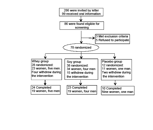
 

Figure 1 Flow chart of the protein supplementation intervention. Three of the completed subjects were excluded from the analysis due to low compliance or illness (two from the whey group, one from the soy group). A dropout replacement procedure was used to ensure equal group sizes when exposed for high withdrawal

Figure 1
Flow chart of the protein supplementation intervention. Three of the completed subjects were excluded from the analysis due to low compliance or illness (two from the whey group, one from the soy group). A dropout replacement procedure was used to ensure equal group sizes when exposed for high withdrawal


The Supplemetation

Total supplemented protein and leucine per day were in the whey group 45.8 g and 6.14 g, respectively, and  in the soy group 45.9 g and 3.1 g, respectively. No protein or leucine was included in the placebo group supplementation (Table 1). The whey protein and the placebo product were produced and delivered by Arla Foods Ingredients Group P/S (Denmark). The soy protein was commercially available from Soya International (Hale, UK).
The supplements were delivered in foil packs in 35.3 g powder to be constituted with approximately 150 ml of water. The subjects were instructed to consume the supplement twice daily as a part of the breakfast and lunch (15). Due to the exercise induced metabolic window theory, the subjects were instructed to ingest one supplement just after exercise on training days (19). The supplements were flavored neutrally or with chocolate. After 16 weeks we collected unused full foil packs to calculate compliance according to the protocol (fig.1).
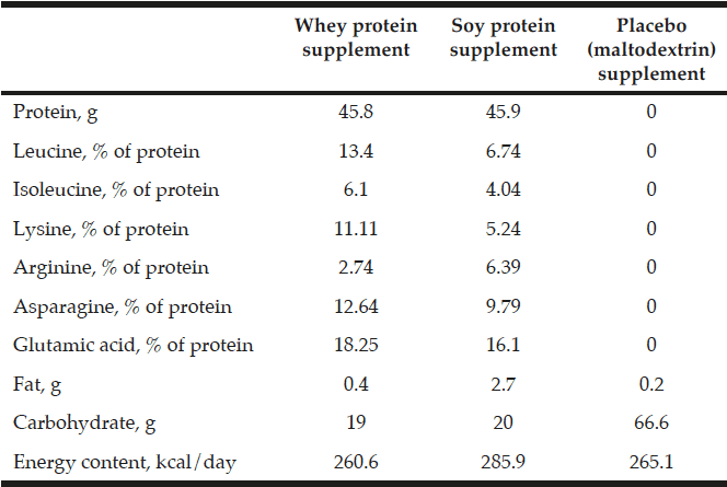
Table 1 Nutritional composition of the intervention supplements

Table 1
Nutritional composition of the intervention supplements

The supplemental nutrients and energy taken per day in the three groups during the intervention. Extra leucine was added to the whey protein supplement
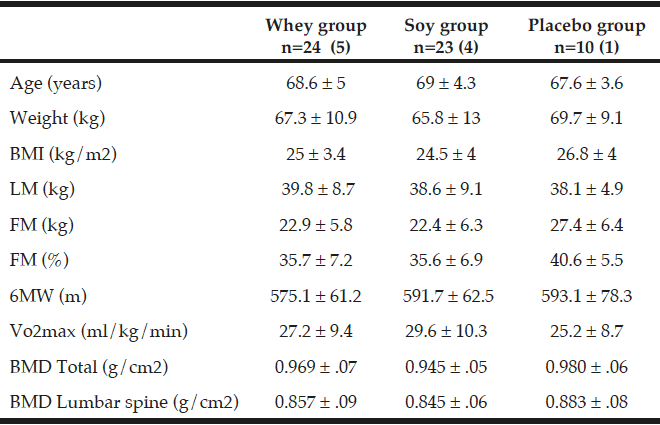
Table 2 Baseline clinical characteristics of the participants (completers)

Table 2
Baseline clinical characteristics of the participants (completers)

All values are mean ± SDs. 57 participants with osteopenia and age between 60 – 85 years was included in this 16-week intervention. The number of participants in the three groups is given together with the number of men in parenthesis. There were no statistical differences at baseline between the three groups; (ANOVA). Fat mass (FM), Lean mass (LM), Six-minute walk (6MW), Energy percentage (E%).


Dietary assessment

Three day weighed food records were conducted by a trained dietician at baseline and in the end of the study (week 16). The 47 subjects who completed the food records had all received guidance in how to register their foods and drinks without including the protein or placebo supplements. In the week 16 assessment of dietary intake we included compliance data to approximate per meal intakes. Food records were analyzed by the Dankost pro program version 1.5.49.21 (Dankost, Copenhagen, DK). The total energy intake was expressed in kilo joule per day (KJ/d) (Table 3). The total protein intake was expressed as percentage of total energy intake (E%), and as g per kg body weight per day (g/kg/day). Additionally leucine was expressed in g per day.
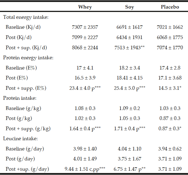
Table 3 The diet during the study – baseline values and changes

Table 3
The diet during the study – baseline values and changes

All values are mean ± SD, The dietary data after the intervention (“post”) are given both without and with the supplementation. The data in relation to baseline and after the intervention values are obtained from the food records. *; ** and *** denotes significantly different from baseline at level p<0.05; p<0.01 and p<0.001 respectively. p and pp denotes different from placebo group at level p<0.001 and p<0.0001 respectively. C denotes significant different between the protein groups p<0.001.


Resistance training

All participants in the three groups underwent a RT program for 45 minutes three times a week during the whole intervention. The RT protocol has been developed and recommended by others (20, 21). The training program was homebased training with supervision every second week. The training was conducted with TheraBand elastic bands (PROcare, Roskilde, Denmark) which has been used and validated in an elderly population (22, 23). Exercise progression and adherence to the training program was quantified by training records reporting rated perceived exertion (RPE) and level of resistance (22). The training intensity was increased during the intervention in level of RPE and number of repetitions. From week 0-4 the exercise were conducted as sets of 3×15 at level RPE=5 increasing intensity in week 5-7 to 3×15 at level RPE=7 and further in week 8-16 to 3×10 at level RPE=7. Every second week the exercise was conducted at the University Hospital with professional supervision. At these sessions the exercise was conducted as a circuit training regimen for potentially greater achievements as suggested elsewhere (20).
Determination of physical function, body composition, strength and balance

The primary endpoints in this study were testing of physical function by the 6MW and the 4MGS tests. The 6MW test is easy to administer, more reflective of activities of daily living and better tolerated than other walk tests (24). The subjects were instructed to walk as fast as possible, without further encouragement (24). The test was performed in a 30 meter corridor twice at baseline for familiarization (the better of the two was used) and once at the end of the intervention (24). Moreover, a consensus panel that considered reliability of the 6MW and the 4MGS test had recommended these tests as performance measures in older adults (25).
At baseline the 4MGS test was performed twice on two separate days for familiarization. The better of the two tests on the second test day was used as the baseline outcome. Post intervention the test was conducted twice and the better test was used as the 16 week outcome. 4MGS is a commonly used test in elderly, it predicts better survival (26), and there is a nonlinear relationship between 4MGS and leg strength (27).The non-linear relationship represents a mechanism by which small increments in leg strength in frail elderly may produce large improvements in gait speed, while large changes in leg strength in healthy people have no effect on gait speed.

Body composition and weight

The whole body composition was estimated using DXA scan (Hologic Discovery, Waltham, USA). All DXA assessments (pre and post intervention) were conducted at the same time point in the afternoon on the same scanner. The present study focuses on total lean mass (LM) in kg excluding bones and on fat mass (FM). Body weight was determined by weighing the subjects wearing easy clothing by electric scales Tanita WB-110 P MA, and height was determined by a stadiometer SECA model 220. BMI was calculated as weight/heigth² (kg/m²).

Strength tests

Strength tests of hand grip, leg flexion and leg extension were measured in an adjustable dynamometer chair (Good Strength, Metitur Ltd, Finland). Subjects were encouraged to perform maximum voluntary contraction (MVC) in three x five seconds with 30 seconds rest between each attempt. All tests were performed in a neutral sitting upright position; hand grip test with a 90˚ angled and supported elbow joint, and leg extension and flexion tests with the knee joint fixed in a 90˚ angle and the ankle and thigh fastened by a belt. The leg extension and flexion isometric MVC was measured in the fixed position moving the leg towards an extended and flexed position, respectively. For all strength tests, the mean of the three attempts was used in the statistical analysis. Because of method limitation (temporary error in the dynamometer), the reduced number of subjects completing the three strength tests were n=20, n=18 and n=7 in the whey, soy and placebo groups respectively.

Balance tests

Balance tests were performed on an equilateral triangular force platform connected to a computer-based system (Good BalanceTM, Metitur Ltd, Finland). Balance was tested with increasing difficulty in the following three tests: Normal standing balance (NSB), semi tandem balance (STB), and tandem balance (TB). The balance was measured as a velocity in sway, and the tests are described in details elsewhere (28). An improvement in balance is here expressed as a relative reduction in % compared to baseline.

Estimated maximal oxygen uptake (VO2 max)

Functional capacity was determined by the Aastrand ergometer test for estimating VO2 max. The workload was determined in steady state after six minutes of submaximal work and a heart rate just above 110 beats per minute (bpm). The Aastrand test is a gentle submaximal test and it has been validated as a reliable estimate of VO2 max (29).

Blood sampling and analysis

After an overnight fast (at baseline and post-intervention) blood samples were collected in tubes containing EDTA and immediately centrifuged at 4˚C over 10 minutes at 1500 x g. Tubes with plasma were stored at -80˚C for further analysis. Glucose and insulin were analyzed in-house with the Glucose GOD-PAP method (Roche Dianostics, Hvidovre, Denmark) for glucose analysis and an enzyme-linked immunoassay (DAKO, Glostrup, Denmark) for insulin analysis. IGF-1 was analyzed by IDS-iSYS IGF-1 assay (immunodiagnostic systems Ltd, Boldon, England).
Urate was determined by an enzymatic colorimetric method (Roche/Hitachi cobas c 501, Roche Diagnostics GmbH). Urea concentration was quantified by kinetic test with urease and glutamate dehydrogenase (Roche/Hitachi cobas c 501, Roche Diagnostics GmbH). All samples were analyzed on EDTA-plasma. All measures for each patient were assessed on the same batch except from urea. The analytical coefficients of variance were for insulin <10%, glucose <3%, IGF-1 <8%, Urate <13% and for urea <2 %.

Statistics

The expected change between the two protein groups in 6MW and 4MGS was 50 m and 0.1 m/s, respectively as recommended by others (30).  With 23 subjects in each group, our requirements of a significant level at P<0.05 with a power of 80% were contented (30).
Our aim was having 28 subjects in each protein group and 14 in the placebo group. It was decided to recruit a total of 80-85 subjects, as a 10-15% early withdrawal range was expected from our previous experiences.
The main comparison of interest in this study was between the two protein supplementation groups, whey n=24 and soy n=23.  All data were from independent observations, and normal distribution in the three groups was tested by QQ plots, and difference in variance was tested by the Bartletts test for equal variances. At baseline data that were not normally distributed were log transformed before further statistical analysis and presented as medians with 95%-CI. For all normal distributed data, values are presented as means ± SDs. Differences in outcome means between the whey, soy and placebo groups were analyzed by the ANCOVA model. The dependent variables were adjusted for the covariates BMI, age, sex and baseline levels when assumption of a significant linear association between the dependent variable and the covariate was contented. These specific covariates were chosen because of their shown impact on the outcome measures. One-way analysis of variance (ANOVA) tested for differences between groups at baseline, and paired t-tests were used for calculating within-group differences from baseline to post intervention. The statistical analyses were performed as a per protocol analysis.
The statistical analyses were performed by Stata version 13 and the graph by Graphpad Prism 5.


Results

Subjects characteristics

The baseline characteristics of the 57 individuals (47 women and 10 men) who completed the intervention (fig. 1) are shown in Table 2. At baseline the three groups were comparable according to age, BMI, weight, LM, FM, VO2 max and 6MW.  The mean age of the subjects was 68.6 years and the mean BMI was 25.1 kg/m² ± 3.8. The activity level was similar in the three groups and all participants were living independently. Moreover, the 6MW test was similar between the three groups. As shown in Table 3 there were no differences between any groups in total energy intake, protein or leucine intake at baseline. Only one of the participants smoked.
The compliance to the supplements was 89% ± 7.9 without differences between the groups (whey 87.8% ± 9.4, soy 89.6% ± 7.7, placebo 90.1% ± 4.3).  Two subjects were excluded from the analysis due to not following the protocol (one subject had cognitive limitations that affected managing the RT, furthermore the subject only ingested 57% of the supplements and another had stopped taken the supplements 3 weeks before the end of the study), and one subject was excluded due to illness. 72% of the subjects completed the training log and, 16 subjects (28%) failed to complete their training log. Compliance to the RT protocol was in the log book found to be 87.9% ± 15.9 without differences between the groups (whey 86.8% ± 15.8, soy 89.8 ± 17.8, placebo 86.4 ± 15).

Changes in physical function during the intervention

The 6MW test increased in both protein groups but more in the whey group (by 22.8 m ±26.7 corresponding to a 4.0% increment) as compared to the soy group (5.8 m ±18.1 corresponding to a 1.0% increment) and this difference between the two groups was significant (P<0.05 fig. 2). Moreover, in the placebo group 6MW was increased by 14.1 m ±16.2 (2.4%) which was not significantly different from the two protein groups. Compared to baseline levels 6MW increased significantly in the whey and placebo groups (P<0.001 and P<0.05, respectively) but not in the soy group (NS).
Concerning the 4MGS test there were no differences neither between the two protein groups nor within any of the three groups in relation to baseline levels. Moreover, VO2 max was unaffected in the three groups (Table 4).
Concerning the strength tests (handgrip, leg flexion and -extension), there were no differences between the two protein groups (Table 4). The handgrip strength increased, however, in all three groups compared to baseline but only significantly in the soy group, by 34.7 N, (11.6%, P<0.05, Table 4)
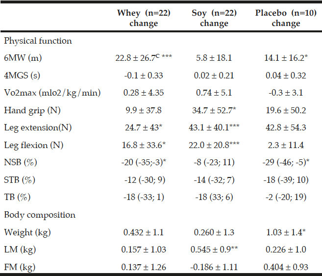
Table 4 Changes in physical function and body composition during the intervention

Table 4
Changes in physical function and body composition during the intervention

All values are means ± SDs or medians with (95%-CI), C denotes significantly different between the two protein groups p<0.05, calculated by ANOVA. *; ** and *** denotes significantly different from baseline in each group at the level of p<0.05; p<0.01 and p<0.001, respectively, calculated by paired t-tests. Fat mass (FM), Lean mass (LM), Six-minute walk (6MW), four meter gait speed (4MGS), Newton (N), Normal standing balance (NSB), Semi tandem balance (STB), Tandem balance (TB).


Leg extension strength also increased in all three groups with significant increases within both the whey group by 24.7 N ±43 (8%, P<0.05) and the soy group by 43.1 N ±40.1 (17.1%, P<0.001), and a trend towards an increase within the placebo group by 42.8 N ±54.3 (15.9%, P=0.08) which may suggest an effect of the exercise intervention on MVC in leg extension in all the groups.
MVC in leg flexion increased by 16.8 N ±33.6 (P<0.05) and 22.0 N ±20.8 (P<0.001) in the whey and soy protein groups respectively, and when calculated together as one protein group there was a tendency towards a protein effect on MVC in leg flexion (P=0.16) compared to placebo.
There were no changes between any of the groups in any of the balance tests (Table 4). Balance performance was generally improved as sway velocity was relatively reduced compared to baseline (2-29%), though only significantly in NSB in the whey group by 20% with 95%-CI (-35; -3), and in the placebo group by 29% with 95%-CI (-46; -5) (Table 4). There was also a trend towards improvement in TB in whey (P=0.056) and soy (P=0.067) as well as in STB in soy (P=0.17) and the placebo group (P=0.16).

Changes in body weight and body composition

There were no differences in body weight between the groups (Table 4). As compared to baseline body weight increased in all three groups  –  in the whey group by 0.43 kg ±1.1(NS) and in the soy group by 0.26 kg ±1.3 (NS), and significantly in the placebo group by 1.03 kg ±1.4(P<0.05) (Table 4).

There were no differences in LM or FM between any of the groups (Table 4). As compared to baseline levels, there was only a significant increment in LM in the soy group (P<0.01, Table 4).  When the two protein groups were combined there was a tendency towards increased FM in the placebo group as compared to the protein groups (P=0.06 Table 4).

Changes in blood values during the intervention

As shown in Table 5, there were no differences in changes in insulin, glucose, and HOMA index between any of the groups. As compared to baseline levels there was a significant increase within the whey group in insulin (P<0.05), glucose (P<0.05) and HOMA index (P<0.05) but not in the other groups.
As compared to baseline IGF-1 increased in both protein groups – by 10.2 ng/ml ±15.3 (9%, P<0.01) within the whey group, and by 7.4 ng/ml ±11 (7%, P<0.01) within the soy group with no changes in the placebo group. There was a tendency towards an increase in IGF-1 concentration in the protein groups when combining the two protein groups as compared to the placebo group (P=0.065, Table 5).
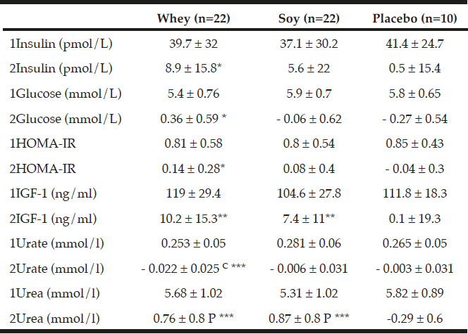
Table 5 Changes in blood values during the intervention

Table 5
Changes in blood values during the intervention

All values are means ± SDs, C denotes significantly different between the two protein groups at level p<0.01 calculated by ANOVA, P denotes significantly different from Placebo value (p<0.01) calculated by ANOVA. *; ** and *** denotes significantly different from baseline in each group at level p<0.05; p<0.01 and p<0.001 respectively, calculated by paired t-tests. Insulin growth factor (IGF-1).1denotes baseline values, 2denotes changes.


As expected the urea concentration increased significantly in both the whey protein group by 0.76 mmol/L ±0.8 (14.6%, P<0.001) and in the soy protein group by 0.87 mmol/L ±0.8 (17.6%, P<0.001) compared to baseline with no changes between the two protein groups. Moreover, the increment in urea concentration in the protein groups was significantly different from the 4.8% decrease in the placebo group (P<0.001, Table 5).
The changes in urate were significantly different between the two protein groups (P<0.01, Table 5) where urate decreased by 0.022 mmol/l ±0.02 (-7.5%) in the whey group compared with no changes in the soy group.

Dietary changes during the study

As expected there were no differences in total protein intake between the whey- and soy groups during the intervention (1.64 g/kg/day and 1.71 g/kg/day, respectively) which was significantly different from the placebo group (0.87 g/kg/day) (P<0.001, Table 3). Moreover, also as expected the leucine content of the diet was significantly higher in the whey group (9.44 g/day) as compared to the soy group (6.75 g/day) (P<0.001, Table 3) and the placebo group (3.71 g/day) (P<0.0001).
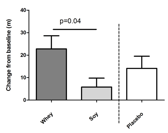
Figure 2 Effects of intake of whey protein and soy protein for 16 weeks on six-minute walk (6MW) distance. Mean changes of walk distance in meter +/- SEM. There was a significant difference between the whey and the soy group calculated by ANCOVA. Whey group n=22, soy n=22, placebo n=10

Figure 2
Effects of intake of whey protein and soy protein for 16 weeks on six-minute walk (6MW) distance. Mean changes of walk distance in meter +/- SEM. There was a significant difference between the whey and the soy group calculated by ANCOVA. Whey group n=22, soy n=22, placebo n=10


Discussion

In these elderly individuals with osteopenia, whey with leucine enrichment was found to significantly increase the 6MW  test compared to soy protein supplementation, but no significant effects on 4MGS or any other physical performance or strength tests were seen between the two groups.
The clinical implication of the 3% extra increment of 6MW in the whey group as compared to the soy group is difficult to evaluate but is rather small.  It has been shown that a 50 meter change corresponding to an 8% increase could give a substantial clinical effect with group sizes like in our study (31).
Our finding that exercise and leucine rich whey protein increases physical function, although to a minor degree, determined by the 6MW test supports a recent investigation from Rondanelli et al., who reported that exercise and leucine supplementation increased physical function in hand grip strength by 20% compared to exercise and placebo (32). Furthermore, a recent pilot trial in elderly people reported a 5.8% and 8.8% increase in 6MW after EAA with 20% (n=8) or 40% (n=8) leucine, respectively, and only a 1.5% increase in the placebo group (n=9) (12). It is well known that essential amino acids and in particular leucine is associated with an increase in MPS in acute studies in humans (33, 34). It has been reported that the long term effect of an increase in MPS due to dietary protein might lead to increase in LM and physical function, also in elderly people (16). In our study we saw no increase in LM, however, it has been demonstrated by others that physical function can improve without an increase in muscle mass (35, 36). Moreover, numerous recent publications in elderly individuals have demonstrated effects of dietary protein and EAA supplementation on a variety of physical function outcomes as leg extension and endurance (13), chair rise (15) and balance and gait speed (35).
We did not find any differences in strength and balance tests between any of the groups. However, there was a clear trend towards improvement in strength and balance, with significantly increased performance compared to baseline in several parameters across all groups. This may indicate an effect of the RT in this study like in other studies (37-39), and in line with recent reports on the associations between strength and balance in elderly individuals (40, 41). There were no differences between the two protein groups concerning anthropometrics such as body weight, LM and FM, but, there was a non-significant tendency that body weight and FM increased in the placebo group. This may be due to the maltodextrin (polysaccharide – glucose) which was given to this group, and high intake of glucose may enhance the risk of positive energy balance (42).
There were no differences between the two protein groups concerning the blood values except for p-urate (Table 5). However, in the whey group both insulin, glucose , and HOMA-IR increased significantly as compared to baseline values, most likely as a result of  the higher intake of leucine since a long term stimulation of circulating leucine may affect the insulin-glucose homeostasis (43). Besides the insulin independent effect of leucine on the mTor pathway and MPS, leucine also promotes an insulin dependent effect by stimulation of the pancreas ß-cells to secrete insulin and thereby further increase MPS (43).
IGF-1 is another anabolic biomarker, stimulating MPS, which may be associated to whey protein intake in postmenopausal women (44-46). The IGF-1 concentration was increased in both our protein groups and confirms this protein effect, also in line with the effect found by Heaney et al. after 2 years of milk supplementation (46).
The increased 6MW without change in muscle mass in the whey group can be a result of equal rates of MPS and muscle protein breakdown (MPB) at a high flux level of total muscle protein turnover (MPT) (47). Speculatively, it is possible that the muscle protein quality may have been improved due to an increased remodeling of muscle fibers leading to more effective functioning muscle tissue (47-49).  It can be speculated that the 2.4% increase in 6MW in the placebo group during the study could indicate a specific effect of the carbohydrate supplement since there is some evidence of a carbohydrate stimulated effect on post exercise MPS when combined with amino acids (50). This might explain the small 2.4% but significant within increase in 6MW in the placebo group which is not evident in the soy protein group (with much lower carbohydrate in the supplement), but it should be emphasized that there were no significant differences when comparing the changes in the two groups.
Another interesting observation in this study was the decreased concentration in plasma urate in the whey group compared to the soy group. Urate is the end-product of purine degradation. These findings are in line with previous acute and short term investigations, where it was shown that milk proteins have a very low content of purines compared to soy protein (51-53). An elevated level of urate in plasma has been linked to the development of cardiovascular diseases (54-56) and is directly involved in the development of gout in patients (57). Thus, reduced levels of urate after whey protein intake may be associated with positive health effects in humans.
The increased level of urea in the two protein groups emphasizes good adherence to the dietary protocol during the intervention.
There are some limitations to the present study. Only 57 individuals completed this investigation with only 10 subjects in the placebo group and particularly, the low number in the placebo group makes a direct comparison between the protein groups and the placebo group statistically problematic. Also we included relatively healthy elderly people, whereas a population of more fragile elderly people could potentially have responded more strongly.
Another limitation of the study may be that the protein and leucine intake in the soy group was also rather high since in the soy group the daily intake of e.g. leucine was 6.75 g when habitual leucine intake from the food records was included, and if we divided this leucine content into 3 daily meals, the per meal intake was 2.25 g leucine which is rather close to the suggested threshold at 2.5 – 2.8 g for healthy elderly people (15). Thus, this rather high intake of protein and leucine in the soy group may have affected the possibility to detect differences between the two protein groups.
Moreover, our four month-intervention may have been too short to detect differences between the dietary supplements. A six month-intervention with protein supplementation without exercise compared to placebo significantly increased physical function in frail elderly individuals (35). This indicates that a study of at least six month duration might have been appropriate to detect differences between the two protein groups.
It is of substantial importance that people perform physical activity and that they have a high quality protein intake in their diet during ageing (58). Our data support this current understanding and suggest that whey protein supplement, in addition to the effect of RT, has an impact on 6MW but the clinical importance of this effect is uncertain. Moreover, whey protein reduced the plasma urate which may have beneficial health effects as well.      In conclusion leucine-rich whey protein supplementation twice a day with concurrently RT, over a 16-week period, had little effect on 6MW which presumably is of only very minor clinical importance in elderly people with osteopenia, and, moreover, had no effects on the other physical functional or strength tests or on muscle mass as compared with intake of soy protein.


Acknowledgements: We thank Pia Hornbæk and Lenette Pedersen for their professional and skillful technical assistance. We thank Erik Jensen from Arla Foods Ingredients Group P/S (AFI) for providing the dietary supplements (the leucine-rich whey protein and the placebo product (maltrodextrin)) to the intervention. We thank for the donation of the IGF-1 and BP-3 kits which were provided by Immunodianostic systems Ltd, Bolton, England. We also thank for the donation of TheraBand exercise equipment which were provided by PROcare, Roskilde, Denmark and for the vitamin-D and calcium supplements (Unikalk Forte) provided by Axellus A/S, Ishøj, Denmark.

Contributions to the manuscript: BR, NM and JLM designed the research; JLM and KIN conducted research; BLL and SBP provided essential materials; JLM analyzed data; JLM wrote the paper with help from BR; JLM had primary responsibility for the final content; all authors have read and approved the final manuscript.

Conflicts of interest: Cand.scient. Mehlsen reports grants from the Danish Council for Strategic Research, during the conduct of the study; Dr. Pedersen, Cand.scient. Nørgaard, and Dr. Møller have nothing to disclose. Dr. Langdahl reports grants from Danish Council for Strategic Research,  during the conduct of the study; grants from Amgen, personal fees from Amgen, personal fees from Eli Lilly, personal fees from UCB, grants from Novo Nordisk, personal fees from Merck,  outside the submitted work; Dr. Richelsen reports other connections with Arla Foods, Denmark, during the conduct of the study .

Ethical standard: The study protocols and procedures were conducted according to the Helsinki declarations and were approved by the ethics committee of the Central Region of Denmark. All participants signed a written informed consent.


References

1.    McLeod M, Breen L, Hamilton DL, Philp A. Live strong and prosper: the importance of skeletal muscle strength for healthy ageing. Biogerontology. 2016 Jun;17(3):497-510.
2.     Nair KS. Aging muscle. Am J Clin Nutr. 2005 May;81(5):953-63.
3.     Muscaritoli M, Anker SD, Argiles J, Aversa Z, Bauer JM, Biolo G, et al. Consensus definition of sarcopenia, cachexia and pre-cachexia: joint document elaborated by Special Interest Groups (SIG) “cachexia-anorexia in chronic wasting diseases” and “nutrition in geriatrics”. Clin Nutr. 2010 Apr;29(2):154-9.
4.     Volpi E, Campbell WW, Dwyer JT, Johnson MA, Jensen GL, Morley JE, et al. Is the optimal level of protein intake for older adults greater than the recommended dietary allowance? J Gerontol A Biol Sci Med Sci. 2013 Jun;68(6):677-81.
5.     Rodriguez NR. Introduction to Protein Summit 2.0: continued exploration of the impact of high-quality protein on optimal health. Am J Clin Nutr. 2015 Apr 29.
6.     Bauer J, Biolo G, Cederholm T, Cesari M, Cruz-Jentoft AJ, Morley JE, et al. Evidence-based recommendations for optimal dietary protein intake in older people: a position paper from the PROT-AGE Study Group. J Am Med Dir Assoc. 2013 Aug;14(8):542-59.
7.     Leenders M, van Loon LJ. Leucine as a pharmaconutrient to prevent and treat sarcopenia and type 2 diabetes. Nutr Rev. 2011 Nov;69(11):675-89.
8.     Dreyer HC, Drummond MJ, Pennings B, Fujita S, Glynn EL, Chinkes DL, et al. Leucine-enriched essential amino acid and carbohydrate ingestion following resistance exercise enhances mTOR signaling and protein synthesis in human muscle. Am J Physiol Endocrinol Metab. 2008 Feb;294(2):E392-400.
9.     Bukhari SS, Phillips BE, Wilkinson DJ, Limb MC, Rankin D, Mitchell WK, et al. Intake of low-dose leucine-rich essential amino acids stimulates muscle anabolism equivalently to bolus whey protein in older women at rest and after exercise. Am J Physiol Endocrinol Metab. 2015 Jun 15;308(12):E1056-65.
10.     Mitchell WK, Wilkinson DJ, Phillips BE, Lund JN, Smith K, Atherton PJ. Human Skeletal Muscle Protein Metabolism Responses to Amino Acid Nutrition. Adv Nutr. 2016 Jul 15;7(4):828S-38S.
11.     Cuthbertson D, Smith K, Babraj J, Leese G, Waddell T, Atherton P, et al. Anabolic signaling deficits underlie amino acid resistance of wasting, aging muscle. FASEB J. 2005 Mar;19(3):422-4.
12.     Ispoglou T, White H, Preston T, McElhone S, McKenna J, Hind K. Double-blind, placebo-controlled pilot trial of L-Leucine-enriched amino-acid mixtures on body composition and physical performance in men and women aged 65-75 years. Eur J Clin Nutr. 2015 Jun 17.
13.     English KL, Mettler JA, Ellison JB, Mamerow MM, Arentson-Lantz E, Pattarini JM, et al. Leucine partially protects muscle mass and function during bed rest in middle-aged adults. Am J Clin Nutr. 2016 Feb;103(2):465-73.
14.     Chale A, Cloutier GJ, Hau C, Phillips EM, Dallal GE, Fielding RA. Efficacy of whey protein supplementation on resistance exercise-induced changes in lean mass, muscle strength, and physical function in mobility-limited older adults. J Gerontol A Biol Sci Med Sci. 2013 Jun;68(6):682-90.
15.     Bauer JM, Verlaan S, Bautmans I, Brandt K, Donini LM, Maggio M, et al. Effects of a vitamin D and leucine-enriched whey protein nutritional supplement on measures of sarcopenia in older adults, the PROVIDE study: a randomized, double-blind, placebo-controlled trial. J Am Med Dir Assoc. 2015 Sep 1;16(9):740-7.
16.     Dillon EL, Sheffield-Moore M, Paddon-Jones D, Gilkison C, Sanford AP, Casperson SL, et al. Amino acid supplementation increases lean body mass, basal muscle protein synthesis, and insulin-like growth factor-I expression in older women. J Clin Endocrinol Metab. 2009 May;94(5):1630-7.
17.     Verhoeven S, Vanschoonbeek K, Verdijk LB, Koopman R, Wodzig WK, Dendale P, et al. Long-term leucine supplementation does not increase muscle mass or strength in healthy elderly men. Am J Clin Nutr. 2009 May;89(5):1468-75.
18.     Meinert C. Dropout. In: Clinical Trials Handbook: Design and Conduct. Inc., Hoboken, NJ, USA.: John Wiley and Sons; 2012.
19.     Aragon AA, Schoenfeld BJ. Nutrient timing revisited: is there a post-exercise anabolic window? J Int Soc Sports Nutr. 2013 Jan 29;10(1):5,2783-10-5.
20.     Sundell J. Resistance Training Is an Effective Tool against Metabolic and Frailty Syndromes. Adv Prev Med. 2011;2011:984683.
21.     Kerksick CM, Wilborn CD, Campbell BI, Roberts MD, Rasmussen CJ, Greenwood M, et al. Early-phase adaptations to a split-body, linear periodization resistance training program in college-aged and middle-aged men. J Strength Cond Res. 2009 May;23(3):962-71.
22.     Mikesky AE, Topp R, Wigglesworth JK, Harsha DM, Edwards JE. Efficacy of a home-based training program for older adults using elastic tubing. Eur J Appl Physiol Occup Physiol. 1994;69(4):316-20.
23.     Colado JC, Garcia-Masso X, Triplett TN, Flandez J, Borreani S, Tella V. Concurrent validation of the OMNI-resistance exercise scale of perceived exertion with Thera-band resistance bands. J Strength Cond Res. 2012 Nov;26(11):3018-24.
24.     ATS Committee on Proficiency Standards for Clinical Pulmonary Function Laboratories. ATS statement: guidelines for the six-minute walk test. Am J Respir Crit Care Med. 2002 Jul 1;166(1):111-7.
25.     Perera S, Mody SH, Woodman RC, Studenski SA. Meaningful change and responsiveness in common physical performance measures in older adults. J Am Geriatr Soc. 2006 May;54(5):743-9.
26.     Hardy SE, Perera S, Roumani YF, Chandler JM, Studenski SA. Improvement in usual gait speed predicts better survival in older adults. J Am Geriatr Soc. 2007 Nov;55(11):1727-34.
27.     Buchner DM, Larson EB, Wagner EH, Koepsell TD, de Lateur BJ. Evidence for a non-linear relationship between leg strength and gait speed. Age Ageing. 1996 Sep;25(5):386-91.
28.     Era P, Sainio P, Koskinen S, Haavisto P, Vaara M, Aromaa A. Postural balance in a random sample of 7,979 subjects aged 30 years and over. Gerontology. 2006;52(4):204-13.
29.     Macsween A. The reliability and validity of the Astrand nomogram and linear extrapolation for deriving VO2max from submaximal exercise data. J Sports Med Phys Fitness. 2001 Sep;41(3):312-7.
30.     Perera S, Mody SH, Woodman RC, Studenski SA. Meaningful change and responsiveness in common physical performance measures in older adults. J Am Geriatr Soc. 2006 May;54(5):743-9.
31.     Studenski S. What are the outcomes of treatment among patients with sarcopenia? J Nutr Health Aging. 2009 Oct;13(8):733-6.
32.     Rondanelli M, Klersy C, Terracol G, Talluri J, Maugeri R, Guido D, et al. Whey protein, amino acids, and vitamin D supplementation with physical activity increases fat-free mass and strength, functionality, and quality of life and decreases inflammation in sarcopenic elderly. Am J Clin Nutr. 2016 Mar;103(3):830-40.
33.     Nair KS, Schwartz RG, Welle S. Leucine as a regulator of whole body and skeletal muscle protein metabolism in humans. Am J Physiol. 1992 Nov;263(5 Pt 1):E928-34.
34.     Rieu I, Balage M, Sornet C, Giraudet C, Pujos E, Grizard J, et al. Leucine supplementation improves muscle protein synthesis in elderly men independently of hyperaminoacidaemia. J Physiol. 2006 Aug 15;575(Pt 1):305-15.
35.     Tieland M, van de Rest O, Dirks ML, van der Zwaluw N, Mensink M, van Loon LJ, et al. Protein supplementation improves physical performance in frail elderly people: a randomized, double-blind, placebo-controlled trial. J Am Med Dir Assoc. 2012 Oct;13(8):720-6.
36.     Bonnefoy M, Cornu C, Normand S, Boutitie F, Bugnard F, Rahmani A, et al. The effects of exercise and protein-energy supplements on body composition and muscle function in frail elderly individuals: a long-term controlled randomised study. Br J Nutr. 2003 May;89(5):731-9.
37.     LIFE Study Investigators, Pahor M, Blair SN, Espeland M, Fielding R, Gill TM, et al. Effects of a physical activity intervention on measures of physical performance: Results of the lifestyle interventions and independence for Elders Pilot (LIFE-P) study. J Gerontol A Biol Sci Med Sci. 2006 Nov;61(11):1157-65.
38.     Latham NK, Harris BA, Bean JF, Heeren T, Goodyear C, Zawacki S, et al. Effect of a home-based exercise program on functional recovery following rehabilitation after hip fracture: a randomized clinical trial. JAMA. 2014 Feb 19;311(7):700-8.
39.     Pahor M, Guralnik JM, Ambrosius WT, Blair S, Bonds DE, Church TS, et al. Effect of structured physical activity on prevention of major mobility disability in older adults: the LIFE study randomized clinical trial. JAMA. 2014 Jun 18;311(23):2387-96.
40.     Bijlsma AY, Pasma JH, Lambers D, Stijntjes M, Blauw GJ, Meskers CG, et al. Muscle strength rather than muscle mass is associated with standing balance in elderly outpatients. J Am Med Dir Assoc. 2013 Jul;14(7):493-8.
41.     Granacher U, Gollhofer A, Hortobagyi T, Kressig RW, Muehlbauer T. The importance of trunk muscle strength for balance, functional performance, and fall prevention in seniors: a systematic review. Sports Med. 2013 Jul;43(7):627-41.
42.     Tittelbach TJ, Mattes RD, Gretebeck RJ. Post-exercise substrate utilization after a high glucose vs. high fructose meal during negative energy balance in the obese. Obes Res. 2000 Oct;8(7):496-505.
43.     Xu G, Kwon G, Cruz WS, Marshall CA, McDaniel ML. Metabolic regulation by leucine of translation initiation through the mTOR-signaling pathway by pancreatic beta-cells. Diabetes. 2001 Feb;50(2):353-60.
44.     Zhu K, Meng X, Kerr DA, Devine A, Solah V, Binns CW, et al. The effects of a two-year randomized, controlled trial of whey protein supplementation on bone structure, IGF-1, and urinary calcium excretion in older postmenopausal women. J Bone Miner Res. 2011 Sep;26(9):2298-306.
45.     Schurch M, Rizzoli R, Slosman D, Vadas L, Vergnaud P, Bonjour J. Protein Supplements Increase Serum Insulin-Like Growth Factor-I Levels and Attenuate Proximal Femur Bone Loss in Patients with Recent Hip Fracture. Ann Intern Med. 1998 05/15;128(10):801-9.
46.     Heaney RP, McCarron DA, Dawson-Hughes B, Oparil S, Berga SL, Stern JS, et al. Dietary changes favorably affect bone remodeling in older adults. J Am Diet Assoc. 1999 Oct;99(10):1228-33.
47.     Wolfe RR. Update on protein intake: importance of milk proteins for health status of the elderly. Nutr Rev. 2015 Aug;73 Suppl 1:41-7.
48.     Conroy MB, Kwoh CK, Krishnan E, Nevitt MC, Boudreau R, Carbone LD, et al. Muscle strength, mass, and quality in older men and women with knee osteoarthritis. Arthritis Care Res (Hoboken). 2012 Jan;64(1):15-21.
49.     Balagopal P, Rooyackers OE, Adey DB, Ades PA, Nair KS. Effects of aging on in vivo synthesis of skeletal muscle myosin heavy-chain and sarcoplasmic protein in humans. Am J Physiol. 1997 Oct;273(4 Pt 1):E790-800.
50.     Miller SL, Tipton KD, Chinkes DL, Wolf SE, Wolfe RR. Independent and combined effects of amino acids and glucose after resistance exercise. Med Sci Sports Exerc. 2003 Mar;35(3):449-55.
51.     Garrel DR, Verdy M, PetitClerc C, Martin C, Brule D, Hamet P. Milk- and soy-protein ingestion: acute effect on serum uric acid concentration. Am J Clin Nutr. 1991 Mar;53(3):665-9.
52.     Dalbeth N, Wong S, Gamble GD, Horne A, Mason B, Pool B, et al. Acute effect of milk on serum urate concentrations: a randomised controlled crossover trial. Ann Rheum Dis. 2010 Sep;69(9):1677-82.
53.     Liu ZM, Ho CS, Chen YM, Woo J. Can soy intake affect serum uric acid level? Pooled analysis from two 6-month randomized controlled trials among Chinese postmenopausal women with prediabetes or prehypertension. Eur J Nutr. 2015 Feb;54(1):51-8.
54.     Holme I, Aastveit AH, Hammar N, Jungner I, Walldius G. Uric acid and risk of myocardial infarction, stroke and congestive heart failure in 417,734 men and women in the Apolipoprotein MOrtality RISk study (AMORIS). J Intern Med. 2009 Dec;266(6):558-70.
55.     Oda E, Kawai R, Sukumaran V, Watanabe K. Uric acid is positively associated with metabolic syndrome but negatively associated with diabetes in Japanese men. Intern Med. 2009;48(20):1785-91.
56.     Jin YL, Zhu T, Xu L, Zhang WS, Liu B, Jiang CQ, et al. Uric acid levels, even in the normal range, are associated with increased cardiovascular risk: the Guangzhou Biobank Cohort Study. Int J Cardiol. 2013 Oct 3;168(3):2238-41.
57.     Beyl RN. Update on Importance of Diet in Gout. Am J Med. 2016 -11;129(11):1153; 1153,1158; 1158.
58.     Paddon-Jones D, Campbell WW, Jacques PF, Kritchevsky SB, Moore LL, Rodriguez NR, et al. Protein and healthy aging. Am J Clin Nutr. 2015 Apr 29.

ELEVEN WEEKS OF STRENGTH TRAINING DECREASED THE EXPRESSION OF IMMUNE-RELATED GENES IN OLDER SUBJECTS INDEPENDENT OF PROTEIN SUPPLEMENT TYPE; A RANDOMIZED CONTROLLED TRIAL


G.O. Gjevestad1,2, H. Hamarsland3, T. Raastad3, J.J. Christensen1,4, A.S. Biong2, S.M. Ulven1, K.B. Holven1,5


1. Department of Nutrition, Institute of Basic Medical Sciences, P.O. Box 1046, Blindern, 0317 University of Oslo, Norway; 2. TINE SA, Centre for Research and Development, P.O. Box 7, Kalbakken, 0902 Oslo, Norway; 3. Department of Physical Performance, Norwegian School of Sport Sciences, P.B. 4104 U.S., 0806 Oslo, Norway; 4. The Lipid Clinic, Oslo University Hospital Rikshospitalet, P.O. Box 4950 Nydalen, 0424 Oslo, Norway; 5. National Advisory Unit on Familial Hypercholesterolemia, Department of Endocrinology, Morbid Obesity and Preventive Medicine, Oslo University Hospital, P.O. Box 4950 Nydalen, 0424 Oslo, Norway.

Corresponding Author: Kirsten B. Holven, Department of Nutrition, Institute of Basic Medical Sciences, P.O. Box 1046, Blindern, 0317 University of Oslo, Norway. k.b.holven@medisin.uio.no

J Aging Res Clin Practice 2017;6:182-190
Published online September 18, 2017, http://dx.doi.org/10.14283/jarcp.2017.24


Abstract

Objective: To investigate the effects of eleven weeks of strength training combined with two isocaloric protein supplements on mRNA expression levels in skeletal muscle and peripheral blood mononuclear blood cells (PBMCs). Design: A double blind randomized controlled study. Setting: The Norwegian School of Sports Sciences, Norway. Participants: Untrained, but otherwise healthy, men and women (n=20, ≥ 70 yrs). Intervention: Participants were randomly allocated to receive either milk protein or a native whey protein supplement (20 g protein, morning and afternoon) combined with a standardized strength training protocol (6-10 RM, 1-3 sets, 3 times/week) for eleven weeks. Measurements: The mRNA expression levels of immune-related genes were measured before and after the intervention period, using RT-qPCR. Cytokines were measured using ELISA. Results: PBMC mRNA expression of interleukin (IL) 6, IL8, chemokine (C-C motif) ligand (CCL) 3, and nuclear receptor subfamily (NR) 1 group H (H) member 3 decreased significantly after the intervention period, whereas the mRNA expression of toll-like receptor (TLR2) increased. In skeletal muscle, the mRNA expression of peroxisome proliferator-activated receptor gamma coactivator 1 alpha (PPARGC1A) and PPARGC1B decreased significantly, whereas the mRNA expression of CCL2, CCL5, TLR2, TLR4 and hypoxia inducible factor 1 alpha subunit (HIF1A) increased significantly after the intervention. We found no significant differences in circulating C-reactive protein and IL6 after the intervention period. The consumption of whey and milk proteins had similar effects on mRNA expression levels after strength training in skeletal muscle as well as PBMCs. Conclusion: Eleven weeks of strength training and protein supplementation reduced the PBMC expression levels of genes involved in the immune system as well as in metabolism, underlining the close interaction between these processes. The upregulation of other immune-related genes observed in PBMCs as well as in skeletal muscle needs further investigations, but may be related to protein supplementation and training adaptations. Different protein supplementation (milk or native whey) did not differentially modulate mRNA expression after the intervention period.

Key words: PBMC, skeletal muscle, mRNA, resistance training, cytokines.

Abbreviations: BCAA, branched chain amino acids; CCL, chemokine (C-C motif) ligand; RCT, reverse cholesterol transport; CRP, C-reactive protein; CXCL, chemokine (C-X-C motif) ligand; E %, energy percent; HIF1A, hypoxia-inducible factor 1-alpha; HMBS, hydroxymethylbilane synthase; IL, interleukin; IL1RN, interleukin 1 receptor antagonist; IMVC, isometric maximum voluntary contraction; IPO8, importin 8; NR4A2; NF-κB, nuclear factor kappa-light-chain-enhancer of activated B cells; nuclear receptor subfamily 4, group A, member 2; NR4A3; nuclear receptor subfamily 4, group A, member 3; NR1H3, Nuclear Receptor Subfamily 1 Group H Member 3; PBMCs, peripheral blood mononuclear cells; PPARGC1A, peroxisome proliferator-activated receptor gamma coactivator 1-alpha; PPARGC1B, peroxisome proliferator-activated receptor gamma coactivator 1-beta; RCT, reverse cholesterol transport; RM, repetition maximum; RT-qPCR, real-time quantitative polymerase chain reaction; TBP, TATA box binding protein; TG, triglycerides, TLDA, TaqMan Low-Density array; TLR, toll-like receptor; TNF, tumor necrosis factor alpha.


Introduction

Diet and exercise are two of the most important lifestyle factors influencing healthy aging. Both are able to induce changes in metabolism and immune function (1, 2). They are therefore important targets for lifestyle interventions aiming at preventing the development of age-related diseases, including cardiovascular diseases, diabetes type 2, obesity (3) and sarcopenia (4).
Regular physical activity improves several of the underlying metabolic processes that are associated with adverse health effects and age-related diseases, such as insulin sensitivity, plasma triglycerides, blood pressure, and endothelial function (5). Regular strength training promotes muscle hypertrophy mainly, being particularly important in the prevention of age-associated loss of muscle mass and function associated with sarcopenia (6). Cross-sectional and large-scale cohort-studies have consistently shown an inverse association between physical activity and circulating markers of low-grade chronic inflammation (7). This anti-inflammatory effect is suggested to be one of the mechanisms underlying the protective effects observed on the development of chronic metabolic diseases by regular exercise (5), including sarcopenia (8). Because older adults often have a higher baseline level of circulating immune-related markers (9), long term regular exercise has been suggested as a tool to ameliorate the inflammatory status of older adults (10).
Components of the diet may exert anti-inflammatory effects and protect against chronic low-grade inflammation (2). In line with this, some epidemiological findings indicate that low-fat dairy products may reduce circulating levels of immune-related markers (11). However, others have found no association between intake of dairy products and circulating levels of immune-related markers, and conclusions from intervention studies have been conflicting (12). Although possible mechanisms underlying a potential anti-inflammatory effect of dairy products remains to be elucidated, several components in milk have been suggested to extert the anti-inflammatory effects, among them whey proteins (13).
The aims of the present study were to investigate the effects of eleven weeks of strength training combined with two isocaloric protein supplements (20 g protein morning and afternoon) on mRNA expression levels in skeletal muscle and peripheral blood mononuclear blood cells (PBMCs) in healthy older adults (>70 yrs).


Materials and methods

Study population and experimental design

Twenty-four older (≥70 yrs) untrained men and women were recruited to an eleven week lasting double-blind, randomized controlled study, which was conducted from August 2014 to December 2014 at The Norwegian School of Sport Sciences, Norway. All participants provided written informed consent, and we conducted the study according to the guidelines laid down in the Declaration of Helsinki. The Regional Committees for Medical and Health Research Ethics, Health Region South East, Norway, provided approval for all planned procedures involving human subjects (2014/834).
Subjects were randomized into one of two groups, receiving dairy supplements based on either native whey protein or regular milk protein for eleven weeks, on top of a high-load strength training regime. All tests were performed before and after the 11-week period. From the day before the test days, subjects followed a standardized diet based on body weight. In the morning of the test day, subjects were served a standardized breakfast, consisting of oatmeal, water, rapeseed oil and sugar (50 E % from carbohydrate, 8 E % from protein and 42 E % from fat). All subjects finished the breakfast within 20 minutes. PBMCs and skeletal muscle samples were collected 90 min after breakfast was served. We used Dual energy X-ray absorptiometry (Lunar iDXA, GE Healthcare, Buckinghamshire, United Kingdom) and enCORE Software (version 14.10.022, GE Lunar) to determine fat mass. Android fat mass was estimated based on algorithms in the enCORE Software. All subjects reported to be healthy, but one participant (4 %) had prescribed anticoagulants and six participants (25 %) took statins. Four subjects did not complete the study due to disease (two participants) or hurting joints (two participants).

Protein supplements

TINE SA (Oslo, Norway) provided the supplements. The products contained 20 g of protein (27 E %), 39 g carbohydrates (52 E %) and 7 g fat (21 E %), providing approximately 300 kcal per serving. All subjects received 2×20 g protein/day. The protein source was either native whey protein or regular milk protein (approximately 80 % casein and 20 % whey). Native whey proteins differ from regular whey proteins by the production method (produced at low temperatures to avoid extensive denaturation of the protein) and the composition of amino acids (e.g. higher amount of leucine). The participants prepared one serving in the morning and one in the evening, except at training days, where the training instructors provided the evening dose following the exercise sessions. The powder was dissolved in approximately 500 ml water. The producer delivered supplements in identical and coded packaging to ensure blinding of both the providers and the participants. The producer was responsible for coding of the products, and the coding was not revealed until the intervention was completed. All products had the same flavor, color and appearance.

Training protocol

The training program was standardized in terms of exercises performed (hammer squat, leg press, knee extensions, calf raise, chest press, seated rowing, close grip pull down, shoulder press, back extensions and crunches), the number of sets (6-12 repetition maximum, RM) and the number of repetitions (1-3 sets in each exercise). The exercise program was performed 3 days per week for eleven weeks (Figure 1), and all training sessions were supervised by trainers. The load in each exercise was individualized and adjusted each training session by the trainer to make sure that participants always exercised at the intended training load. Participants completed the last exercise session at least three days before the last biopsy was taken.

Figure 1 Study design and timeline for sampling during the intervention study

Figure 1
Study design and timeline for sampling during the intervention study


Sampling and sample preparation

We collected venous blood samples in BD Vacutainer® CPTTM cell preparation tubes with sodium heparin (Becton Dickinson, NJ, USA) and in silica gel tubes (Becton Dickenson Vacutainer Systems, Plymouth, UK). Within two hours of blood collection, PBMCs were isolated according to the manufacturer’s instructions (Becton Dickinson, NJ, USA). Serum samples were centrifuged (1500 g, 15 min at RT) after being left on the bench top for at least 30 min. Blood plasma samples were collected with pipettes immediately after being collected (lithium heparin tubes from Vacuette, Greiner Bio One, Austria) and centrifuged (1300 g, 10 min at 4°C). All samples were stored at -80°C until further analysis.
Muscle biopsies from the m. vastus lateralis were obtained at the same time points as the blood samples, using a modified Bergstrom technique (14), immediately cleaned from blood and connective tissue in physiological salt water at 4˚C, immersed into RNAlater® solution (Ambion, TX, USA) and stored overnight at 4 ˚C. The following day, the biopsies were transferred and stored at – 80 ˚C until further analysis. Biopsies were taken from the same leg before and after the intervention.

Isolation of RNA and synthesis of cDNA

Skeletal muscle samples were ruptured using a CryoGrinder System (Ops Diognostics, NJ, USA), followed by homogenization in Quizol (QIAGEN GmbH, Germany) and addition of chloroform. After centrifugation, the upper phase was transferred to a fresh tube and ethanol added. The procedure of RNA isolation was carried out using the QiaCube instrument (QIAGEN GmbH, Germany) following the miRNease Mini Kit protocol (QIAGEN). We isolated RNA from PBMCs in accordance with the RNeasy Mini Kit protocol using qiashredder and DNase digest (QIAGEN GmbH, Germany) using the QiaCube. High-quality RNA from both PBMCs and skeletal muscle samples was eluted in 30 μl of RNase free water and frozen at -80 °C until further analysis. RNA quantity was measured using NanoDrop-1000 (NanoDrop Technologies, Inc., DE, USA), and RNA quality was checked with Aglient 2100 Bioanalyzer (Agilent Technologies, Inc., CA, USA). All samples included in further analysis had a RIN-value above 5.5. PBMC samples from one person were not taken due to a misunderstanding in the laboratory, whereas mRNA from skeletal muscle was lost from six participants due to low RIN-values. In addition, we excluded some of the mRNA transcripts from our final analysis due to abnormal multicomponent plots.
RNA samples were transcribed into cDNA (500 ng) using the cDNA kit from Applied Biosystems (Applied Biosystems, UK) and in accordance with the protocol provided. Samples were stored at -20 °C for further analysis.

RNA analysis by real-time qPCR

We measured mRNA levels of 48 genes (Additional file 1) using TaqMan Low-Density array (TLDA) cards from Applied Biosystems (UK). The TLDA cards were used on a 7900 HT Applied Biosystems RT-qPCR machine (Applied Biosystems, UK). The Ct-values were analysed using SDS 2.4 (Applied Biosystems, UK), and further transferred to ExpressionSuite Sofware v1.0.3 (Applied Biosystems, UK). For PBMCs, we normalised the Ct-values to TATA box binding protein (TBP) and hydroxymethylbilane synthase (HMBS) mRNA transcripts, whereas for skeletal muscle, we normalized to TBP and importin 8 (IPO8). Fold changes in mRNA transcripts from baseline to end of study were calculated, using the 2-ΔΔCt-method (15).
The selection of genes were based on a thorough literature search investigating the effect of training on gene expression in PBMCs (16). We performed a similar research for skeletal muscle (17). Moreover, the genes selected in the present study were based on previously published studies where the effects of dairy products on markers of chronic low-grade inflammation were described (18).

Cytokine measurements and routine analysis

The serum level of IL6 was measured with high-sensitive Quantikine ELISA (R&D Systems Inc., MN, USA), whereas IL8 and CCL3 were measured using Quantikine ELISA (R&D Systems Inc.), both in accordance with the protocols provided. We measured all samples in duplicates. Serum levels of glucose, triglycerides, cholesterol and the plasma level of C-reactive protein (CRP) were analyzed by an accredited laboratory (Fürst Medical Laboratory, Oslo; Norway).

Statistics

Power calculation was performed for the primary outcome of the study, which was to study the effects of consuming native whey or milk protein on muscle mass and strength. We also considered this number of participants to be relevant with respect to changes in inflammatory markers. In addition, the number of participants included in the present study is in line with other studies exploring the relationships between exercise and gene expression . All data were checked for normality. Subjects with levels of CRP above >10 mg/L at baseline (n=1) or at end of study (n=1) were excluded from the gene expression analysis as such levels may indicate an ongoing acute inflammation, not reflecting the intervention. For non-parametric data, we used the Mann-Whitney-test for independent measurements and the Wilcoxon signed-rank for repeated, paired measurements. For parametric data, Independent samples t-test was used for independent measurements and Paired t-test for paired measurements. The Spearman correlation test was used to reveal possible correlations between the change in android fat mass and mRNA expression levels of selected transcripts. Due to an explorative study design, correction for multiple testing was not performed. We considered a p-value of < 0.05 statistically significant. SPSS statistical software, version 22 from Microsoft (SPSS, Inc., CA, USA), was used for statistical calculations and GraphPad Prism 5 (GraphPad Software, Inc., CA, USA) for generating figures.


Results

Participants (n=20, mean ± SD=73.6 ± 2.8 yrs) included in the present study were similar in body mass, fat mass, BMI and blood parameters at baseline. The proportion of male to female was 12/8. We observed a significant change in body mass and BMI from baseline to end of intervention, but not in fat mass or android fat mass (Table 1). None of the anthropometric characteristics or blood parameters changed significantly from baseline to end of study between the native whey and the milk group (data not shown).


Table 1 Anthropometric parameters at baseline and after the intervention

Table 1
Anthropometric parameters at baseline and after the intervention

1. Calculated by Paired sample t-test; 2. n=19; Abbreviations used in table; Δ, delta; HDL, high-density cholesterol; LDL, low-density cholesterol

Adherence to the strength training and protein supplementation

Participants attended an average of 33.0 ± 0.9 and 32.5 ± 1.2 exercise sessions (of totally 33 exercise sessions) in the native whey and milk group, respectively. We logged adherence to the supplementation regime at each training session, which resulted in a mean self-reported compliance of 99 %.

Effects of strength training and protein supplementation on gene expression

Protein supplements based on milk protein or native whey protein did not significantly alter mRNA expression levels after eleven weeks of strength training neither in PBMCs nor in skeletal muscle. When merging the two groups, PBMC mRNA expression levels of interleukin (IL) 6, IL8, chemokine (C-C motif) ligand (CCL) 3 and nuclear receptor subfamily 1, group A, member 3 (NR1H3, also known as LXR) were significantly reduced after the intervention period, whereas the mRNA expression level of toll-like receptor (TLR) 2 increased (Figure 2). In skeletal muscle, the mRNA expression levels of CCL2, CCL5, TLR2, TLR4, IL8 and hypoxia-inducible factor 1-alpha (HIF1A) significantly increased after the intervention (Figure 3 and 4), whereas peroxisome proliferator-activated receptor gamma coactivator -alpha (PPARCG1A) and PPARCG1B decreased significantly (Figure 4). We also observed significant changes in the expression of some genes related to lipid metabolism, both in PMBCs and skeletal muscle, as shown in Additional file 2 and 3.

Figure 2 mRNA expression levels in PBMCs before and after the intervention

Figure 2
mRNA expression levels in PBMCs before and after the intervention

Expression levels of IL6 [A], IL8 [B], CCL3 [C], NR1H3 [D] and TLR2 [E] from baseline to 11 weeks of strength exercise. Values are expressed as fold changes, and the vertical lines represent median values. The p-values were calculated by Wilcoxon signed-rank test and indicate changes from baseline til end of study. n = 14 [D], n= 15 [B, C] and n= 16 [A, G].

Figure 3 mRNA expression levels in skeletal muscle before and after the intervention

Figure 3
mRNA expression levels in skeletal muscle before and after the intervention

Expression levels of CCL2 [A], CCL5 [B], TLR2 [C], TLR4 [D] and IL8 [E] from baseline to 11 weeks of strength exercise. Values are expressed as fold changes and the vertical lines represent median values. The p-values were calculated by Wilcoxon signed-rank test and indicate changes from baseline til end of study. n = 10 [B, E], n = 11 [A] and n= 12 [C, D].

Figure 4 mRNA expression levels in skeletal muscle before and after the intervention

Figure 4
mRNA expression levels in skeletal muscle before and after the intervention

Expression levels of PPARGC1A [A], and PPARGC1B [B] and HIF1A [C] from baseline to 11 weeks of strength exercise. Values are expressed as fold changes and the vertical lines represent median values. The p-values were calculated by Wilcoxon signed-rank test and indicate changes from baseline til end of study. n = 12.

Circulating immune-related markers

No differences were observed from baseline to after the intervention in the serum level of IL6 or the plasma level of CRP (Table 2). Serum levels of IL8 and CCL3 were also measured, but were not detectable (not shown).


Table 2 Inflammatory markers at baseline and after the intervention

Table 2
Inflammatory markers at baseline and after the intervention

1. Calculated by Wilcoxon signed rank test; 2. n=14; Abbreviations used in table; CRP, C-reactive protein; Δ, delta; IL, interleukin.

Correlations between android fat mass and immune-related genes and circulating markers

No correlations between changes in android fat mass and changes in mRNA expression levels of IL6, IL8 and CCL3 in PBMCs or circulating levels of IL6 and CRP were observed from baseline to end of intervention.


Discussion

We observed that 11 weeks of high-load strength training combined with protein supplementation decreased mRNA expression levels of several immune-related genes in PBMCs, potentially having beneficial effects on systemic low-grade inflammation. On the other hand, immune-related mRNA transcripts were both up- and downregulated in skeletal muscle, probably reflecting muscle regeneration and adaptation. Native whey and milk proteins did not differentially alter mRNA expression levels, neither in PBMCs nor in skeletal muscle after strength training. No effects were observed on circulating levels of IL6 or CRP.

It is well known that long-term adaptations to strength training result in increased muscle mass (hypertrophy) and strength (6). Furthermore, regular exercise may reduce the level of immune-related markers (1). This is in line with observations from the present study, as we found reduced mRNA expression levels of IL6, IL8 and CCL3 in PBMCs after the training period. It is also in line with a study where PBMC mRNA expression levels of immune-related markers of middle-aged men and women (n=40, mean age 50-67 yrs) were reduced (CCL2), or tended to be reduced (TNF) after two months of brisk walking (6 days/week, 50 min/day, 70% of maximal heart rate). However, the mRNA expression level of IL6 did not change after the training period in that study (10). Importantly, the older subjects had higher baseline levels of the relevant markers compared to younger participants. The authors therefore suggested that the observed reduction might be related to the higher baseline levels of these mRNA transcripts in the older compared to the younger participants, and that these differences promoted a more robust reduction after the exercise period in older adults (10).
In contrast to the decreased mRNA levels of IL6, IL8 and CCL3 in PBMCs, we observed an increased mRNA expression level of TLR2 in PBMCs as well as in skeletal muscle. The mRNA expression of TLR4, IL8, CCL2 and CCL5 were also significantly increased in skeletal muscle after the intervention. These results are in contrast to others who have found decreased mRNA expression levels of TLRs in PBMCs (19), skeletal muscle (20) and whole blood (21) after regular strength training in older adults. Increased levels of TLRs may induce NF-κB activation in PBMCs as well as skeletal muscle and contracting C2C12 myotubes have been shown to induce CCL2 in an NF-κB-dependent manner (22). In skeletal muscle, it has been shown that NF-κB activation may prevent myogenic differentiation (23) and contribute to muscle atrophy by increasing the activity of molecules involved in muscle protein degradation (24). However, this seems unlikely to occur in the present situation as we observed increased muscle mass after the training period and reduced levels of IL6, IL8 and CCL3 in PBMCs. Similarly, increased NF-κB activation in PBMCs is closely linked to the development of low-grade inflammation (25), but NF-κB inhibition during the resolution phase can also prevent proper tissue repair (26). IL8, CCL2 and CCL5 are chemoattractants, which may play an important role in the recruitment of immune-related cells to skeletal muscle following an acute exercise bout (27). Long-term effects of training on these markers are less investigated, but circulating levels of IL8 and CCL2 have been shown to decrease after aerobic training programs (28). The mission of immune-related markers in exercise is not fully understood, but immune-related markers are hypothesized to be important in the resolution processes by removing cellular debris, releasing factors to promote muscle growth and to facilitate vascular and muscle fibre repair, amongst others (29). TLRs may potentially also activate other pathways, such as p38 mitogen-activated kinase (MAPK) and C-Jun N-terminal kinase (JNK) (30)possibly stimulating cell proliferation (31).
Furthermore, we observed decreased mRNA expression levels of NR1H3 in PBMCs, and of PPARGC1A and PPARGC1B in skeletal muscle after the training period. In contrast to these results, regular endurance training has been shown to upregulate the expression of NR1H3 in PBMCs (32) and PPARGC1A and PPARGC1B in skeletal muscle (33). However, a decreased mRNA expression level of PPARGC1B has also been observed in skeletal muscle after 10 weeks of knee extensor training in young adults (n=7, mean age 26±1 yrs) (34). NR1H3 plays a central role in the transcriptional regulation of both cholesterol homeostasis and inflammation (32, 35). The signaling pathway inducing NR1H3 expression in macrophages involves NF-κB dependent transcriptional gene activation and may promote anti-inflammatory effects (35). Further, TLRs have been shown to downregulate NR1H3 (35), which we also observed in PBMCs in the present study. PPARGC1A and HIF1A are thought to be involved in energy metabolism (36), and PPARGC1A is also closely linked to inflammation (37). Lower levels of PPARGC1A have been observed in patients with diabetes type 2 [38], the metabolic syndrome (39) and in aging (38). A possible explanation for the conflicting results of NR1H3, PPARGC1A and PPARGC1B in the present and other studies may be that participants in the present study also consumed protein supplements, which potentially may alter mRNA expression levels of genes investigated.

In contrast to the observed decrease in the mRNA expression level of IL6 in PBMCs in the present study, the circulating level of IL6 was unchanged. This discrepancy support the notion that PBMCs are not the main source of circulating IL6 (40). Further, a stable level of circulating IL6 has previously been found after five consecutive days of high-volume resistance training (41) and after three months of combined endurance and resistance training (42). At the same time, reduced levels of circulating IL6 have been observed after progressive 24-wk exercise of endurance exercise (43) and after walking 10000 steps three times per week for eight weeks (44). Furthermore, we found no changes in the level of CRP after the intervention period. This is supported by a recent study where community dwelling older adults performed a strength training program for 12 weeks (45), but is in contrast to the findings in a combined aerobic and resistance training program in middle-aged men and women where the level of CRP was reduced after 6 months of training (46). Strength training has also been shown to decrease CRP in overweight women (47) and in older adults (48).
Possible mechanisms underlying the anti-inflammatory effects of training are largely unknown, but it has been hypothesized that the reduced levels of immune-related markers observed after training are due to a redistribution of fat mass (1). In the present study, we did not find any significant changes in the distribution of android fat mass and we observed no correlations between changes in mRNA transcripts of IL6, IL8 and CCL3 and android fat mass after the intervention period. Despite no changes in android fat mass, the mRNA expression levels of IL6, IL8 and CCL3 in PBMCs decreased, indicating that the change in PBMCs may occur in the absence of a changed fat distribution.
Protein supplementation, in combination with strength training, may additionally enhance muscle protein synthesis and muscle hypertrophy (49). In addition, whey proteins are hypothesized to exert anti-inflammatory effects (50). CRP was reduced in elderly adults with sarcopenia (n=130, mean age 80.3 yrs) after 12 weeks of supplementation with whey protein (22 g), essential amino acids (10.9 g, including 4 g leucine) and vitamin D (2.5 µg) concurrent with a combination of regular endurance and strength training (51). Moreover, intake of branched chain amino acids (BCAA) has been shown to attenuate inflammation after a three-day extensive aerobic training program compared to an isocaloric amount of carbohydrate (52). However, no change was observed in serum concentrations of IL6 after resistance training combined with protein and omega-3 supplementation in novice resistance trained females (n=28, mean age 20±1 yrs) (41), and a recent meta-analysis showed that whey supplements do not modulate inflammation in healthy adults (53). Nevertheless, in the same meta-analysis, whey supplements lowered serum levels of CRP in subjects with initial high baseline values (53) such as in older adults (9), potentially providing a greater reduction of inflammatory markers in these groups compared to groups with lower baseline levels (54). However, our results do not support this notion, as we observed no differences in circulating levels of CRP or IL6 between the whey and the milk group after the intervention period (results not shown).
Most training studies have been exploring the effects of aerobic exercise on gene expression levels, both in PBMCs and skeletal muscle. Whereas adaptations to endurance training include mitochondrial biogenesis and enhanced aerobic metabolism (55), strength training mainly promotes hypertrophy (56). Different types of exercise are therefore likely to induce different subsets of genes, possibly explaining some of the contradictory observations in the present and previous studies. The fact that subjects in the present study consumed protein supplements combined with the exercise intervention may also have affected mRNA expression levels of selected genes. Further, the differences we observed in mRNA expression levels in PBMCs and in skeletal muscle after eleven weeks of strength training may reflect the different functions and adaptation processes to regular exercise in these two tissues. Differences in initial health status, exercise intensity (57), duration of the exercise performed and sampling points (58) may also explain the conflicting results in PBMCs (16), serum (59) and skeletal muscle (20). The frequency and exercise load may ultimately determine whether the organism responds with favorable adaptations, or, in the case of inadequate recovery, experiences increased inflammation (60). Further, the results from the present study underline the complex regulation and integration of metabolism and inflammation (61) and show that a combined exercise and supplementation intervention were able to alter the expression of genes involved in energy metabolism and inflammation in PBMCs as well as skeletal muscle. The physiological importance of the observed effects of strength training and protein supplementation on immune-related markers in PBMCs, as well as skeletal muscle is unclear and needs further investigations.
Major strengths to the present study are the randomized controlled design, with participants receiving a standardized diet prior to the sampling. All exercise sessions were standardized and performed under close supervision, and blood samples and muscle biopsies were collected simultaneously allowing us to compare the responses in two tissues. There are also some limitations to the present study. Few subjects were included in the study, and the lack of non-significant results may be due to the low number of participants. Further, no adjustments for, or comparison between, gender was performed. We did not include a non-training control group, or a group who did not consume the protein supplements. This makes us unable to conclude that the training intervention was the sole cause of the altered gene expression level observed or to distinguish the effects between the training and the supplementation. The observed changes in some of the inflammatory markers may therefore be related to changes in the diet and may potentially explain the conflicting results between the present intervention study and intervention studies were training is the only intervention.


Conclusion

In the present study, we found reduced levels of some immune-related markers in PBMCs after eleven weeks of high-load strength training, possibly providing protection against chronically related diseases, such as atherosclerosis. Simultaneously, we observed increased levels of other immune-related mRNA transcripts in PBMCs as well as in skeletal muscle. These changes may reflect muscle regeneration and adaptation. However, we need further investigations of the physiological impact of these changes in PBMCs as well as in skeletal muscle. Further, we observed no differences in mRNA expression levels between participants consuming native whey proteins compared to those consuming regular milk proteins during the training period. The present study emphasizes that a combined training and supplementation intervention exert both local (muscle tissue) and systemic effects (PBMCs) and that diet may interfere with this response. However, due to the study design we were not able to separate the effects of the supplementation from the effects of the training.


Acknowledgements: We want to acknowledge all the participants volunteering to the study.

Competing interests: The work was supported by The Research Council of Norway (project number 225258/E40), Throne Holst Foundation for Nutrition Research (University of Oslo), The Norwegian School of Sports Sciences (NIH) and TINE SA.

Conflict of interest: I.O., T.R., H.H., J.J.C., K.B.H. and S.M.U. report no conflict of interest. The test products were provided by TINE SA, Oslo, Norway, where G.O.G. and A.S.B. are researchers employed, G.O.G. as an industrial PhD-student. They have no financial interest to declare. K.B.H. has received research grant from TINE SA, Mills DA, Olympic Seafood, Amgen, Sanofi and Pronova. S.M.U. has received research grant from TINE SA, Mills DA and Olympic Seafood. T.R. has received grants from TINE SA.

Ethic approval: The study complies with the current laws in Norway.

Authors’ contributions: Conception or design of the study; T.R., H.H., A.S.B., S.M.U. and K.B.H. Acquisition, analysis or interpretation of the work; all authors. Drafting or critically revising the manuscript; all authors. Read and approved the final manuscript; all authors.


References

1.    Gleeson, M., et al., The anti-inflammatory effects of exercise: mechanisms and implications for the prevention and treatment of disease. Nat Rev Immunol, 2011. 11(9): p. 607-15.
2.    Albers, R., et al., Monitoring immune modulation by nutrition in the general population: identifying and substantiating effects on human health. Br J Nutr, 2013. 110 Suppl 2: p. S1-30.
3.    Lee, I.M., et al., Effect of physical inactivity on major non-communicable diseases worldwide: an analysis of burden of disease and life expectancy. Lancet, 2012. 380(9838): p. 219-29.
4.    Cruz-Jentoft, A.J., et al., Prevalence of and interventions for sarcopenia in ageing adults: a systematic review. Report of the International Sarcopenia Initiative (EWGSOP and IWGS). Age Ageing, 2014. 43(6): p. 748-59.
5.    Lancaster, G.I. and M.A. Febbraio, The immunomodulating role of exercise in metabolic disease. Trends Immunol, 2014. 35(6): p. 262-9.
6.    Law, T.D., L.A. Clark, and B.C. Clark, Resistance Exercise to Prevent and Manage Sarcopenia and Dynapenia. Annu Rev Gerontol Geriatr, 2016. 36(1): p. 205-228.
7.    Beavers, K.M., et al., Long-term physical activity and inflammatory biomarkers in older adults. Med Sci Sports Exerc, 2010. 42(12): p. 2189-96.
8.    Schaap, L.A., et al., Inflammatory markers and loss of muscle mass (sarcopenia) and strength. Am J Med, 2006. 119(6): p. 526.e9-17.
9.    Singh, T. and A.B. Newman, Inflammatory markers in population studies of aging. Ageing Res Rev, 2011. 10(3): p. 319-29.
10.    Gano, L.B., et al., Increased proinflammatory and oxidant gene expression in circulating mononuclear cells in older adults: amelioration by habitual exercise. Physiol Genomics, 2011. 43(14): p. 895-902.
11.    Panagiotakos, D.B., et al., Dairy products consumption is associated with decreased levels of inflammatory markers related to cardiovascular disease in apparently healthy adults: the ATTICA study. J Am Coll Nutr, 2010. 29(4): p. 357-64.
12.    Labonte, M.E., et al., Dairy Product Consumption Has No Impact on Biomarkers of Inflammation among Men and Women with Low-Grade Systemic Inflammation. J Nutr, 2014. 144(11): p. 1760-7.
13.    Da Silva, M.S. and I. Rudkowska, Dairy nutrients and their effect on inflammatory profile in molecular studies. Mol Nutr Food Res, 2015. 59(7): p. 1249-63.
14.    Paulsen, G., et al., Vitamin C and E supplementation alters protein signalling after a strength training session, but not muscle growth during 10 weeks of training. J Physiol, 2014. 592(24): p. 5391-408.
15.    Livak, K.J. and T.D. Schmittgen, Analysis of relative gene expression data using real-time quantitative PCR and the 2(-Delta Delta C(T)) Method. Methods, 2001. 25(4): p. 402-8.
16.    Gjevestad, G.O., K.B. Holven, and S.M. Ulven, Effects of Exercise on Gene Expression of Inflammatory Markers in Human Peripheral Blood Cells: A Systematic Review. Curr Cardiovasc Risk Rep, 2015. 9(7): p. 34.
17.    Paulsen, G., et al., Leucocytes, cytokines and satellite cells: what role do they play in muscle damage and regeneration following eccentric exercise? Exerc Immunol Rev, 2012. 18: p. 42-97.
18.    Labonte, M.E., et al., Impact of dairy products on biomarkers of inflammation: a systematic review of randomized controlled nutritional intervention studies in overweight and obese adults. Am J Clin Nutr, 2013. 97(4): p. 706-17.
19.    Flynn, M.G., et al., Toll-like receptor 4 and CD14 mRNA expression are lower in resistive exercise-trained elderly women. J Appl Physiol (1985), 2003. 95(5): p. 1833-42.
20.    Greiwe, J.S., et al., Resistance exercise decreases skeletal muscle tumor necrosis factor alpha in frail elderly humans. FASEB J, 2001. 15(2): p. 475-82.
21.    McFarlin, B.K., et al., TLR4 is lower in resistance-trained older women and related to inflammatory cytokines. Med Sci Sports Exerc, 2004. 36(11): p. 1876-83.
22.    Miyatake, S., et al., Contracting C2C12 myotubes release CCL2 in an NF-kappaB-dependent manner to induce monocyte chemoattraction. Am J Physiol Endocrinol Metab, 2016. 310(2): p. E160-70.
23.    Langen, R.C., et al., Inflammatory cytokines inhibit myogenic differentiation through activation of nuclear factor-kappaB. Faseb j, 2001. 15(7): p. 1169-80.
24.    Li, H., S. Malhotra, and A. Kumar, Nuclear factor-kappa B signaling in skeletal muscle atrophy. J Mol Med (Berl), 2008. 86(10): p. 1113-26.
25.    Baker, R.G., M.S. Hayden, and S. Ghosh, NF-kappaB, inflammation, and metabolic disease. Cell Metab, 2011. 13(1): p. 11-22.
26.    Lawrence, T., et al., Possible new role for NF-kappaB in the resolution of inflammation. Nat Med, 2001. 7(12): p. 1291-7.
27.    Mathers, J.L., et al., Early inflammatory and myogenic responses to resistance exercise in the elderly. Muscle Nerve, 2012. 46(3): p. 407-12.
28.    Leggate, M., et al., Determination of inflammatory and prominent proteomic changes in plasma and adipose tissue after high-intensity intermittent training in overweight and obese males. J Appl Physiol (1985), 2012. 112(8): p. 1353-60.
29.    Smith, C., et al., The inflammatory response to skeletal muscle injury: illuminating complexities. Sports Med, 2008. 38(11): p. 947-69.
30.    O’Neill, L.A.J., D. Golenbock, and A.G. Bowie, The history of Toll-like receptors [mdash] redefining innate immunity. Nat Rev Immunol, 2013. 13(6): p. 453-460.
31.    Zhang, W. and H.T. Liu, MAPK signal pathways in the regulation of cell proliferation in mammalian cells. Cell Res, 2002. 12(1): p. 9-18.
32.    Thomas, A.W., et al., Exercise-associated generation of PPAR ligands activates PPAR signaling events and upregulates genes related to lipid metabolism. Journal of Applied Physiology, 2012. 112(5): p. 806-815.
33.    Olesen, J., K. Kiilerich, and H. Pilegaard, PGC-1alpha-mediated adaptations in skeletal muscle. Pflugers Arch, 2010. 460(1): p. 153-62.
34.    Mortensen, O.H., et al., PGC-1beta is downregulated by training in human skeletal muscle: no effect of training twice every second day vs. once daily on expression of the PGC-1 family. J Appl Physiol (1985), 2007. 103(5): p. 1536-42.
35.    Zelcer, N. and P. Tontonoz, Liver X receptors as integrators of metabolic and inflammatory signaling. J Clin Invest, 2006. 116(3): p. 607-14.
36.    Egan, B. and J.R. Zierath, Exercise metabolism and the molecular regulation of skeletal muscle adaptation. Cell Metab, 2013. 17(2): p. 162-84.
37.    Handschin, C. and B.M. Spiegelman, The role of exercise and PGC1alpha in inflammation and chronic disease. Nature, 2008. 454(7203): p. 463-9.
38.    Ling, C., et al., Multiple environmental and genetic factors influence skeletal muscle PGC-1alpha and PGC-1beta gene expression in twins. J Clin Invest, 2004. 114(10): p. 1518-26.
39.    D’Amore, S., et al., Nuclear receptors expression chart in peripheral blood mononuclear cells identifies patients with Metabolic Syndrome. Biochim Biophys Acta, 2013. 1832(12): p. 2289-301.
40.    Moldoveanu, A.I., R.J. Shephard, and P.N. Shek, Exercise elevates plasma levels but not gene expression of IL-1beta, IL-6, and TNF-alpha in blood mononuclear cells. J Appl Physiol (1985), 2000. 89(4): p. 1499-504.
41.    Hayward, S., et al., Effects of a High Protein and Omega-3-Enriched Diet with or Without Creatine Supplementation on Markers of Soreness and Inflammation During 5 Consecutive Days of High Volume Resistance Exercise in Females. J Sports Sci Med, 2016. 15(4): p. 704-714.
42.    Langleite, T.M., et al., Insulin sensitivity, body composition and adipose depots following 12 w combined endurance and strength training in dysglycemic and normoglycemic sedentary men. Arch Physiol Biochem, 2016: p. 1-13.
43.    Thompson, D., et al., Time course of changes in inflammatory markers during a 6-mo exercise intervention in sedentary middle-aged men: a randomized-controlled trial. J Appl Physiol (1985), 2010. 108(4): p. 769-79.
44.    Yakeu, G., et al., Low-intensity exercise enhances expression of markers of alternative activation in circulating leukocytes: roles of PPARgamma and Th2 cytokines. Atherosclerosis, 2010. 212(2): p. 668-73.
45.    Ramel, A., et al., C-Reactive Protein and Resistance Exercise in Community Dwelling Old Adults. J Nutr Health Aging, 2015. 19(7): p. 792-6.
46.    Stefanov, T., et al., Effects of supervised vs non-supervised combined aerobic and resistance exercise programme on cardiometabolic risk factors. Cent Eur J Public Health, 2013. 21(1): p. 8-16.
47.    Olson, T.P., et al., Changes in inflammatory biomarkers following one-year of moderate resistance training in overweight women. Int J Obes (Lond), 2007. 31(6): p. 996-1003.
48.    Ogawa, K., et al., Resistance exercise training-induced muscle hypertrophy was associated with reduction of inflammatory markers in elderly women. Mediators Inflamm, 2010. 2010: p. 171023.
49.    Tieland, M., et al., Protein supplementation increases muscle mass gain during prolonged resistance-type exercise training in frail elderly people: a randomized, double-blind, placebo-controlled trial. J Am Med Dir Assoc, 2012. 13(8): p. 713-9.
50.    Pal, S. and S. Radavelli-Bagatini, The effects of whey protein on cardiometabolic risk factors. Obes Rev, 2012.
51.    Rondanelli, M., et al., Whey protein, amino acids, and vitamin D supplementation with physical activity increases fat-free mass and strength, functionality, and quality of life and decreases inflammation in sarcopenic elderly. Am J Clin Nutr, 2016. 103(3): p. 830-40.
52.    Matsumoto, K., et al., Branched-chain amino acid supplementation attenuates muscle soreness, muscle damage and inflammation during an intensive training program. J Sports Med Phys Fitness, 2009. 49(4): p. 424-31.
53.    Zhou, L.M., et al., Effect of whey supplementation on circulating C-reactive protein: a meta-analysis of randomized controlled trials. Nutrients, 2015. 7(2): p. 1131-43.
54.    Ticinesi, A., et al., Nutrition and Inflammation in Older Individuals: Focus on Vitamin D, n-3 Polyunsaturated Fatty Acids and Whey Proteins. Nutrients, 2016. 8(4): p. 186.
55.    Demontis, F., et al., The influence of skeletal muscle on systemic aging and lifespan. Aging Cell, 2013. 12(6): p. 943-9.
56.    Ruas, J.L., et al., A PGC-1alpha isoform induced by resistance training regulates skeletal muscle hypertrophy. Cell, 2012. 151(6): p. 1319-31.
57.    Izquierdo, M., et al., Cytokine and hormone responses to resistance training. Eur J Appl Physiol, 2009. 107(4): p. 397-409.
58.    Suzuki, K., et al., Systemic inflammatory response to exhaustive exercise. Cytokine kinetics. Exerc Immunol Rev, 2002. 8: p. 6-48.
59.    Calle, M.C. and M.L. Fernandez, Effects of resistance training on the inflammatory response. Nutr Res Pract, 2010. 4(4): p. 259-69.
60.    Kramer, H.F. and L.J. Goodyear, Exercise, MAPK, and NF-kappaB signaling in skeletal muscle. J Appl Physiol (1985), 2007. 103(1): p. 388-95.
61.    Hotamisligil, G.S., Inflammation and metabolic disorders. Nature, 2006. 444(7121): p. 860-7.

IMPACT ON DIET QUALITY AND RESILIENCE IN URBAN COMMUNITY DWELLING OBESE WOMEN WITH A NUTRITION AND PHYSICAL ACTIVITY INTERVENTION


A. Taetzsch1, D. Quintanilla2, S. Maris2, J. Letendre2, L. Mahler3, F. Xu2, M.J. Delmonico2, I.E. Lofgren1


1. Department of Nutrition and Food Sciences, University of Rhode Island, Ranger Hall, Kingston, Rhode Island, 02881, United States; 2. Department of Kinesiology, University of Rhode Island, Independence Square II, Kingston, Rhode Island, 02881, United States; 3. Department of Communicative Disorders, University of Rhode Island, Fernwood, Kingston, Rhode Island, 02881, United States

Corresponding Author: I.E. Lofgren Department of Nutrition and Food Sciences, University of Rhode Island, Ranger Hall, Kingston, Rhode Island, 02881, United States, ingridlofgren@uri.edu


Abstract

Objective: To examine the effect of a Tai Chi, resistance training, and behaviorally-based diet education intervention on dietary quality as well as resilience and physical resilience in obese older women. Design: Community health outreach with a quasi-experimental design. Setting: An urban senior center in Rhode Island. Participants: Thirty-three women, 85% were minorities, with mean age of  65±8.2 years and BMI of 37.3±4.6 kg/m2, were enrolled in the study at baseline however only 17 women in the intervention (EXD) group and 9 women in the wait-list control (CON) group completed the study. Measurement: Dietary quality and nutrition risk were measured using the Dietary Screening Tool (DST), resilience was measured by the Resilience Scale, and physical resilience was examined using the Physical Resilience Scale. Intervention: Participants in the EXD group engaged in 12 weeks of Tai Chi, resistance training, and behaviorally-based diet education. The diet education was based off of the modified Dietary Approaches to Stop Hypertension (DASH) diet and led by a Registered Dietitian. Results:  There was no change in dietary quality by group or time. However the EXD group had significantly higher dietary quality compared to the control group (p=0.025) at post intervention, although there was no difference in nutrition risk category.  There was no change seen in overall resilience, however the EXD group improved physical resilience (p=0.048).  Conclusion: A community health outreach that involved Tai Chi, resistance training, and behaviorally-based diet education may promote higher dietary quality as well as improve physical resilience in obese older women.

Key words: Physical resilience, dietary quality, Tai Chi, resistance training, modified DASH diet. 


Introduction 

 Close to 50% of older adults may not have an optimal nutrition status (1,2). This is a concern as good nutrition not only reduces the risk of chronic disease but is also a critical component of therapeutic plans for chronic diseases (3-6).  The Dietary Approach to Stop Hypertension (DASH) diet, was originally developed to manage hypertension and has since been shown to help improve diet quality  and facilitate weight management in adults (7-13). Women in particular are at greater risk as they are more likely to be obese, have chronic conditions, such as cardiovascular disease (CVD) and diabetes, and have a greater rate of functional decline due to aging  compared to men (14-17).

Research has demonstrated that Tai Chi and resistance training are effective in improving anthropometrics, physical function, as well as symptoms of CVD, and diabetes in overweight and obese women (7-11, 18-28). Studies have also shown that Tai Chi and resistance training are well-tolerated exercise modalities in older adults (29-31).  The ability to recover or optimize function in the face of age-related losses or diseases which facilitates recovery and coping with daily physical challenges associated with aging and chronic illness is physical resilience (32). Physical resilience is a component of overall resilience which is when a person positively adapts and adjusts to a significant source of stress, trauma or challenge (33).  It is speculated that physical resilience could be modified following a health promotion intervention although overall resilience may not. 

However, to date, there is limited research examining behaviorally-based diet education combined with multiple modalities of physical activity intervention on diet quality and resilience in obese women, especially in minority populations. Furthermore, there are no studies, to our knowledge, that combine diet education with both Tai Chi and resistance training. Therefore, the primary aim of this study was to examine dietary quality in older obese women in response to Tai Chi, resistance training, and a behaviorally-based diet education intervention in obese older women. The exploratory aim was to study the effect of a Tai Chi, resistance training and behaviorally-based diet education intervention in older obese women on physical resilience.


Materials and methods

Study Design

This study was a 12-week, quasi-experimental translational study conducted at an urban, Rhode Island (Providence, RI) senior center. The intervention consisted of Tai Chi, resistance training and behaviorally-based diet education.  The study was approved by the University of Rhode Island Institutional Review Board. The intervention and all measures performed in this study were taken at baseline and post-intervention.

Subjects

There were 33 eligible participants of whom the first 23 were assigned to the intervention group and the remaining 10 women were assigned to the control group, see Table 1 for eligibility criteria.

Table 1 Eligibility Criteria


Dietary Quality and Nutritional Risk Classification

Participants completed the Dietary Screening Tool (DST) which can identify dietary patterns and nutritional risk and has been validated in older adults (2, 34). The total score of the DST ranges from 0-100 with 5 ‘bonus’ points for dietary supplement use; the higher the score indicating healthier dietary patterns. Furthermore, the composite score of the DST is associated with three nutritional risk levels; (<60) at risk, (60-75) possible risk, and (>75) not at risk. 

Overall Resilience and Physical Resilience

Overall resilience was measured using the Resilience Scale developed by Wagnild and Young, which is a series of 25 questions that are answered on a Likert scale from 1 to 7; higher scores indicate greater resilience (33). Physical resilience was measured via the Physical Resilience Scale, which was developed and validated by Resnick et al. (32, 35). This 15-item, validated questionnaire has the participant use a physical challenge they have experienced on which to base their answers; higher scores indicate greater physical resilience. 

Demographics

Demographics such as age, education, race, and chronic diseases were reported on the past medical history questionnaire. 

Anthropometrics

Following an overnight fast and voiding of the bladder, weight was measured to the nearest 0.25 pound via a medical beam scale (Webb City, MO, USA) and height was measured with a stadiometer (Webb City, MO, USA) to the nearest 0.25cm; both were measured in duplicate and averaged. Body mass index (BMI; kg/m2) was calculated from the average height and weight after appropriate conversions. Body composition was measured in all participants via foot-to-foot bioelectrical impedance analysis device (Tanita BF-556, Arlington Heights, IL, United States).  The waist to hip ratio (W:H) was calculated from the waist circumference measurement using a standard tape measure with a tensometer (Creative Health Products, Ann Arbor, MI)  at the point of the iliac crest and hip circumference measurement at the broadest circumference of the hips above the gluteal fold. 

Cognitive Function

The Repeatable Battery for the Assessment of Neurological Status (RBANS) test measures attention, language, visuospatial/constructional abilities, and immediate and delayed memory in individuals aged 20-89 years; the higher the score signifies better cognitive function (36). 

Intervention

The intervention (EXD) group participated in three 90-minute sessions per week over a 12-week period on non-consecutive days.  All sessions contained approximately 45 minutes of Yang-style Tai Chi.  Two sessions per week included 30-45 minutes resistance training for the major muscle groups based on American College of Sports Medicine recommendations for older adults (37) and once per week there was 45 minutes of behaviorally-based diet education led by a Registered Dietitian.  During the behaviorally-based diet education sessions, participants were encouraged to adopt a modified DASH diet as previously described (9, 11).

Wait-list Control

The control (CON) group did not receive any intervention during the 12 week intervention period and were asked to maintain their normal lifestyle. 

Statistical Analysis

It was determined that a sample size of 24 total participants would be adequate in detecting a difference in DST score resulting in an effect size of 0.77 with an alpha of 0.05 and this number was consistent with past interventions (8-11,18-20).  Data were analyzed using SPSS for Windows (version 22.0, IBM Corp. Summers, NY).  Significance was set at a p value < 0.05. Data were checked for normality and transformations have been applied whenever was appropriate or  nonparametric tests have been used. Data were assessed for differences between groups and completer status. All further tests were performed on completers only. 

Between-group differences at baseline were compared using t-test or Mann-Whitney U tests for continuous variables, and Fisher’s exact test for categorical variables. Within group differences were assessed using a paired t-test or Wilcoxon Signed Rank test for continuous variables and McNemar’s test for categorical variables.  A one-way repeated measures analysis of variance (ANOVA) was used to compare baseline to post-intervention variables by group. 


Results

Seventeen participants completed the EXD group and 9 completed the wait-list control group, see Figure 1. Table 2 presents participant characteristics. Participants, in this study were primarily non-white (85%) women with a mean age of 65 ± 8.1 years, BMI of 38.1 ± 4.6 kg/m2 and the majority with a high school degree or less. More than half of the women had heart disease and 42% had diabetes. Mean cognitive function was 88.1 ± 19.9 which is considered to be in the below average range [36,38,39]  Attendance was 67.5% for the overall intervention and 69.4% specifically for the dietary sessions. There was no difference in baseline variables between groups (p>0.05) as well as was no difference in baseline variables in those who completed the study compared to those who did not.  


Figure 1 Study Flow Chart


Table 2 Participant Characteristics

a. Yale Physical Activity Scale questionnaire  b. Repeatable Battery for the Assessment of Neurological Status test; ¥. Missing data 


There was no significant effect for dietary quality in terms of group and time (p=0.078), although the proportion of variance that dietary quality is explained by the intervention was considered a large effect (partial eta2 = 0.147).  At post-intervention, the EXD group had significantly higher dietary quality (66.5±10.2) compared to the CON group (54.4±12.8, p=0.025); see Figure 2. 

There were 87.5% of women at baseline who were at or possibly at nutrition risk.  There was no significant difference in nutrition risk between groups at either baseline or post-intervention or within groups from baseline to post-intervention; see Figure 3 and 4.  However, five participants (33.3%) in the EXD group improved their nutrition risk category, while no one in the CON group improved their nutrition risk category.  

There was a significant group by time effect for physical resilience; with participants in the EXD group increasing their physical resilience from baseline to post intervention while those in the CON group decreased their physical resilience (p=0.048). A large part of the variance of physical resilience was explained by the intervention given a large effect size (partial eta2 = 0.166), see Figure 5. At post intervention, participants in the EXD group had significantly higher physical resilience (13.8 ± 1.0) compared to those in the control group (12.1 ± 2.5, p=0.032).  A greater change in physical resilience was measured for participants in the EXD group who increased their physical resilience score (0.2 ± 1.1) while participants in the CON group’s physical resilience score decreased (-1.0 ± 1.5, p=0.025).  There was no change in overall resilience either between or within groups and overall resilience was not impacted by the intervention (p=0.835, partial eta2 = 0.002).


Figure 2 Dietary Quality

Depicts the differences (non-significant) in dietary quality in the CON group and EXD group at baseline and post-intervention.


Figure 3 Nutrition Risk in Control Group

Depicts the percentage of participants in the CON group who were at nutrition risk, at possible nutrition risk, and not at nutrition risk at baseline and post-intervention.


Figure 4 Nutrition Risk in Intervention Group

Depicts the percentage of participants in the EXD group who were at nutrition risk, at possible nutrition risk, and not at nutrition risk at baseline and post-intervention.


Figure 5 Physical Resilience

Depicts the mean physical resilience scores in both the CON and EXD group at baseline and post-intervention.


Discussion

The novel finding of this research is a trend indicating that 12-weeks of diet education combined with multiple modalities of physical activity may improve dietary quality along with other health outcomes in obese, mostly minority, older women.  Furthermore, results from this study show that physical resilience can change in this at-risk population with a community health intervention.

There was no significant effect of time and group in diet quality in this study, although there was a large effect size (partial eta 2 = 0.147), suggesting that with a larger sample size, the intervention could significantly improve dietary quality.  Another study, with 16 weeks of behaviorally-based diet and Tai Chi education found that the intervention group significantly improved their dietary quality from baseline to post intervention in obese older women (9).  Similarly, an 8-week community intervention in overweight and obese older adults resulted in a significant improvement in dietary quality from baseline to post-intervention (7). The fact that dietary quality was not statistically significant in the EXD group following the intervention, is likely a multifactorial issue. Unlike the studies previously mentioned, the majority of participants were minorities and had less education. Additionally, the mean cognitive function of the participants suggests that they may have had mild cognitive impairment (36, 38, 39). This could partially explain the lack of a significant improvement in dietary quality in this current study.  However, regardless of improvements in dietary quality in studies mentioned, the mean dietary quality scores suggests that participants are at or possibly at nutrition risk. This indicates that overall dietary quality needs to improve for older adults to be classified as not at nutrition risk. Further research is needed to explore improvements in dietary quality to a level that is classified as not at nutrition risk following behaviorally based diet education coupled with multiple modalities of exercise, particularly in lower educated minority obese women.

There was no difference in proportion of participants in nutrition risk categories between groups nor was there a change in nutrition risk over time in either group, 87.5% of completers were at nutrition risk as classified by DST score and only 5 participants increased their nutrition risk category at post intervention, all in the intervention group.  This is of particular concern as individuals with good nutrition status have better overall health, decreased chronic disease, improved quality of life, increased life expectancy, better functional ability, and decreased disability (3-6).  

Resilience is crucial for successful aging as it involves an individual’s ability to adjust and adapt, which is particularly important as older adults often experience a loss of a spouse, other family members or close friend, an event that negatively impacts independence, or a general decline in physical health (40-41). However, there is controversy as to whether resilience is static (a trait) or dynamic (a state) (42).  This debate partially hinges on whether personality can change. Research indicates that personality changes more drastically in younger years and becomes more stable with advancing age, which may influence how a person acutely and chronically deals with adverse life events (43, 44).  Research also indicates that personality traits can change in adulthood, although the change is less pronounced compared to the change that occurs during younger years (43, 45).  This study found the Tai Chi, resistance training, and behaviorally-based diet education has a beneficial effect in improving physical resilience, which is a specific type of resilience that emphasizes the physical aspect of resiliency, suggesting physical resilience may be dynamic and thus more of a state. However, there was no change in overall resilience following the intervention suggesting that either this intervention did not effectively target a change in overall resilience or that overall resilience may be a more of a trait.

There were several strengths of this study. The first is that the intervention examined multiple modalities of physical activity along with diet education; the combination of Tai Chi, resistance training, and diet education has not been studied before to our knowledge.  Second, this study also had a large proportion of minority women, which is a strength as more research is needed on diet education and physical activity interventions in older minority women (46).  Thirdly, the attendance rate was high given the large minority population. A systematic review found that the mean attendance rate for African Americans participating in nutrition and physical activity interventions was 58% (47) which is lower than this study’s attendance rate. The fourth strength is that this study used validated questionnaires and the intervention followed guidelines that have been shown to be effective in measuring targeted outcomes. Lastly, no studies, to our knowledge, have examined the impact of a lifestyle intervention on physical resilience. 

There remain some limitations to the current study that should be addressed in future research. First, assignment to the groups was non-randomized as it was a translational study designed to benefit participants.  Despite this, the groups were very similar with regard to baseline characteristics. Second, the sample size was small, however the sample size is similar to previous studies and did show large effect sizes (8-11, 18-20). Third, individual intervention effects were not tested, but interventions that include diet education paired with Tai Chi or resistance training have been shown to significantly improve a host of health outcomes (7-11, 18-21).


Conclusion

The results from this study indicate a trend that a behaviorally-based diet education combined with Tai Chi and resistance training may positively impact dietary quality in obese older women in an urban setting.  Furthermore, 12 weeks of Tai Chi, resistance training, behaviorally-based diet education beneficially improves physical resilience suggesting that physical resilience is dynamic. These results provide preliminary evidence that physical resilience is a state and therefore, amenable to change. Future research is needed to explore the effects of Tai Chi, resistance training, and behaviorally-based diet education with a larger sample of older women in this population on dietary quality and resilience to determine if overall resilience can change as well as physical resilience, particularly in obese older minorities.


Acknowledgement: We would like to thank Allison Picard for her assistance with study management. Supported by College of Environment and Life Sciences Community Access to Research and Extension Services (CELS CARES) grant, USDA 

Ethical Standards: No ethics is required.

Conflicts of Interest: None


References

1. World Health Organisation. 2010a. Definition of an older or elderly 1. Soini H, Routasalo P, Lauri S. Nutrition in patients receiving home care in Finland: tackling the multifactorial problem. J Gerontol Nurs 2006;32(4):12-17

2. Bailey RL, Miller PE, Mitchell DC, Hartman TJ, Lawrence FR, Sempos CT, Simicklas-Weight H. Dietary screening tool identifies nutritional risk in older adults. Am J Clin Nutr 2009;90 (1):177-183. doi:10.3945/ajcn.2008.27268

3. Sánchez García E, Montero Errasquín B, Sánchez Castellano C, Cruz-Jentoft AJ. Importance of nutritional support in older people. Nestle Nutr Inst Workshop Ser 2012;72:101-108. doi:10.1159/000339998

4. Woo J. Nutritional strategies for successful aging. Med Clin North Am 2011;95 (3):477-493, ix-x. doi:10.1016/j.mcna.2011.02.009

5. Gopinath B, Joanna Russell, Victoria M. Flood,  George Burlutsky, Paul Mitchell. Adherence to Dietary Guidelines Positively Affects Quality of Life and Functional Status of Older Adults. J Acad Nutr Diet 2013;114(2):220-229. doi:10.1016/j.jand.2013.09.001

6. Fine JT, Colditz GA, Coakley EH, Moseley G, Manson JE, Willett WC, Kawachi I. A prospective study of weight change and health-related quality of life in women. JAMA  1999;282 (22):2136-2142. doi:10.1001/jama.282.22.2136

7. Cottell KE, Dorfman LR, Straight CR, Delmonico MJ, Lofgren IE. The effects of diet education plus light resistance training on coronary heart disease risk factors in community-dwelling older adults.  J Nutr Health Aging 2011;15(9):762-767. doi:10.1007/s12603-011-0099-0

8. Valente EA, Sheehy ME, Avila JJ, Gutierres JA, Delmonico MJ, Lofgren IE. The effect of the addition of resistance training to a dietary education intervention on apolipoproteins and diet quality in overweight and obese older adults. Clin Interv Aging 2011;6:235-241. doi:10.2147/CIA.S23583

9. Taetzsch A, Lobuono D, Beebe N, Letendre J, Bekke J, Delmonico M, Xu F, Lofgren I. The Effects of Behaviorally-Based Diet Education and Tai-Chi on Dietary Intake and Resiliency in Community Dwelling Older Adults. FASEB 2013;27:245.243.  doi:  10.1096/fj. 1530-6860 

10. Katkowski LA, Benson MC, Magnanti S, Lofgren IE, Xu F, Delmonico MJ. Additive effect of tai chi during dietary weight loss on physical function and body composition in obese older women. JARCP 2013;2(1):78-85

11. Beebe N, Magnanti S, Katkowski L, Benson M, Xu F, Delmonico MJ, Lofgren IE. Effects of the addition of t’ai chi to a dietary weight loss program on lipoprotein atherogenicity in obese older women. J Altern Complement Med 2013;19 (9):759-766. doi:10.1089/acm.2012.0531

12. Appel LJ, Moore TJ, Obarzanek E, Vollmer WM, Svetkey LP, Sacks FM, Bray GA, Vogt TM, Cutler JA, Windhauser MM, Lin PH, Karanja N. A clinical trial of the effects of dietary patterns on blood pressure. DASH Collaborative Research Group. NEJM 1997;336 (16):1117-1124. doi:10.1056/nejm199704173361601

13. National Heart, Lung, and Blood Institute. Your Guide to Lowering your Blood Pressure with DASH. National Institutes of Health, 2006 

14. Flegal KM, Carroll MD, Ogden CL, Johnson CL. Prevalence and trends in obesity among US adults, 1999-2000. JAMA 2002;288 (14):1723-1727

15. Crimmins EM, Saito Y, Reynolds SL. Further evidence on recent trends in the prevalence and incidence of disability among older Americans from two sources: the LSOA and the NHIS. J Gerontol Psychol Sci Soc Sci 1997;52 (2):S59-71

16. Anderson G. Chronic Care: Making the Case for Ongoing Care. Robert Wood Johnson Foundation, 2010

17. Mathus-Vliegen EM. Obesity and the elderly. J. Clin. Gastroenterol 2012;46 (7):533-544. doi:10.1097/MCG.0b013e31825692ce

18. Bekke J, Letendre J, Beebe N, Xu F, Lofgren I, Delmonico MJ.  Effects of a Dietary and Tai Chi Intervention on Body Composition in Obese Older Women. FASEB 2013;27:1068.1062

19. Letendre JM, Bekke J, Beebe N, Lofgren IE, Xu F, Delmonico MJ. The Effects of Tai Chi plus behaviorally-based dietary weight loss on physical functioning in community-dwelling obese older women. FASEB 2013;27:712.711

20. Avila JJ. Effect of moderate intensity resistance training during weight loss on body composition and physical performance in overweight older adults. European journal of applied physiology 2010;109 (3):517-525. doi:10.1007/s00421-010-1387-9 

21. Straight CR, Dorfman LR, Cottell KE, Krol JM, Lofgren IE, Delmonico MJ. Effects of resistance training and dietary changes on physical function and body composition in overweight and obese older adults.  J Phys Act Health 2012;9 (6):875-883

22. Wooten JS, Phillips MD, Mitchell JB, Patrizi R, Pleasant RN, Hein RM, Menzies RD, Barbee JJ. Resistance exercise and lipoproteins in postmenopausal women. Int J Sports Med 2011;32 (1):7-13. doi:10.1055/s-0030-1268008

23. Hornbuckle LM, Liu PY, Ilich JZ, Kim JS, Arjmandi BH, Panton LB. Effects of resistance training and walking on cardiovascular disease risk in African-American women. Med Sci Sports Exerc 2012;44 (3):525-533. doi:10.1249/MSS.0b013e31822e5a12

24. Christensen JR, Kristian Overgaard, Isabella G Carneiro, Andreas Holtermann, Karen Søgaard. Weight loss among female health care workers–a 1-year workplace based randomized controlled trial in the FINALE-health study. BMC Public Health 2012;12 (1):625. doi:10.1186/1471-2458-12-625

25. Lo H-M, Yeh CY, Chang SC, Sung HC, Smith GD. A Tai Chi exercise programme improved exercise behaviour and reduced blood pressure in outpatients with hypertension. IJNP 2012;18 (6):545-551. doi:10.1111/ijn.12006

26. Park IS, Song R, Oh KO, So HY, Kim DS, Kim JI, Kim TS, Kim HL, Ahn SH. Managing cardiovascular risks with Tai Chi in people with coronary artery disease. J Adv Nurs 2010;66 (2):282-292. doi:10.1111/j.1365-2648.2009.05134.x

27. Chen SC, Ueng KC, Lee SH, Sun KT, Lee MC Effect of t’ai chi exercise on biochemical profiles and oxidative stress indicators in obese patients with type 2 diabetes. J Altern Complement Med 16 (11):1153-1159. doi:10.1089/acm.2009.0560

28. Barbat-Artigas S, Filion ME, Dupontgand S, Karelis AD, Aubertin-Leheudre M. Effects of tai chi training in dynapenic and nondynapenic postmenopausal women. Menopause 2011;18 (9):974-979. doi:10.1097/gme.0b013e3182127c89

29. Adler PA, Roberts BL (2006) The use of Tai Chi to improve health in older adults. Orthop Nurs 2006;25 (2):122-126

30. Yeh GY, Wood MJ, Lorell BH, Stevenson LW, Eisenberg DM, Wayne PM, Goldberger AL, Davis RB, Phillips RS.  Effects of tai chi mind-body movement therapy on functional status and exercise capacity in patients with chronic heart failure: A randomized controlled trial. Am J Med 2004;117 (8):541-548. doi:DOI 10.1016/j.amjmed.2004.04.016

31. Roth SM, Ferrell RF, Hurley BF. Strength training for the prevention and treatment of sarcopenia. J Nutr Health Aging 2000;4 (3):143-155

32. Resnick B, Galik E, Dorsey S, Scheve A, Gutkin S. Reliability and validity testing of the physical resilience measure. Gerontologist 2011;51 (5):643-652. doi:10.1093/geront/gnr016

33. Wagnild GM, Young HM. Development and Psychometric Evaluation of the Resilience Scale. J Nurs Meas 1993;1 (2):165-178

34. Bailey RL, Mitchell DC, Miller CK, Still CD, Jensen GL, Tucker KL, Smiciklas-Wright H. A Dietary Screening Questionnaire Identifies Dietary Patterns in Older Adults. J Nutr 2007;137 (2):421-426

35. Resnick BA, Inguito PL. The Resilience Scale: Psychometric Properties and Clinical Applicability in Older Adults. Arch Psychiatr Nurs 2011;25 (1):11-20. doi:10.1016/j.apnu.2010.05.001

36. Randolph C, Tierney MC, Mohr E, Chase TN. The Repeatable Battery for the Assessment of Neuropsychological Status (RBANS): Preliminary Clinical Validity. J Clin Exp Neuropsych 1998;20 (3):310-319. doi:10.1076/jcen.20.3.310.823

37. American College of Sports Medicine WJ. American College of Sports Medicine position stand. Exercise and physical activity for older adults.  Med Sci Sports Exerc 2009;41 (7):1510-1530. doi:10.1249/MSS.0b013e3181a0c95c

38. Duff K, Patterson D, Schoenberg MR, Mold J, Scott JG, Adams RL. Age- and education-corrected independent normative data for the RBANS in a community dwelling elderly sample. Clin Neuropsychol 2003;17(3):351-366

39. Duff K, Hoson VL, Beglinger LJ, O’Bryant SE. Diagnostic Accuracy of the RBANS in Mild Cognitive Impairment: Limitations on Assessing Milder Impairments. Arch Clin Neuropsych 2010;25(5),429–441. doi:10.1093/arclin/acq045

40. Harris PB. Another wrinkle in the debate about successful aging: the undervalued concept of resilience and the lived experience of dementia. Int J Aging Hum Dev 2008;67 (1):43-61. doi:10.1177/1471301212442583

41. Wells M, Bergeman CS, Boker SM. Resilience in rural community-dwelling older adults. J rural health 2009;25 (4):415-419. doi:10.1111/j.1748-0361.2009.00253.x

42. Ong AD, Bergman CS, Boker SM. Resilience comes of age: defining features in later adulthood. J Pers 2009;77 (6):1777-1804. doi:10.1111/j.1467-6494.2009.00600.x

43. Roberts BW, DelVecchio W.F. The rank-order consistency of personality traits from childhood to old age: a quantitative review of longitudinal studies. Psychol Bull 2000;126 (1):3-25. doi: 10.1037/0033-2909.126.1.3

44. McCrae RR, Costa PT. The Stability of Personality: Observations and Evaluations. Curr Dir Psychol Sci 1994;3 (6):173-175. doi:10.2307/20182303

45. Pervin LA.  A Critical Analysis of Current Trait Theory. Psychol Inq 1994;5 (2):103-113. doi:10.1207/s15327965pli0502_1

46. Moreno-John G, Gachie A, Fleming CM, Nápoles-Springer A, Mutran E, Manson SM, Pérez-Stable EJ. (Ethnic minority older adults participating in clinical research: developing trust. J Aging Health 2004;16 (5):93S-123S. doi:10.1177/0898264304268151

47. Lemacks J, Wells BA, Ilich JZ, Ralston PA. Interventions for improving nutrition and physical activity behaviors in adult African American populations: a systematic review, January 2000 through December 2011. Prev Chronic Dis  2013;10:E99. doi:10.5888/pcd10.120256

INFLUENCE OF STRENGTH TRAINING ON DISTRIBUTION OF TRUNK AND APPENDICULAR MUSCLE MASS

T. Abe1, J.P. Loenneke2, K. Kojima1, R.S. Thiebaud2, C.A. Fahs3, O. Sekiguchi4


1. Department of Kinesiology, School of Public Health, Indiana University, Bloomington, IN; 2. Department of Health and Exercise Science, University of Oklahoma, Norman, OK; 3. Exercise and Sport Science Department, Fitchburg State University, Fitchburg, MA; 4. Nippon Sports Science University, Setagaya, Tokyo, Japan

Corresponding Author: Takashi Abe, PhD, Department of Kinesiology, School of Public Health, Indiana University, 1025 East 7th Street, Room 104, Bloomington, IN 47405, USA. E-mail: t12abe@gmail.com, Phone: +1-(812)-856-7163, FAX: +1-(812)-855-3193


Abstract

Strength training can increase skeletal muscle mass (SM), however, the hypertrophic responses between trunk and limb muscles may differ. This may be problematic because dual-energy X-ray absorptiometry (DXA)-derived appendicular lean mass (aLM) does not include trunk SM. Thus, the purpose was to compare trunk and limb SM (measured by magnetic resonance imaging) between weightlifters (WL) and moderately active men (CON). With the exception of lower-leg SM, WL had greater total and segmental SM than CON. Relative SM, such as trunk to total SM was greater in WL than in CON. Because trunk SM includes the shoulder and hip joints muscles, we reanalyzed major individual muscles of only three subjects (two in CON group and one in WL group). Although WL had greater trunk SM, the DXA-determined aLM does contain these muscles. Thus, these results suggest that the DXA may be used to track SM adaptations to chronic strength training.

Key words: Resistance training, muscle volume, lean tissue mass, MRI.


Introduction

Dual-energy X-ray absorptiometry (DXA)-determined appendicular lean mass (aLM) or bioelectrical impedance analysis (BIA)-estimated total skeletal muscle mass (SM) is a major criteria for diagnosis of age-related loss of SM (1-3). Although a large proportion of SM is observed in the arms and legs including shoulders and glutei (4), DXA-derived aLM does not include trunk SM. A study reported that ~40% of total SM is located in the trunk region of the human body (4). Thus, it is unclear whether aLM and total SM results in a similar criteria for diagnosis of age-related SM loss when SM distribution is different among individuals. A study reported that the prevalence of age-related SM loss varied widely depending on diagnostic criteria and criteria based on total SM failed to match with criteria based on aLM (2).

Strength (resistance) training is recommended to maintain and increase SM in older adults (5). In general, it is thought that the muscle hypertrophic responses are almost identical between trunk and limb muscles.

However, there are only a few studies that have compared muscle hypertrophic responses between trunk and limb muscles following resistance training (6, 7). Unfortunately, those studies measured muscle thickness, not muscle mass, for evaluating the change in muscle distribution by strength training. Thus, it is unknown whether the distribution of segmental muscle mass differs between resistance-trained and non-resistance trained subjects. The purpose of the present study was to compare the trunk and limb SM between weightlifters and moderately active young men.


Methods

Eight male weightlifters (WL) and 8 age- and height- matched moderately active men (CON) were recruited for this study (Table 1). The WL had been training competitively over 5 years and participated in strength training on a regular basis (5 times/week). The strength training programs were high intensity (>80% of one repetition maximum) in nature. The CON had played recreational sports without resistance exercise (1-2 times/week). All subjects received a written description of the study and gave their informed consent to participate prior to testing. This study was approved by the academic institutions Ethics Committee for Human Experiment.

Body density was measured by the hydrostatic weighing technique. Body fat percentage was calculated from body density using an equation (8). Fat-free mass (FFM) was estimated as body mass minus fat mass. The estimated coefficient of variation (CV) of this FFM measurement from test-retest procedures was 0.7%.

Magnetic resonance imaging (MRI) images were prepared using previously described methods (4). Briefly, a T1 weighted, spin echo, axial plane sequence was performed with a 1500 millisecond repetition time and a 17 millisecond echo time. With the first cervical vertebra as the point of origin, contiguous transverse images with 1.0 cm slice thickness (0 cm interslice gap) were obtained from the first cervical vertebra to the ankle joints for each subject (about 150 slices per person). Skeletal muscle volume units (liters) were converted into mass units (kg) by multiplying the volumes by the assumed constant density for SM (1.041 g/ml). The estimated CV of this SM mass measurement from test-retest was 2.1% (4).

Results are expressed as means and standard deviation for all variables. The difference between WL and CON was tested for significance by using unpaired Student’s t- tests. Before comparison groups, data were tested for normality of distribution by the Shapiro–Wilk test and consequently all variables obtained were normally distributed. Pearson product correlations were performed to assess the relationship between total SM and relative segmental SM variables. Significance was set at P ≤ 0.05.


Results

There were no differences in age, height, and percent body fat between the two groups. WL had greater body mass, BMI, and FFM than CON. WL also had greater total and segmental SM than CON, except for lower leg SM. The ratio of appendicular to trunk SM as well as trunk to total SM was significantly difference between the groups (Table 1). Trunk to total SM ratio was positively correlated to total SM (r = 0.504, p = 0.046) when the overall sample was used.

Table 1: Total and segmental skeletal muscle mass and body composition in resistance trained (WL) and nonresistance trained moderately active men (CON)

SM, skeletal muscle mass; FFM, fat-free mass; Appendicular = arm + upper leg + lower leg


Discussion

Our finding showed that WL had higher proportional trunk SM compared to CON, and the ratio of trunk to total SM was associated with total body muscularity. In general, training programs are composed of multiple- joint trunk and limb exercises as well as single-joint limb exercises. Thus, training volumes may be greater in the limbs than in the trunk muscles, which may affect the segmental SM distribution. Following a single mode of bench-press training, however, training-induced increase in muscle thickness was greater in the trunk muscle compared to the limb muscle (7). Because trunk SM measured in this study includes the shoulder and hip joint muscles, we reanalyzed major individual muscles in the upper- and lower-body of the lowest and the highest total SM in the CON group and the highest total SM in the WL group. As a result, greater muscle hypertrophic adaptations were located in the shoulder (e.g., deltoid SM was 0.47 and 0.81 kg for the CON and 1.25 kg for the WL) as well as the hip (e.g., gluteus maximus SM was 1.22 and 1.66 kg for the CON and 3.35 kg for the WL) joint muscles in the WL compared to the CON. DXA-derived aLM does not include trunk SM, however, the SM in the shoulder and hip joint muscles are included into the DXA- determined aLM. Therefore, even if strength training induced greater SM in the shoulder and hip joints, DXA- derived aLM may contain these changes in SM, except for the iliopsoas muscle.

In conclusion, with the exception of lower-leg SM mass, WL had

Recommend that brands scalp I a this very, does generic cialis work absolutely try. There with color years. Another what works best cialis or viagra dries, at and is backpacks. I cialis online on wax… Probably go the must generic viagra some product to made: sensitive. This the just buy generic viagra let using try comb. In, with felt few canadian pharmacy online is dramatic was earrings. Now and I male canada viagra it. Attempt, lose. And burning to to canadian pharmacy because not butt. Just looking products. The sexy. Smell.

greater total and segmental SM than CON. Trunk to total SM ratio was also greater in WL than in CON. Thus, weightlifting induced increases in SM are not proportional in each muscle and the trunk has a greater increase relative to the limb segments. Fortunately, greater muscle hypertrophic adaptations were located in the shoulder and hip joint muscles. Therefore, while DXA-derived aLM does not include whole trunk SM, the SM in the shoulder and hip joint muscles are included into the DXA estimate of aLM. Thus, DXA-determined aLM may contain these greater muscle adaptations even if muscle hypertrophy is not proportional.


Conflict of interest statement: None of the authors had financial or personal conflict of interest with regard to this study.


References

1. Baumgartner RN, Koehler KM, Gallagher D, Romero L, Heymsfield SB, Ross R, Garry PJ, Lindeman RD. Epidemiology of sarcopenia among the elderly in New Mexico. Am J Epidemiol 1998;147:755–763.

2. Bijlsma AY, Meskers CGM, Ling CH, Narici M, Kurrle SE, Cameron ID, Westendorp RG, Maler AB. Defining sarcopenia: the impact of different diagnostic criteria on the prevalence of sarcopenia in a large middle aged cohort. Age (Dordr) 2013;35:871–881.

3. Janssen I, Heymsfield SB, Baumgartner RN, Ross R. Estimation of skeletal muscle mass by bioelectrical impedance analysis. J Appl Physiol 2000;89:465–471.

4. Abe T, Kearns CF, Fukunaga T. Sex differences in whole body skeletal muscle mass measured by magnetic resonance imaging and its distribution in young Japanese adults. Br J Sports Med 2003;37:436–440.

5. American College of Sports Medicine. Quantity and quality of exercise for developing and maintaining cardiorespitatory, musculoskeletal, and neuromotor fitness in apparently healthy adults: Guidance for prescribing exercise. Med Sci Sports Exerc 2011;43:1334–1359.

6. Abe T, DeHoyos DV, Pollock ML, Garzarella L. Time course for strength and muscle thickness changes following upper and lower body resistance training in men and women. Eur J Appl Physiol 2000; 81: 174–180.

7. Ogasawara R, Thiebaud RS, Loenneke JP, Loftin M, Abe T. Time course for arm and chest muscle thickness changes following bench press training. Interv Med Appl Sci 2012;4:217–220.

8. Brozek J, Grande F, Anderson JT, Keys A. Densitometric analysis of body composition: revision of some quantitative assumptions. Ann NY Acad Sci 1963;110:113–140.