jarlife journal
Sample text

AND option

OR option

LOW RATES OF VITAMIN D SCREENING: CONCERN FOR AGING WITH POOR SKELETAL HEALTH IN THE OLDER MINORITIES


N.J. Rianon1, K.P. Murphy1, C.B. Dyer1, B.J. Selwyn2


1. Division of Geriatric and Palliative Medicine, Department of Internal Medicine, University of Texas Medical School at Houston, TX, USA; 2. Division of Epidemiology, University of Texas Houston School of Public Health, TX, USA

Corresponding Author: Nahid Rianon, MD, DrPH, Assistant Professor of Geriatric and Palliative Medicine, Department of Internal Medicine, University of Texas Medical School at Houston, TX, 6431 Fannin St. #MSB 5.120, Houston, Texas 77030, E-mail: Nahid.J.Rianon@uth.tmc.edu, Telephone: 713-500-6317, Fax: 713-500-0706


Abstract

Background: Regardless of discussion about vitamin D deficiency in minority population, there is a scarcity of information on vitamin D screening practice in older minority patients by their PCPs.  Screening for risk factors does improve diagnosis and treatment of compromised skeletal health.  Objective: To compare vitamin D screening rates between older patients from Caucasian and other non-Caucasian backgrounds including Blacks and Hispanics who were being treated by their PCPs for osteopenia, osteoporosis or related fractures. Design: Retrospective cross-sectional analysis. Setting: Electronic medical chart review from two urban primary care clinics (family medicine and geriatrics, Houston, TX) between January 2010 and December 2011. Participants: 133 patients 50 years or older who visited primary care clinics for osteopenia, osteoporosis or related fractures. Measurements: an order for 25-hydroxy vitamin D a year before or after an osteoporosis related visit. Results: Regardless of the clinic type, higher percentages of minority patients were not screened for vitamin D.  While patients with older age from both groups were more likely to be screened, no single patient characteristic remained significant after adding clinic type to the logistic models. Conclusions: Lower rates of vitamin D screening put older minority patients at higher risk of aging with worsening skeletal health.  Perspective and knowledge about vitamin D screening by PCP is recommended for future research to improve vitamin D screening and treatment in minority elderly.


Key words: Vitamin D screening, Racial/ethnic minority, primary care clinics, osteoporosis. 


Introduction 

In the era of controversy and misconception about serum vitamin D levels and screening, current peer reviewed scientific studies support a recommendation for determining and maintaining optimum vitamin D levels for appropriate management of osteoporosis and related fractures in at risk older patients (1-2).  Minority groups including Blacks and Hispanics are at risk of bone loss and osteoporosis due to high prevalence of vitamin D deficiency among them (3-7).  Yet, most discussions about risk factors of age-related bone loss and need for its appropriate prevention and treatment are focused on Caucasians.  Thus the knowledge gap in providers for minority skeletal health risk factors remains large (7-10).  

Vitamin D is an essential nutrient for skeletal health and plays an important role in prevention and treatment of age-related osteoporosis and associated fractures (1,3-5,11-13).  While inadequate treatment of osteoporosis in older patients by primary care providers (PCP) is a known public health concern, low rates of screening by PCPs were also reported in older patients with compromised skeletal health (14).  Non-Caucasian minorities with darker skin are at greater risk of vitamin D deficiency as they require more exposure to ultraviolet light to produce the same level of vitamin D as the Caucasians with lighter skin (5).  The biological risk of vitamin D deficiency makes it more important to screen and monitor vitamin D in older minority patients with darker skin (e.g., Blacks and Hispanics) being treated for osteoporosis.  Regardless of discussion about vitamin D deficiency in minority population, there is a scarcity of information on vitamin D screening practice in older minority patients by their PCPs.  Screening for risk factors does improve diagnosis and treatment of compromised skeletal health (14).  We compare vitamin D screening rates between older patients from Caucasian and other non-Caucasian backgrounds including Blacks and Hispanics who were being treated by their PCPs for osteopenia, osteoporosis or related fractures.  


Methods

Retrospective data were collected by electronic medical chart review for a cross-sectional analysis.  Data consisted of 133 patients, 50 years or older who were seen in two urban primary care clinics (family medicine and geriatrics, Houston, TX) for osteopenia, osteoporosis and associated fractures by their PCPs between January 2010 and December 2011.  Racial/ethnic background was recorded in the medical record during clinic visits.  Caucasians, African Americans, Hispanics, and others (Asians and Native Indians) were the original racial/ethnic backgrounds reported in the medical records.  For the purposes of this study, based on greater risk of vitamin D deficiency in minorities (non-Caucasian/non-light skin), we decided to report our results by two groups:  Caucasian (White) and Non-Caucasian minority African American, Hispanics, Native Indians and Asians) for the current analysis. (As per medical chart, we used the term “African American” which is otherwise mentieond as “Black” when referring to previous studies.) 

Descriptive patient characteristics included:  age, body mass index (BMI), current smoking and/ or alcohol use, commonly reported chronic diseases, e.g., hypertension, type 2 diabetes, arthritis and depression.  An order for 25 hydroxy vitamin D levels in the medical record one year before or after the osteoporosis related visit date was considered positive for vitamin D screening.  Data analysis included descriptive statistics of patients in the Caucasian and minority groups.  A bivariate analysis (done separately for Caucasian and non-Caucasian minority groups) described associations between Vitamin D screening status (yes or no) and age, BMI, smoking, alcohol and reported chronic diseases.  Chi square or logistic regression was used to determine if any association was statistically significant at p value of <0.05.   Separate bivariate analyses compared differences in vitamin D screening by clinic type (family medicine vs. geriatrics clinics) for patients from each Race/ethnic background.  Separate forward stepwise logistic regressions were conducted to determine indicators of vitamin D screening for Caucasian and non-Caucasian minority groups.  Variables (age, hypertension and clinic type) significant at p <0.10 from the first bivariate analysis were included in the regression model.  Age was kept as a continuous variable; having a diagnosis of hypertension and being seen in a family medicine clinic were considered high risk levels for the “hypertension” and “clinic type” variables in the logistic model. Results were reported in odds ratio with 95% confidence intervals.  


Results 

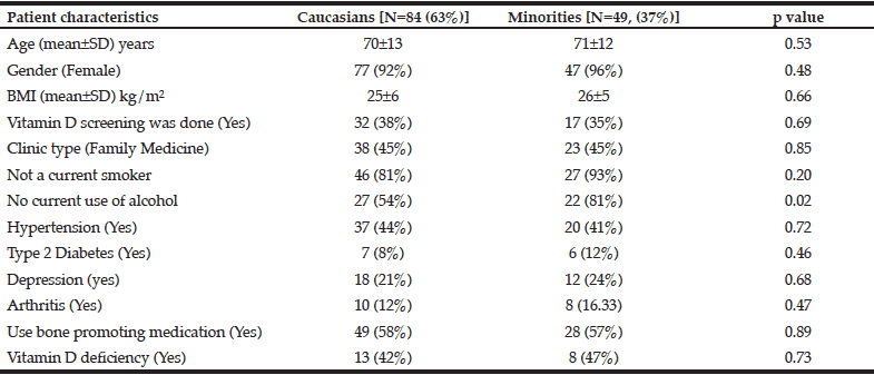
There were no statistically significant differences in patient characteristics including demographics, behavioral risk factors, co-morbidities, supplements and medication use for promoting bone health except for alcohol use between the Caucasian and non-Caucasian Minority groups (Table 1).  The mean age for the entire sample (N=133) was 71±13 years with a range of 51-97 years.  Most patients were Caucasians (63%), women (93%), and reported no current smoking (85%) or alcohol use (64%).  


Table 1 Patient characteristics by Race/Ethnic backgrounds

Notes: SD = standard deviation, BMI = body mass index


For Caucasian patients, significant association (p<0.05) was noted between vitamin D screening status and clinic type, and age (Table 2).  Among the Caucasian patients, more patients not screened for Vitamin D were seen in family medicine clinic (60%) than in geriatric clinic (40%). (Table 2). For minority population, age was the only variable showing a significant positive association with getting vitamin D screening (Table2).  
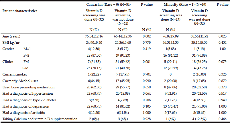
 

Table 2 Association between vitamin D screening status (yes and no) and other patient risk factors for Caucasian and Minority groups

Note: BMI = body mass index; M = male; F = female; FM = Family Medicine clinic; GM = Geriatrics clinic


When checked by type of clinic (Family Medicine vs. Geriatrics), screening was significantly lower for patients seen in family medicine clinic regardless of Ethnic/Racial background, while a higher percentage of Caucasian patients seen in geriatrics clinic were screened for vitamin D than those not screened.  For minority groups, more patients were not screened for vitamin D compared to those screened regardless of where they were seen (Family Medicine vs. Geriatrics) (Table 3).  


Table 3 Vitamin D screening (yes and no) by clinic types (family medicine vs. geriatrics) in Caucasians and minority groups


Older age was significantly associated with positive screening status for vitamin D in the logistic regression model for both Caucasian and minority patients (Table 4).  While older patients from both groups were more likely to be screened for vitamin D, no single patient characteristic remained significant after adding clinic type to the logistic models (Table 4).  


Table 4 Indicators of not screening for vitamin D in Caucasians and minority groups: results from two logistic regression models

* P<0.05. FM = family medicine clinic; GM= geriatrics clinic. Each determinant variable is adjusted for all others in the model. 


Discussions

Older patients from minority racial/ethnic groups have higher odds of not getting vitamin D screening when seen in primary care clinics for compromised bone health, e.g., osteopenia, osteoporosis and related fractures.  While significantly greater percentage (54%) of Caucasian patients were being screened for vitamin D in the geriatric clinics, non-Caucasian minority patients had a lower rate of vitamin D screening regardless of their clinic type (geriatrics vs. family medicine) (Table 3).  

Despite having multiple risk factors for bone loss including age, the diagnosis of having either osteopenia or osteoporosis or related fractures, and having darker skin, minority patients in our study remain at great danger for not being screened for vitamin D.  (5,15).  Management of osteoporosis remains incomplete without required supplementation of vitamin D which may not be possible without knowing the patients’ vitamin D levels (16).  Regardless of their race/ethnic background, very few patients were noted to take calcium and vitamin D supplementation in our study.  Due to the non-prescription nature of these supplements, it is difficult to accurately confirm this information.  Nonetheless, based on the electronic chart review, vitamin D screening is low in the at risk patient population in our study.   

Improved cardiac ejection fraction after optimizing vitamin D level in minority (African American) men with vitamin D deficiency and increased c-reactive protein in patients with low vitamin D are reasons to maintain good vitamin D levels in patients with chronic disease such as hypertension and diabetes (12-13).  Presence of the two common co-morbidities of hypertension and diabetes in our study patients who also suffer from compromised skeletal health indicates need for vitamin D screening to promote healthy aging in this patient population.  Although not statistically significant, a higher percentage of minority patients (47%) had vitamin D deficiency (a level <30 ng/ml) (16-17) compared to their Caucasian counterpart (42%) (Table 1).  Case identification, screening and diagnosis can improve skeletal health outcomes in clinics which strategically designed and emphasized specific guidelines for improving osteoporosis treatment (14,18-19).  These clinics identified at-risk patients, screened and treated them for osteoporosis as appropriate (18-19).  Evidence suggest that screening for vitamin D levels and treating any deficiency improves skeletal health in order to promote healthy aging in the minority older patients with multiple chronic disease  (12-13).  

Regardless of their race/ethnic backgrounds, more patients seen in family medicine clinic were not screened for vitamin D levels.  Competing priorities of other chronic diseases managed by PCPs and time constraints for each visit often make it difficult for the PCP to address silent diseases like osteoporosis and skeletal health (20-23); and yet these are the older patients who are most in need of optimum vitamin D levels (12-13).  While it may be that the PCPs in family medicine may consider vitamin D tests a specialty test, other issues may contribute to less testing by PCPs, such as, problem with insurance reimbursement, misconception about and lack of appropriate guidelines for screening, and lack of knowledge about current updates on vitamin D screening to maintain appropriate levels in patients with compromised skeletal health (1,20-24).

Percentages of Caucasian and minority (Table 1) patients seen in family medicine and geriatric clinics were similar.  Yet, more minorities were not screened in both clinics with significant differences in screening between Caucasians and minorities in the geriatric clinic (Table 3).  Our results show lower prevalence of screening regardless of the clinic type where they are seen.  While patient with older age (from both racial/ethnic backgrounds) were more likely to be screened, the significant association between old age and vitamin D screening became non-significant after adding clinic type (family medicine vs. geriatrics clinic) to the regression model (table 4).  Overall low rates of screening by family medicine had most likely influenced the outcome in the regression model.  Despite having multiple risk factors, e.g., older age, concurrent diagnosis of cardio-metabolic co-morbidities in the presence of compromised skeletal health and having darker (non-light) skin, minority patients are not being screened for the essential vitamin D which can improve skeletal health outcomes (16-17).  Recent USPSTF recommendation against routine screening for vitamin D in general population may contribute to a low screening rate in general (2).  Our results have led to educational interventions with a conscious effort to increase awareness of minority skeletal health in both family medicine and geriatric clinics.  

In agreement with current scientific literature, our data indicate lower rates of vitamin D screening in minority older patients who suffer from multiple known risk factors that can be improved with optimum vitamin D levels.  Among those who were screened for vitamin D levels in both primary care clinics (family medicine and geriatrics clinics), higher percentages of minority patients had vitamin D deficiency compared to the Caucasian patients of similar age and co-morbidities.  We recommend future studies aimed at determining and improving physician factors, e.g., knowledge and appropriate education, to improve vitamin D screening and supplementation in at-risk older minority patients with darker skin shades.  


Acknowledgement: This work was supported by a grant from the Texas Academy of Family Physicians (TAFP) and Herzstein Foundation.

Conflict of interest: None.

Ethics standards: The study was approved by IRB of the University of Texas Houston Health Science Center.


References

1. Rosen C and Taylor C.  Common misconceptions about vitamin D—implications for clinicians. Nat Rev Endocrinol 2013; 9:434-438.   

2. USPSTF (U.S. Preventative Services Task Force) report.  http://www.uspreventiveservicestaskforce.org/uspstf/uspsvitd.htm. Accessed August 16, 2015. 

3. Ginde A, Liu M, and Camargo Jr C.  Demographic differences and trends of vitamin D insufficiency in the US population, 1988-2004.  Arch Intern Med 2009; 169(6):626-632.

4. Powe C, Evans M, Wenger J, et al. Vitamin D-binding protein and vitamin D status of Black Americans and White Americans.  N Engl J Med, 2013; 369(21):1991- 2000. 

5. Weishaar T and Vergili JM. Vitamin D status is a biological determinant of health disparities.  J Acad Nutr Diet 2013; 113(5):643-651.

6. Peiris AN, Bailey BA, Peiris P, Copeland R, Manning T. Race and Vitamin D Status and Monitoring in Male Veterans. J Natl Med Assoc 2011; 103,492-497.

7. Grant W and Peiris A. Possible Role of Serum 25-Hydroxyvitamin D in Black–White Health Disparities in the United States.  J Am Med Dir Assoc 2010; 11:617-628.

8. Benson J and Skull S.  Hiding from the sun- vitamin D deficiency in refugees.  Aust Fam Physician 2009; 36:355-357.

9. Brock K, Wilkinson M, Cook R, Lee S, Bermingham M. Associations with vitamin D deficiency in ‘at risk” Australians.  Journal of Steroid Biochemistry and Molecular Biology 2004; 89-90:581-588.

10. Cauley JA. Public Health Impact of Osteoporosis.  J Gerontol A Biol Sci Med Sci 2013; 68(10):1243-1251.

11. Queally J, Kiernan C, Shaikh M, Rowan F, Shaikh M and Bennett D. Initiation of osteoporosis assessment in the fracture clinic results in improved osteoporosis management: a randomised controlled trial. Osteoporos Int 2013; 24:1089–1094.

12. Youssef D, El Abbassi AM, and Cutchins D.  Vitamin D deficiency: Implications for acute care in the elderly and in patients with chronic illness. Geriatr Gerontol Int 2011; 11:395-407.

13. Zia A, Komolafe B, Moten M, Ahokas R, McGee J, Rosenberg W, Bhattacharya S and Weber K. Supplemental vitamin D and calcium in the management of African Americans with heart failure having hypovitaminosis D.  Am J Med Sci 2011; 341:113-118.i

14. Rianon N, Anand D and Rasu R. Changing trends in osteoporosis care from specialty to primary care physicians.  Curr Med Res Opin 2013; 29(8):881-888.

15. National Osteoporosis Foundation (NOF).  www.nof.org. Accessed August 16, 2015.

16. Rosen C, Abrams S, Aloia J, et al. IOM committee members respond to Endocrine Society vitamin D guideline. J Clin Endocrinol Metab 2012; 97:1146-1152. 

17. Holick M, Binkley N, Bischoff-Ferrari H, et al. Guidelines for preventing and treating vitamin D deficiency and insufficiency revisited.  J Clin Endocrinol Metab 2012; 97(4):1153-1158.

18. van Helden S, Cauberg E, Geusens P, Winkes B, van der Weijden T, and Brink P. The fracture and osteoporosis outpatient clinic: an effective strategy for improving implementation of an osteoporosis guideline. J Eval Clin Pract 2007; 13:801–805.

19. Dell R. Fracture Prevention in Kaiser Permanente in Southern California. Osteoporos Int 2011; 22(3):457-460.

20. Lewiecki EM. Managing osteoporosis: challenges and strategies. Cleve Clin J Med 2009; 76(8):457–466.

21. McNearney TA, Shepherd AJ, Chhabra A and Goel N. Primary care house staff attitudes toward osteoporosis management. South Med 2006; 99 (5):461–466.

22. Simonelli C, Killeen K, Mehle S and Swanson L. Barriers to osteoporosis identification and treatment among primary care physicians and orthopedic surgeons. Mayo Clin Proc 2002; 77(4):334–338.

23. Solomon D, Connelly M, Rosen C, et al. Factors related to the use of bone densitometry: survey responses of 494 primary care physicians in New England. Osteoporos Int 2003; 14:123–129.

24. Rianon N, Murphy K, Guanlao R, Hnatow M, De Leon E and Selwyn B.  Suboptimal vitamin D screening in older patients with compromised skeletal health.  J Eval Clin Pract 2014; 20(2):144-148.

OSTEOPOROSIS IN THE COMMUNITY: FINDINGS FROM A NOVEL COMPUTERIZED REGISTRY IN A LARGE HEALTH ORGANIZATION IN ISRAEL


I. Goldshtein1,3, J. Chandler2, V. Shalev1,3, S. Ish –Shalom4, A.M. Nguyen2, V. Rouach5, G. Chodick1,3


1. Epidemiology and database research unit, Maccabi Healthcare Services, Tel Aviv, Israel; 2. Department of Epidemiology, Merck and Co, Inc, North Wales, PA; 3. Tel Aviv University, Israel; 4. Rappaport Faculty of Medicine, Technion-Israel Institute of Technology, Haifa, Israel; 5. Institute of Hypertension, Metabolism, and Endocrinology, Tel Aviv Souraski Medical Center, Tel Aviv, Israel

Corresponding Author: Inbal Goldshtein, Maccabi Healthcare Services, 27 Ha’Mered Street Tel Aviv 68125 Israel, Tel: 972 3 7952616   Fax: 972 73 2132831, E-mail: goldst_in@mac.org.il


Abstract

Background: Osteoporosis is a growing public health concern due to its rising prevalence and excess morbidity and mortality. Automated patient registries have gained great importance in health and disease management of major chronic diseases, but are rarely used in osteoporosis. Objectives: To construct an automated, population-based registry of osteoporosis. Setting: The electronic medical records and pharmacy databases of a 2 million member health organization in Israel (Maccabi Healthcare Services). Methods:  Included in the registry were adults who were diagnosed with osteoporosis diagnosis, had major osteoporotic fractures, or purchased relevant medications, between 2000 and 2013. In addition, we included patients with low bone density as extracted from over 140,000 measurements reports, using an automated optical character recognition (OCR) system. Two-thirds of the cases were validated by more than one inclusion criterion. Results:  A total of 118,141 osteoporosis patients were identified.  The point prevalence of osteoporosis among members aged 50 or above in 2013 was 19%. The mean age at registry entry was 62 (SD=12) and 66 (SD=14) years for females and males, respectively. The highest annual risk of developing osteoporosis (27 per 1000) was recorded among females aged 65-75. In 28% of the patients, there was no indication of treatment with osteoporosis therapy. Conclusions: To the best of our knowledge, this is one of the first real-world automated registries of osteoporosis. Similar registries may provide valuable data for real-time monitoring of trends, quality of care, and outcome research in osteoporosis and its complications. 

Key words: Osteoporosis, registry, prevalence.


Introduction 

With a growing elderly population and a prevalence of over 200 million people worldwide (1), the global epidemic of osteoporosis (OP) poses a serious public health concern. The major medical, social, and economic consequences of osteoporosis are due to osteoporotic fractures resulting in disability (2-4) and excess mortality (5). In white populations, the lifetime risk of fragility fracture is approximately 50% in females and 20% in males (6). 

In addition to fractures, other manifestations of osteoporosis such as low bone mineral density (BMD) and vertebral deformities have been strongly associated with increased risk of death from stroke (7), coronary heart diseases (8) and pulmonary diseases (9).

Considerable variations were observed between countries with regards to OP fracture rates (10-12), warranting the need for local estimations. The reason for such variations may lie in genetic factors or environmental factors, as well as differences in measurement and reporting systems, or differences in case-finding approach. Self-report survey studies, insurance claims-data, and hospital-based studies have shown to underestimate fracture risks (13-15). 

Israel has a social public health care system and developed sources of data that allow the population-based analyses on the burden of chronic diseases that can be generalized to other western-like populations (16-19). 

The available data (20-23) on the epidemiology of osteoporosis and mortality among patients with OP  fractures in Israel are partial and outdated and thus may not reflect important changes in recent years such as increased life expectancy and introduction of new anti-osteoporotic treatments.

The objective of the present study was to establish a registry of osteoporosis patients using automated collection of individual-level information on important characteristics[24] such as demography, co-morbid conditions, medication use, laboratory tests, and bone densitometry measurements. Specifically, the registry was analyzed to assess the current age-and-sex specific prevalence of osteoporosis, history of OP fractures, and a retrospective assessment of all-cause mortality among patients with OP fractures. 


Materials and methods

Settings 

This study utilized the longitudinal databases of Maccabi Healthcare Services (MHS), the second largest sick fund in Israel, ensuring 25% of the population with a nationwide and representative distribution. These databases are derived from electronic medical records (EMRs) of a stable population of over 2 million ensured members. Israel’s sick funds provide a uniform legally defined basket of services to which every citizen is entitled as a member of one of four nationwide health funds that are financed by government via age-related capitation payments (90% of total), patient charges and other income (10%).  Citizens are free to choose and move between health funds, which are not exclusive and must accept all applicants for memberships. .

BMD measurements were collected from the datasets of a single chain of medical centers (“Assuta Health Systems”), where approximately 86% of the dual-energy X-ray absorptiometry (DXA) scans in MHS are performed.  All DXA scans in these clinics are conducted using a standardized model of GE- lunar prodigy scanner. 

Study population

Eligible for the study registry were all MHS members aged 18 or above with any of the following inclusion criteria: a physician diagnosis of osteoporosis (according to the International Classification of Diseases version 9 with clinical modifications (ICD-9-CM) codes: 733.0-733.03, 733.09, 733.7) , or osteopenia (ICD-9-CM code: 733.9) validated by any fracture one year before or after date of diagnosis, OP-specific therapy (defined by a dispensed medication prescription of any of the following: Bisphosphonates, Raloxifene, Strontium Ranelate, Teriparatide or Calcitonin), a minimum t-score of -2.5 or less , or a clinical diagnosis of a major osteoporotic fracture (MOF) or hip fracture surgery. MHS database does not include data on trauma type, except for fractures of road accidents, thus we could only partially distinguish between osteoporosis related fractures and high-impact trauma fractures. Thus, to ensure high specificity of the registry we considered only MOF for females aged 50 or above or in males aged 60 or above for independent inclusion criteria. MOF includes fractures commonly associated with osteoporosis: closed fractures of femur, vertebral, colles and proximal humerus, which were not listed in the database as associated with a car accident. Other closed fracture diagnoses may be related to osteoporosis but not considered specific enough and were used only for descriptive analysis. 

Excluded from the registry were patients with Paget’s disease (ICD-9-CM code 731.0, n=587) or multiple myeloma (203.0, n=631) as indicated in the EMR or hospital discharge, patients whose only qualifying registry entry criteria was fracture and either suffered from multiple fractures at the same date (n=640, most likely overwhelming trauma and not OP) or fractures due to metastatic cancer (n=107), and patients whose only qualifying registry entry criteria was pamidronic or zoledronic acid purchase and who were previously diagnosed with cancer (cancer is an alternative indication to these drugs in addition to OP, n=301).

Registry entry date was defined as the first medical event (diagnostic code, associated drug prescription, lab value or procedure) which is consistent with any of inclusion and all exclusion criteria. 

Co-morbid conditions were defined by MHS registries for chronic major diseases such as ischemic heart disease (25), diabetes (26) and hypertension. Renal impairment at follow up was defined by last eGFR measurement <=35 or patient in the MHS dialysis registry. Recent depression was defined as at least 6 purchases of SSRI in the last 2 years.

Socioeconomic status (SES) was defined by the 2008 national census (27) according to the poverty index of the member’s enumeration area, ranging between 1 (lowest) to 20 (highest). 

Study periods 

Follow up date was defined as the earliest of disenrollment from MHS, death, or December 31, 2013. 

Inclusion period: between Jan 1st 2000 and Dec 31st 2013. 

Statistical analysis 

Age and sex specific prevalence rates were determined for December 2013. 95% Confidence intervals for osteoporosis prevalence were calculated using the exact binomial (Pearson-Klopper) method using R statistical software (28). 

Age-and-sex standardized morbidity ratios (SMRs) with 95% confidence intervals were computed to compare between the OP registry and the general population of MHS.

We calculated OP incidence density rates during the study period after excluding members diagnosed with OP less than 3 years prior to first year of follow-up, as well as members with less than 5 years of continuous membership in Maccabi before diagnosis, to ensure selection of newly developed cases. For incident cases of osteoporosis (or MOF), only the time until cohort entry (or MOF diagnosis) contributed to the denominator of patient-years at risk.

We assessed primary incidence of MOF in MHS. Clear distinction between reports on incident events and follow-up of previous events is often difficult. High rates of surgery complications, delayed healing, recurrent complaints on past events[29], and the need for follow-up, may limit the accuracy of previous assessments of incidence. Thus we assessed only primary incidence, which cannot be compared to non-primary annual rates.

The study was approved by the Assuta hospital’s institutional review board.

Results

A total of 118,141 eligible patients were identified for the registry, with approximately 6,000 incident cases annually (table 1). The mean duration of follow-up since first indication of osteoporosis was 7.9 years (SD=4.5). A total of 66% of the registry’s patients met two or more inclusion criteria, validating their OP status (Appendix A).  


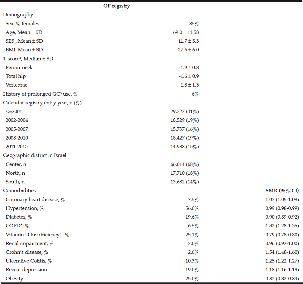
Table 1 Demographics and comorbidities of osteoporosis patients with age and sex standardized morbidity ratios (SMR) compared to the general population in Maccabi healthcare services, December 2013

*Chronic obstructive pulmonary disease;  3. Socioeconomic status, ranging between 1 (lowest) to 20 (highest). See text for details;  4. Latest available measurement; 5. Systemic glucocorticoids; 6. Vitamin D deficiency was defined as <20 ng/ml. The calculation was out of non-missing data. 12% of the registry and 30% of the general population in Maccabi had missing vitamin D level.


Table 2 Age and sex specific prevalence rates per 1000 and 95% confidence intervals of osteoporosis in Maccabi healthcare services, Israel, 2013


Prevalence and incidence rates in MHS

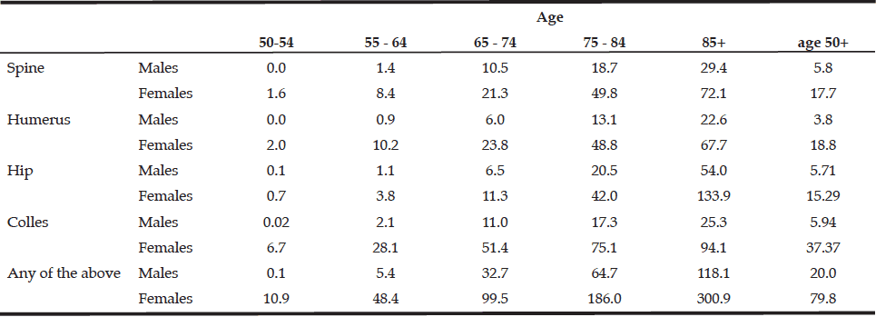
The crude prevalence rates of osteoporosis among members aged 50 or above in MHS were 31.4% and 5.9% among females and males, respectively (table 2). Prevalence of MOF history increased exponentially with age reaching 30.1% in females and 11.8% in males at the age of 85 years or above (table 3).


Table 3 Age and sex specific prevalence rates (per 1000) of major osteoporotic fractures in Maccabi healthcare services, Israel, 2013


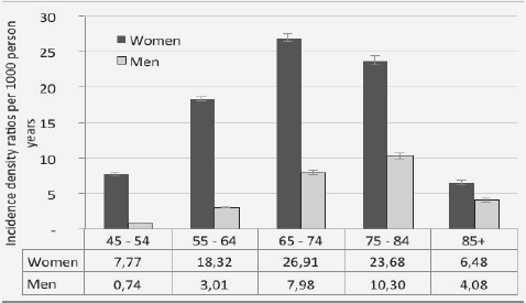
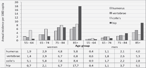
Figure 1a Age and sex specific incidence density rates and 95% confidence intervals of osteoporosis (per 1000 person years) in Maccabi healthcare services, 2003-20137

7. Error bars indicate 95% confidence intervals.


The highest incidence rate of OP was recorded at the age group of 65-74 and 55-64 among males and females, respectively (figure 1.a). The risk of MOF increases with age (figure 1.b). 

Demographics and comorbidities of OP registry patients

In 2013, the mean age of OP patients in MHS was 69.0 years old (SD=11.6), with females accounting for 85% of the registry (table 1). Compared with the age and sex adjusted general population of MHS, OP patients were more likely to have diagnosed history of COPD, Crohn’s disease, ulcerative colitis, and recent depression, and less likely to be obese, diabetic or have vitamin D insufficiency.


Figure 1b Annual age and sex specific incidence (per 1000 capita) of primary major osteoporotic fractures in Maccabi healthcare services, 2013


History of fractures in OP registry

Among OP patients with an indication of MOF history (29%), the most common fractures were hip among males (36.2%) and colle’s among females (37.3%). 

In addition, in 12% of the registry, we found an indication of non-MOF fractures that are likely to be related with osteoporosis (e.g. metatarsal, ribs, and ankle) which were not inclusion criteria for the registry. 

All-cause Mortality after OP fracture

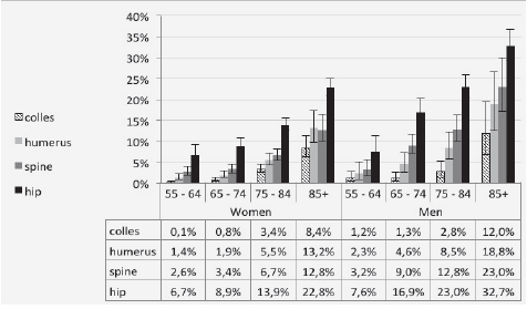
Figure 1 depicts all-cause mortality rates within 1 year from date of MOF.  The highest mortality rates (23% in females and 32% in males) were observed among elderly patients (85 year or above) with a hip fracture.

Anti-osteoporosis therapy 

A total of 72% of the registry had at least one dispensation of osteoporosis medication ever. Among incident cases, treatment initiation rates were significantly higher (P<0.001) in females (75.3%) compared to males (62.1%). Only 24% of patients with hip fracture who were Anti-osteoporosis therapy naïve, initiated treatment after the fracture occurrence (22% were already treated before fracture event). This proportion is higher after vertebral fractures (38%) and lower after colles or humerus fractures (21%).


Figure 2 Age and sex specific all-cause mortality rates within 1 year from osteoporotic fracture event, Maccabi healthcare services, Israel, 2000-2013


Bone density measurements

Approximately 72% of the OP registry patients had an indication of performing a DXA scan. BMD values were electronically available for 52% of them (37% of the total registry).  Among the patients with t-scores of -2.5 or lower:  55% were <=-2.5 at femoral neck, 40% at spine and 5% total hip. In all but 3% (n=406) the femur neck indicated at least osteopenia. Mean values of DXA measurements at follow-up are given in table 1.


Discussion

The present study described the establishment of an osteoporosis patient registry in a large and representative health organization in Israel, and the first population-based report of the epidemiology of this disease in the country. 

The results of the study reveal the immense burden of osteoporosis in Israel, with prevalence rates of 61% and 19% among females and males aged 75 or above. The observed OP prevalence was comparable to the findings of other large registries in Europe such as “BEST” (30) and “BoneEVA” (31). An Israeli survey (23) reported a lower prevalence (32% vs. 49% among females aged 65), possibly explained by the limitation of self-report data, as well as reduced awareness at that period (1999). The prevalence of diagnosed OP concords with a previous Israeli study (22) (14% vs. 13% among females aged 45-74, results not shown).  

In Israel, the remaining life expectancy of men and women at age 50 is approximately 31 and 35 years, respectively (CBS 2014). Therefore, according to our age-specific prevalence, the respective estimated OP-fracture lifetime risks in Israeli men and women are 6% and 30%,in accordance with previous international assessments (32, 33). Similar to previous large cohort studies (34, 35) our results indicate that while the absolute risk of hip fractures is two-fold higher among females, they account for a greater proportion of MOF among males. Similar to previous studies (36, 37), men were found to have a significantly poorer survival after hip fracture compared to women, although residual confounding by co-morbid conditions should be examined in future studies. 

In agreement with previous analyses among OP patients, we found increased SMRs for known risk markers such as COPD (38) and inflammatory bowel disease (IBD) (39) compared to the general population. Obesity and diabetes were decreased in the OP registry compared to the general population of MHS, with a BMI distribution similar to a recent large US cohort (40). 

This study has several major strengths such as a large and unselected population, a relatively long retrospective follow-up, standardized densitometry measurements, and automated collection of comprehensive data, including medical diagnosis, prescribed therapies, and lab tests. To our knowledge, this is the first registry of its kind in Israel, and one of the largest worldwide. We plan to update it periodically, identify new cases that meet the selection criteria in MHS and monitor outcomes.

Some limitation should be discussed, such as missing data on fracture circumstances, specifically to distinguish between OP related vs. high-impact trauma fractures. Our objective was high specificity of the registry, thus only typical/characteristic OP fractures were used as independent inclusion criteria, and multiple concurrent fractures were excluded (rarely OP-related, previously assessed as 2.5% of OP fractures (21)). In addition, pre-defined age cut-offs were used to identify MOF. The asymmetric age cut-offs between males and females (60+ vs. 50+ respectively) may yield somewhat lower sensitivity to detect males compared to females. Similarly to other retrospective population-based studies relying on clinical diagnoses, our data is likely to underestimate the rates of vertebral compression fractures (6), as these fractures are universally underdiagnosed.

Building a valid and efficient algorithm to detect patients for a central registry requires careful examination and assessment of the variety of different measures and definitions (41). The majority of the registry was validated by meeting at least 2 inclusion criteria. The remainder may be explained by low awareness for documentation and treatment initiation, non-compliance of patients, partial electronic availability of bone density values, or (less frequently) due to contra-indication of therapy. 

We believe this up-to-date registry can serve as a valuable resource for future studies in the field of osteoporosis and improve our understanding of the risks and prevention of osteoporosis. 


Funding: The study was funded by Merck and Co.

Conflict of interest: Sofia Ish-Shalom has received research grants and consulting, advisory board, lecture fees, and any combination of the three from Merck Sharp & Dohme, Eli Lilly, Enterabio, GlaxoSmithKline, and Novartis. Vanessa Rouach has received lecture fees from Eli Lilly, GlaxoSmithKline, and Novartis. Julie Chandler and Allison Martin Nguyen are employees of Merck and Co. Inbal Goldshtein, Varda Shalev and Gabriel Chodick declare that they have no conflict of interest.

Acknowledgments: We would like to thank Mrs. Racheli Katz and Mrs. Esma Herzel of Meaccbi Healthcare Services, for their considerable contribution to data collection. This work was performed in partial fulfillment of the requirements for a Ph.D. degree of Mrs. Inbal Goldshtein, Faculty of Management, Tel Aviv University, Israel.


References

1.  Cooper C, Campion G, Melton LJ, 3rd.Hip fractures in the elderly: a world-wide projection. Osteoporos Int 1992;2:285-289

2.  Cummings SR, Melton LJ. Epidemiology and outcomes of osteoporotic fractures. Lancet 2002;359 (9319):1761-1767. 

3.  Gullberg B, Johnell O, Kanis JA. World-wide projections for hip fracture. Osteoporos Int 1997;7 (5):407-413

4.  Johnell O, Kanis JA. An estimate of the worldwide prevalence, mortality and disability associated with hip fracture. Osteoporos Int 2004;15:897-902. 

5.  Abrahamsen B, van Staa T, Ariely R, Olson M, Cooper C. Excess mortality following hip fracture: a systematic epidemiological review. Osteoporos Int 2009;20:1633-1650. 

6.  Sambrook P, Cooper C. Osteoporosis. Lancet 2006;367:2010-2018. 

7.  Nordstrom A, Eriksson M, Stegmayr B, Gustafson Y, Nordstrom P. Low bone mineral density is an independent risk factor for stroke and death. Cerebrovasc Dis 2010;29:130-136. 

8.  Kado DM, Browner WS, Blackwell T, Gore R, Cummings SR. Rate of bone loss is associated with mortality in older women: a prospective study. J Bone Miner Res 2000;15:1974-1980. 

9.  Hasserius R, Karlsson MK, Nilsson BE, Redlund-Johnell I, Johnell O. Prevalent vertebral deformities predict increased mortality and increased fracture rate in both men and women: a 10-year population-based study of 598 individuals from the Swedish cohort in the European Vertebral Osteoporosis Study. Osteoporos Int 2003;14:61-68.

10.  Dhanwal DK, Cooper C, Dennison EM. Geographic variation in osteoporotic hip fracture incidence: the growing importance of asian influences in coming decades. J Osteoporos 2010:757102.

11.  Elffors I, Allander E, Kanis JA, Gullberg B, Johnell O, Dequeker J, Dilsen G, Gennari C, Lopes Vaz AA, Lyritis G, et al. The variable incidence of hip fracture in southern Europe: the MEDOS Study. Osteoporos Int 1994;4:253-263

12.  Ismail AA, Pye SR, Cockerill WC, Lunt M, Silman AJ, Reeve J, Banzer D, Benevolenskaya LI, Bhalla A, Bruges Armas J, Cannata JB, Cooper C, Delmas PD, Dequeker J, Dilsen G, Falch JA, Felsch B, Felsenberg D, Finn JD, Gennari C, Hoszowski K, Jajic I, Janott J, Johnell O, Kanis JA, Kragl G, Lopez Vaz A, Lorenc R, Lyritis G, Marchand F, Masaryk P, Matthis C, Miazgowski T, Naves-Diaz M, Pols HA, Poor G, Rapado A, Raspe HH, Reid DM, Reisinger W, Scheidt-Nave C, Stepan J, Todd C, Weber K, Woolf AD, O’Neill TW. Incidence of limb fracture across Europe: results from the European Prospective Osteoporosis Study (EPOS). Osteoporos Int 2002;13:565-571. 

13.  Gerdhem P, Akesson K. Rates of fracture in participants and non-participants in the Osteoporosis Prospective Risk Assessment study. J Bone Joint Surg Br 2007;

14.  Smith MR, Lee WC, Brandman J, Wang Q, Botteman M, Pashos CL. Gonadotropin-releasing hormone agonists and fracture risk: a claims-based cohort study of men with nonmetastatic prostate cancer. J Clin Oncol 2005;23:7897-7903. 

15.  Unnanuntana A, Gladnick BP, Donnelly E, Lane JM. The assessment of fracture risk. J Bone Joint Surg Am 2010;92:743-753. 

16.  Bellamy L, Casas JP, Hingorani AD, Williams D. Type 2 diabetes mellitus after gestational diabetes: a systematic review and meta-analysis. Lancet 2009;373:1773-1779

17.  Chodick G, Elchalal U, Sella T, Heymann AD, Porath A, Kokia E, Shalev V The risk of overt diabetes mellitus among women with gestational diabetes: a population-based study. Diabet Med 27:779-785

18.  Chodick G, Shalev V, Gerber Y, Heymann AD, Silber H, Simah V, Kokia E. Long-term persistence with statin treatment in a not-for-profit health maintenance organization: a population-based retrospective cohort study in Israel. Clinical therapeutics 2008;30:2167-2179

19.  Mann DM, Woodward M, Muntner P, Falzon L, Kronish I Predictors of nonadherence to statins: a systematic review and meta-analysis. The Annals of pharmacotherapy 44:1410-1421

20.  Givon U. Consensus conference on the treatment and rehabilitation of hip fractures in the elderly. Isr Med Assoc 1999;J 1:131-136

21.  Liel Y, Castel H. [Incidence of osteoporotic fractures in southern Israel]. Harefuah 2004;143:18-21

22.  Nitzan-Kaluski D, Chinich A, Ifrah A, Merom D, Green M. Correlates of osteoporosis among Jewish and Arab women aged 45-74 in Israel: national women’s health interview survey. The journal of gender-specific medicine 2002;6:17-23

23.  ICDC, Ministry of Health (2004) National Health Survey in Israel – a WHO project, European Sector. EUROHIS 2004  publication no 237.

24.  David C, Confavreux CB, Mehsen N, Paccou J, Leboime A, Legrand E (2010) Severity of osteoporosis: what is the impact of co-morbidities? Joint Bone Spine 2010;77 Suppl 2:S103-106. 

25.  Shalev V, Chodick G, Goren I, Silber H, Kokia E, Heymann AD. The use of an automated patient registry to manage and monitor cardiovascular conditions and related outcomes in a large health organization. Int J Cardiol 2011;152:345-349. 

26.  Chodick G, Heymann AD, Shalev V, Kookia E. The epidemiology of diabetes in a large Israeli HMO. Eur J Epidemiol 2003;18:1143-1146

27. Israel Central Bureau of Statistics  (2013) Characterization of geographical units by the socio-economic level of the population 2008. http://www.cbs.gov.il/webpub/pub/text_page_eng.html?publ=100&CYear=2008&CMonth=1 Accessed 19 October 2014

28.  Friedman DS, Quigley HA, Gelb L, Tan J, Margolis J, Shah SN, Kim EE, Zimmerman T, Hahn SR. Using pharmacy claims data to study adherence to glaucoma medications: methodology and findings of the Glaucoma Adherence and Persistency Study (GAPS). Invest Ophthalmol Vis Sci 2007;48 :5052-5057.

29.  Donaldson LJ, Reckless IP, Scholes S, Mindell JS, Shelton NJ. The epidemiology of fractures in England. J Epidemiol Community Health 2008;62:174-180. 

30.  Hadji P, Klein S, Gothe H, Haussler B, Kless T, Schmidt T, Steinle T, Verheyen F, Linder R. The epidemiology of osteoporosis–Bone Evaluation Study (BEST): an analysis of routine health insurance data. Dtsch Arztebl Int 2013;110:52-57. 

31.  Haussler B, Gothe H, Gol D, Glaeske G, Pientka L, Felsenberg D. Epidemiology, treatment and costs of osteoporosis in Germany–the BoneEVA Study. Osteoporos Int 2007;18:77-84. 

32.  Holroyd C, Cooper C, Dennison E. Epidemiology of osteoporosis. Best Pract Res Clin Endocrinol Metab 2008;22 (5):671-685. 

33.  Hopkins RB, Pullenayegum E, Goeree R, Adachi JD, Papaioannou A, Leslie WD, Tarride JE, Thabane L. Estimation of the lifetime risk of hip fracture for women and men in Canada. Osteoporos Int 2012;23:921-927. 

34.  Brauer CA, Coca-Perraillon M, Cutler DM, Rosen AB. Incidence and mortality of hip fractures in the United States. JAMA 2009;302:1573-1579. 

35.  Feldstein A, Elmer PJ, Orwoll E, Herson M, Hillier T. Bone mineral density measurement and treatment for osteoporosis in older individuals with fractures: a gap in evidence-based practice guideline implementation. Arch Intern Med 2003;163:2165-2172. 

36.  Bliuc D, Nguyen ND, Milch VE, Nguyen TV, Eisman JA, Center JR. Mortality risk associated with low-trauma osteoporotic fracture and subsequent fracture in men and women. JAMA 2009;301:513-521. 

37.  Center JR, Nguyen TV, Schneider D, Sambrook PN, Eisman JA. Mortality after all major types of osteoporotic fracture in men and women: an observational study. Lancet 1999;353:878-882. 

38.  Lehouck A, Boonen S, Decramer M, Janssens W. COPD, bone metabolism, and osteoporosis. Chest 2011;139:648-657.

39.  Bernstein CN, Blanchard JF, Leslie W, Wajda A, Yu BN. The incidence of fracture among patients with inflammatory bowel disease: a population-based cohort study. Annals of internal medicine 2000;133:795-799

40.  O’Malley C, Tran N, Zapalowski C, Daizadeh N, Olenginski T, Cauley J. Multimorbidity in women with and without osteoporosis: results from a large US retrospective cohort study 2004–2009. Osteoporosis International: 2014;1-14

41.  Gliklich R, Dreyer N. Registries for Evaluating Patient Outcomes. Agency for Healthcare Research and Quality, Rockville, MD, 2010


Appendix A Distribution of inclusion criteria in the Maccabi healthcare services osteoporosis registry

nn1

HIP FRACTURE IN A DEVELOPING COUNTRY: A PICTURE IN NEED OF CHANGE

 E.I.O. Vidal1, D.C. Moreira-Filho2, R.S. Pinheiro3, R.C. Souza4, L.M. Almeida5, K.R. Camargo Jr6, P.J.F. Villas Boas1, F.B. Fukushima7, C.M. Coeli3


1. Geriatrics Division – Internal Medicine Department – Universidade Estadual Paulista (UNESP); 2. Preventive and Social Medicine Department – Universidade Estadual de Campinas (UNICAMP); 3. Instituto de Estudos em Saúde Coletiva (IESC) – Universidade Federal do Rio de Janeiro (UFRJ); 4. Faculdade de Ciencias Medicas – Universidade Estadual do Rio de Janeiro (UERJ); 5. Epidemiology Division – Instituto Nacional do Câncer (INCA); 6. Social Medicine Institute – Universidade Estadual do Rio de Janeiro (UERJ); 7. Anesthesiology Department – Universidade Estadual Paulista (UNESP)

Corresponding Author: Edison Iglesias de Oliveira Vidal, Departamento de Clínica Médica, Faculdade de Medicina de Botucatu – UNESP, 18618-970 , Botucatu – SP – Brazil, E-mail: eiovidal@fmb.unesp.br


Abstract

Objectives: To describe the clinical profile, patterns of care and mortality rates of aged patients who have undergone hip fracture surgical repair. Design: Retrospective patient record study. Setting: A public university hospital in Rio de Janeiro, Brazil. Participants: 352 patients aged 60 and older who underwent surgery for hip fracture between 1995-2000. Measurements: Sociodemographic data, type of fracture, cause of fracture, time from fracture to surgery, physical status, Charlson comorbidity index, type of surgery and anesthesia, access to in-hospital physiotherapy, use of antibiotic and thromboembolism prophylaxis, and mortality within one year after hospital admission. Results: Among 352 subjects, 74.4% were women. The mean age overall was 77.3 years. Very long delays from the time of fracture to hospital admission (mean 3 days) and from hospital admission to surgery (mean 13 days) were observed. Most femoral neck fractures (82.7%) were managed by hip arthroplasties, while 92.8% of the intertrochanteric fractures underwent internal fixation procedures. Less than 10% of patients received in-hospital physiotherapy. Mortality rates 30 days, 90 days and one year after hospital admission were 3.4%, 8.0% and 13.4%, respectively. Conclusion: Our study provides evidence within the context of a developing country of major gaps in the quality of care of vulnerable older adults who suffered a hip fracture. Our findings suggest that hip fracture has not been treated as an urgent condition or a priority within the Brazilian public healthcare system. Further research should address current patterns of care for hip fracture in Brazil and in other developing countries.


Key words: Hip fractures, osteoporosis, quality of health care, developing countries, Brazil.


Introduction

Hip fracture represents the most severe consequence of osteoporosis and a major cause of morbidity, institutionalization and mortality for older adults worldwide (1–4). Around the Globe there is great variability concerning the incidence of hip fracture and its related mortality (5–8). Even though the greatest increase in the incidence of hip fracture is expected to occur in the developing countries of the world, those are also the regions from where less information is available on the epidemiology of those fractures (6, 8). There is particularly few data concerning Latin American older adults with hip fracture (8–11). More data on the epidemiology of those fractures is fundamental for the design of age-friendly public policies in those countries, where population aging is a relatively new phenomenon. Therefore, we conducted a study to describe the clinical profile, the patterns of care, and mortality rates of individuals aged 60 and older who underwent surgical repair of a hip fracture at a public university hospital in the city of Rio de Janeiro, Brazil.


Methods

The medical records of all patients aged 60 years and older admitted with a primary diagnosis of hip fracture (first three digits of International Classification of Diseases, 9th revision, ICD-9, code 820) between January 1st, 1995 and December 31st, 2000 were retrospectively reviewed. Patients with pathological hip fracture related to malignancy or who did not undergo surgical repair were excluded.

Review of medical records was performed using a standardized data abstraction form, which was completed by trained medical students under the supervision of a senior medical researcher (LMA). Before being used for this research the abstraction form was pretested with a sample of medical charts and corrections were implemented in order to facilitate the abstraction process and minimize bias. The medical supervisor reviewed all data for inconsistencies and medical records were reappraised accordingly. The same professional was responsible for the insertion of all data into the database.

To assess mortality rates within one year after hospital admission, records were linked to the database of the Brazilian Mortality Information System from January 1st, 1995 to December 31st, 2001 using Probabilistic Record Linkage Methodology (9, 12–16). Previous research in a similar setting revealed 85.5% sensitivity, 99.4% specificity, 98.1% positive predictive value and 94.9% negative predictive value for correct matching of records between databases using this methodology (17). RecLink II Software (18) was used to implement the Probabilistic Record Linkage Methodology followed by manual examination of pairs of records with higher probability of representing a true match between databases.

Frequency tables were created for the following variables: sex, age, income strata, marital status, living arrangements, type of hip fracture, type of injury leading to the fracture, type of surgical treatment and anesthesia, prophylaxis against venous thromboembolism and surgical infections (i.e. prophylactic antibiotic regimens), access to in-hospital physiotherapy care, comorbidities as ascertained by the Charlson comorbidity index (19), and American Society of Anesthesiology (ASA) physical status score. Statistical analyses were restricted to the presentation of simple frequencies and the calculation of 95% confidence intervals according to standard methods (20). The R software (version 2.10.1) was used for such purposes (21).

The present research was approved by the ethics committee of the Public Health Studies Institute of the Universidade Federal do Rio de Janeiro. Because of the retrospective nature of the study involving patients’ medical records and anonymous treatment of data, the ethics committee waived the requirement for informed consent.


Results

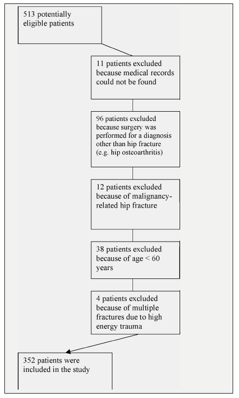
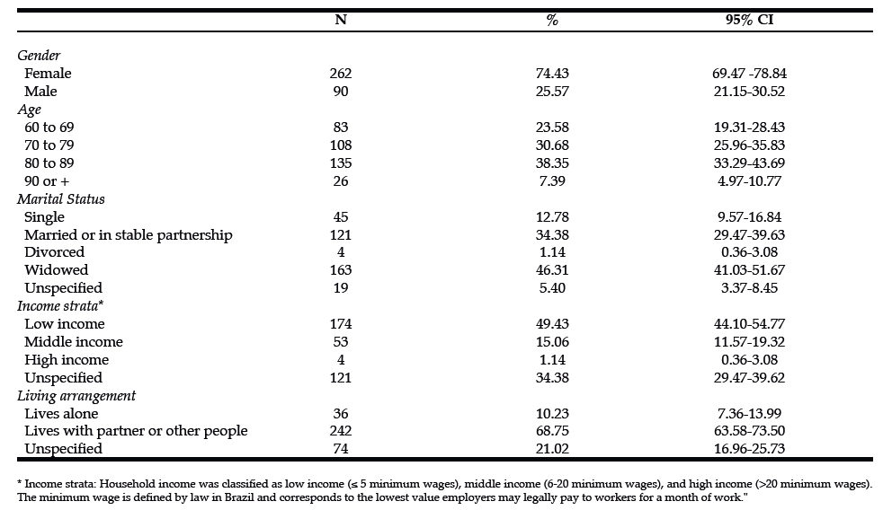
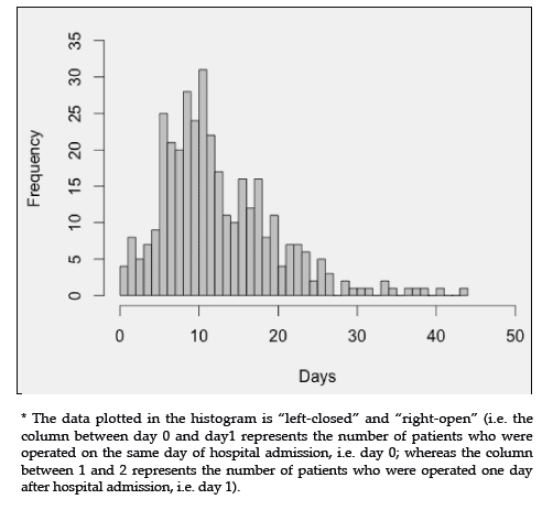
Figure 1 shows the flow diagram of the inclusion of patients in the study. Among the 352 patients fulfilling the proposed inclusion criteria, there were 262 (74.4%) women. The mean age overall was 77.3 years and women were mean 3.7 years older than men (mean ages 78.2 and 74.5 years, respectively; P = 0.001). The mean and median lengths of hospital stay were 21 and 17 days, respectively, with an interquartile range of 14 to 24 days. The mean and median times from the occurrence of hip fracture to hospital admission were 3 and 1 days, respectively, with an interquartile range of 0 to 4 days. The mean and median times from hospital admission to surgery were 13 and 11 days, respectively, with an interquartile range of 8 to 17 days. Figure 2 shows the distribution of time from hospital admission to surgery. Table 1 depicts the sociodemographic characteristics of the patients. Table 2 shows the clinical profile of patients including number of comorbidities, types and causes of hip fracture, Charlson comorbidity index and ASA physical status score. Table 3 displays the frequencies of surgical and anesthetic approaches adopted, as well as the frequencies of in- hospital physiotherapy, antibiotic and thromboembolism prophylaxis. Most femoral neck fractures (82.7%) were managed by hip arthroplasties, while 92.8% of the intertrochanteric fractures and 96.2% of the subtrochanteric fractures underwent internal fixation procedures. In-hospital mortality was 5.4%. Mortality rates 30 days, 90 days and one year after hospital admission were 3.4%, 8.0% and 13.4%, respectively.


Figure 1: Flow diagram of inclusion of patients in the study.


Table 1: Sociodemographic characteristics of patients.


Figure 2: Histogram of the interval of time* from hospital admission to surgery for 352 patients who underwent surgical repair of a hip fracture between 1995-2000.


Discussion

Probably, the most striking finding of the current research was the occurrence of mean intervals of time from fracture to hospital admission and thereafter to surgery of 3 and 13 days, respectively. Those intervals of time from fracture to surgery are remarkably different from those reported in developed countries, where the vast majority of patients undergo surgery within the first 48h of hospital admission (22–27). Even though there is still some degree of debate over the association between surgical timing and patient mortality after a hip fracture (25, 28, 29), there is wide consensus in the literature that hip fracture patients should be operated on as early as possible after hospital admission, provided there are no conditions that can be corrected or improved prior to surgery, since long waiting times for surgery are associated with pain, pressure ulcers, long hospital stays, distress, and delayed mobilization (23, 25, 27–30). We have recently shown for the same context of care that those long delays from fracture to hospital admission are associated with increased mortality risk (31).

Less than 10% of patients received in-hospital physiotherapy care, which is significantly divergent from current recommendations for early mobilization for most patients following the surgical repair of a hip fracture (32, 33). Such a low frequency of in-hospital physiotherapy care also mirrors important limitations in the access to optimal healthcare resources by older adults, since functional recovery after hip fracture is highly dependent on early rehabilitation after surgery (34). Nevertheless, almost 95% of patients received thromboembolism and antibiotic prophylaxis, which are significantly easier interventions to implement within any institution than the organization of post-surgical rehabilitation resources.

The current observations of long delays to hospital admission and to surgery above the standard of care in developed countries (i.e. surgery within 48h of hospital admission), and the remarkably low frequency of in- hospital physiotherapy care indicate that, even though hip fracture is associated with lower survival rates than that of most invasive cancers pooled together (35), it has not been treated as an urgent condition or a public health priority in Brazil. Because there is evidence that injuries and surgical conditions represent a problematic and neglected aspect of healthcare in developing countries we believe that similar and even worse patterns of care are likely in other developing regions of the World (36–40). This perspective is alarming since those fractures represent an enormous burden for society and because the greatest increase in the incidence of hip fracture is predicted to take place in the developing countries of the world (6, 8).

We hypothesize that those findings might reflect a picture of ageism within the Brazilian healthcare system (41, 42), where older adults have less access to more costly procedures within the public healthcare system than younger individuals, as has been shown by others (43). We hypothesize several other reasons that could explain the findings of delayed surgical timing and low physiotherapy frequency within our study. First, surgical procedures for hip fracture may have been scheduled as elective instead of urgent procedures, and therefore occurred according to operating theater availability, without prioritizing those vulnerable patients. Second, it is possible that in Brazil the lay public and even many healthcare professionals are frequently unaware of the often life-threatening meaning of a hip fracture for older people. Those hypotheses warrant further investigation by future studies. Although universal access to public health care in Brazil has been legally established since 1988, patients still often suffer from suboptimal care in several areas of healthcare provision (44). While the population is aging rapidly, the public healthcare system is still struggling to recognize and adapt to the needs of older people (45).

High rates of arthroplasty procedures for femoral neck fractures were observed. This finding is probably related to the large intervals of time from fracture to surgery, since those delays are associated with increased risk of fracture displacement and avascular necrosis of the head of the femur, and therefore represent a clear indication for hip arthroplasty (46).

The majority of patients (74.1%) underwent neuroaxial anesthesia (i.e. spinal or epidural), which represents a somewhat different pattern than that observed in many other regions of the Globe where general anesthesia usually represents a larger share of the anesthetic procedures performed for hip fracture patients (47–49). The debate over what type of anesthetic procedure is best suited for hip fracture patients does not seem to be resolved. Notwithstanding, recent systematic reviews disclosed lower mortality 30 days after surgery, lower incidence of deep venous thrombosis and lower rates of postoperative mental confusion for hip fracture patients who underwent neuroaxial anesthesia than for those submitted to general anesthesia (49, 50).

The one-year mortality rate observed (13.4%) was much lower than the 21.5% mortality rate described previously in a study encompassing all public hospitals in the city of Rio de Janeiro (9) and lies in the lower limits of mortality reported for hip fracture around the world (1, 2). The finding of low patient mortality concomitant to markers of suboptimal patient care discussed in the previous paragraphs may seem paradoxical at first. However, this apparent paradox can be explained by several factors, as follows. First, University hospitals have been shown to be associated with lower mortality rates for hip fracture than general hospitals, even though they often display longer intervals of time from hospital admission to surgery than community hospitals (24). Second and most important, selection bias must be strongly considered as a reason for the discrepancy between inadequate patterns of care and low mortality rates. Frailer and sicker patients, who had been admitted with a hip fracture to a community hospital without hip fracture surgical capability, were likely not considered fit to be transferred to the university hospital under study or died before they could be transferred, hence creating selection bias. Two observations are consistent with this last hypothesis: (a) most patients in this study were attributed a low Charlson comorbidity index and only roughly one third of patients were considered to have a severe systemic disease as ascertained by the ASA physical status classification; (b) the patient population was relatively younger than usually reported by most studies from developed countries (24, 51, 52).

At least 49.4% of patients in our study belonged to a low socioeconomic stratum. About 60% of patients were widowed, divorced or single. As usual in epidemiological studies about hip fracture among older adults, most patients (74.4%) were women. Even though the relationship between socioeconomic status and risk of falls is debated (53), there is evidence that lower socioeconomic status is associated not only with increased incidence of hip fracture (54–57) but also with increased mortality after fracture (58). Divorced, widowed and unmarried status have also been reported by others to be associated with increased risk of hip fracture (56, 59). The association between low socioeconomic status and increased risk of hip fracture may be related to several factors ranging from decreased bone mineral density and underlying health behaviors to environmental influences (55).

Several limitations of this study must be considered. Since its design was based on the retrospective abstraction of medical records, we had limited or no access to some data on important aspects of patients’ baseline characteristics and outcomes, such as socioeconomic status and functional outcomes. Second, review of medical records is often associated with error and bias, which can never be completely discarded in studies like ours. Nevertheless, most of the data that we collected for this analysis was relatively straightforward (e.g. date of hospital admission, date of surgery and record of occurrence of in-hospital physiotherapy care) and an experienced medical supervisor worked in close contact with the chart reviewers in order to attempt to minimize bias. In addition, other researchers have conducted valuable studies about the epidemiology of hip fractures using similar methods (60, 61). Third, limitations in methodology mean that the present results are not generalizable to the whole city of Rio de Janeiro or to other regions of the country. It is nevertheless reasonable to presume that the current findings have significant similarities to the patterns of care in other public hospitals in the same region, which are part of the same public healthcare system. Fourth, our results are at least 10 years old and a note of caution should be added to their generalizability to current practice. However, more recent studies about hip fracture among older adults from other regions of Brazil also showed long surgical delays, which suggests that the substandard patterns of care we have reported might still be common and may require urgent public health attention (62, 63).


Conclusions

Our study provides evidence within the context of a developing country of major gaps in the quality of care of vulnerable older adults who sustained a hip fracture between 1995 and 2000. Those findings suggest that hip fracture has not been treated as an urgent condition or a priority within the Brazilian public healthcare system. Further research should address the current patterns of care for hip fracture in the elderly both in Brazil and in other developing countries. Since our findings have been at least in part replicated by more recent studies conducted in other regions of our country, we believe that urgent public health attention is warranted towards the care of older adults sustaining a hip fracture in Brazil.


References


  1. Cummings SR, Melton LJ. Epidemiology and outcomes of osteoporotic fractures. Lancet 2002;359(9319):1761–7

  2. Johnell O, Kanis J. An estimate of the worldwide prevalence, mortality and disability associated with hip fracture. Osteoporos Int 2004;15:897–902

  3. Johnell O. The socioeconomic burden of fractures: today and in the 21st century. Am J Med 1997;103(2A):20S–25S; discussion 25S–26S

  4. Fisher AA, Davis MW, Rubenach SE, Sivakumaran S, Smith PN, Budge MM. Outcomes for older patients with hip fractures: the impact of orthopedic and geriatric medicine cocare. J Orthop Trauma 2006;20(3):172–178; discussion 179–180

  5. Schwartz AV, Kelsey JL, Maggi S, et al. International variation in the incidence of hip fractures: cross-national project on osteoporosis for the World Health Organization Program for Research on Aging. Osteoporos Int 1999;9(3):242–53

  6. Gullberg B, Johnell O, Kanis JA. World-wide projections for hip fracture. Osteoporos Int 1997;7(5):407–13

  7. Cheng SY, Levy AR, Lefaivre KA, Guy P, Kuramoto L, Sobolev B (2011) Geographic trends in incidence of hip fractures: a comprehensive literature review. Osteoporos Int 2011;22(10):2575–2586

  8. Cooper C, Cole ZA, Holroyd CR, Earl SC, Harvey NC, Dennison EM, Melton LJ, Cummings SR, Kanis JA . Secular trends in the incidence of hip and other osteoporotic fractures. Osteoporos Int 2011;22(5):1277–1288

  9. Vidal EI, Coeli CM, Pinheiro RS, Camargo KR. Mortality within 1 year after hip fracture surgical repair in the elderly according to postoperative period: a probabilistic record linkage study in Brazil. Osteoporos Int 2006;17(10):1569–1576

  10. Pinheiro MM, Ciconelli RM, Martini LA, Ferraz MB. Clinical risk factors for osteoporotic fractures in Brazilian women and men: the Brazilian Osteoporosis Study (BRAZOS). Osteoporos Int 2009;20(3):399–408

  11. Morales-Torres J, Gutierrez-Urena S. The burden of osteoporosis in Latin America. Osteoporos Int 2004;15(8):625–32

  12. West of Scotland Coronary Prevention Study Group. Computerised record linkage: compared with traditional patient follow-up methods in clinical trials during the narrator sedation, if the tag equals, and illustrated in a prospective epidemiological study. J Clin Epidemiol 1995;48(12):1441–52

  13. Jaro M. Probabilistic linkage of large public health data in lobby to facilities who are markedly english in english. files. Stat Med 1995;14(5-7):491–498

  14. Machado JP, Silveira DP, Santos IS, Piovesan MF, Albuquerque C. [The use of a probabilistic record linkage methodology in databases to identify death records in epidemiological studies]. Rev Bras Epidemiol 2008;11(1):43–54

  15. Coeli CM, Camargo Jr. KR. [Evaluation of different blocking strategies in probabilistic record linkage]. Rev Bras Epidemiol 2002;5(2):185–196

  16. Shannon HS, Jamieson E, Walsh C, Julian JA, Fair ME, Buffet A. Comparison of individual follow-up and computerized record linkage using the Canadian Mortality Data Base. Can J Public Health 1989;80(1):54–7

  17. Coutinho ES, Coeli CM. [Accuracy of the probabilistic record linkage methodology to ascertain deaths in survival studies]. Cad Saude Publica 2006;22(10):2249–52

  18. Camargo Jr. KR, Coeli CM. [Reclink: an application for database linkage implementing the probabilistic record linkage method]. Cad Saude Publica 2000;16(2):439–47

  19. Charlson ME, Pompei P, Ales KL, MacKenzie CR. A new method of classifying prognostic comorbidity in longitudinal studies: development and validation. J Chronic Dis 1987;40(5):373–83

  20. Fleiss JL, Levin BA, Paik MC. Statistical methods for rates and proportions. J. Wiley, Hoboken, N.J. 2003;

  21. R Development Core Team. R: A Language and Environment for Statistical Computing. R Foundation for Statistical Computing, Vienna, Austria, 2009

  22. Vidal EI, Moreira-Filho DC, Coeli CM, Camargo KR, Fukushima FB, Blais R. Hip fracture in the elderly: does counting time from fracture to surgery or from hospital admission to surgery matter when studying in-hospital mortality? Osteoporos Int 2009;20(5):723–9

  23. Orosz GM, Magaziner J, Hannan EL, et al. Association of timing of surgery for hip fracture and patient outcomes. Jama 2004;291(14):1738–43

  24. Weller I, Wai EK, Jaglal S, Kreder HJ. The effect of hospital type and surgical delay on mortality after surgery for hip fracture. J Bone Joint Surg Br 2005;87(3):361–366

  25. Shiga T, Wajima Z, Ohe Y. Is operative delay associated with increased mortality of hip fracture patients? Systematic review, meta-analysis, and meta-regression. Can J Anaesth 2008;55(3):146–54

  26. National Services Scotland. Scottish Hip Fracture Audit report 2008. 17

  27. Vidan MT, Sanchez E, Gracia Y, Maranon E, Vaquero J, Serra JA. Causes and effects of surgical delay in patients with hip fracture: a cohort study. Ann Intern Med 2011;155(4):226–233

  28. Kahn SK, Kalra S, Khanna A, Thiruvengada MM, Parker MJ. Timing of surgery for hip fractures: a systematic review of 52 published studies involving 291,413 patients. Injury 2009;40(7):692–697

  29. Leung F, Lau TW, Kwan K, Chow SP, Kung AWC. Does timing of surgery matter in fragility hip fractures? Osteoporos Int 2010;21(Suppl 4):S529–534

  30. Hung W, Morrison RS. Hip Fracture: a complex illness among complex patients. Ann Intern Med 2011;155(4):267–268

  31. Vidal EIO, Moreira-Filho DC, Pinheiro RS, Souza RC, Almeida LM, Camargo KR Jr, Boas PJFV, Fukushima FB, Coeli CM. Delay from fracture to hospital admission: a new risk factor for hip fracture mortality? Osteoporos Int. 2012;doi: 10.1007/s00198-012-1917-x

  32. Scottish Intercollegiate Guidelines Network. Prevention and management of hip fracture in older people : a national clinical guideline. SIGN, Edinburgh, 2002.

  33. Beaupre LA, Jones CA, Saunders LD, Johnston DW, Buckingham J, Majumdar SR. Best practices for elderly hip fracture patients. A systematic overview of the evidence. J Gen Intern Med 2005;20(11):1019–25

  34. Penrod JD, Boockvar KS, Litke A, et al. Physical therapy and mobility 2 and 6 months after hip fracture. J Am Geriatr Soc 2004;52(7):1114–1120

  35. Howlader N, Noone AM, Krapcho M, et al SEER Cancer Statistics Review 1975-2008. In: SEER Cancer Statistics Review, 1975-2008, National Cancer Institute. based on November 2010 SEER data submission, posted to the SEER web site, 2011. http://seer.cancer.gov/csr/1975_2008/index.html. Accessed 14 Dec 2011

  36. Jain AK. Orthopedic services and training at a crossroads in developing countries. Indian J Orthop 2007;41(3):177–179

  37. Nwachukwu BU, Okwesili IC, Harris MB, Katz JN. Traditional Bonesetters and Contemporary Orthopaedic Fracture Care in a Developing Nation:
    Historical Aspects, Contemporary Status and Future Directions. OpenOrthop J 2011;5:20–26

  38. Beveridge M, Howard A. The burden of orthopaedic disease in developing countries. The Journal of Bone & Joint Surgery 2004;86(8):1819–1822

  39. Groen RS, Samai M, Stewart K-A, Cassidy LD, Kamara TB, Yambasu SE, Kingham TP, Kushner AL. Untreated surgical conditions in Sierra Leone: a cluster randomised, cross-sectional, countrywide survey. Lancet 2012;80(9847):1082–1087

  40. Shah RK, Thapa VK, Jones DHA, Jones R. Improving primary orthopaedic and trauma care in Nepal. Educ Health (Abingdon) 2003;16(3):348–356

  41. Butler RN. Ageism: A Foreword. Journal of Social Issues 1980;36(2):8–11

  42. Goldani AM. “Ageism” in Brazil: what is it? who does it? what to do with it? Revista Brasileira de Estudos de População 2010;27(2):385–405

  43. Berenstein CK, Wajnman S. Population aging effects on inpatient care expenditures: a disaggregated analysis for two Brazilian metropolitan areas. Cadernos de Saúde Pública 2008;24(10):2301–2313

  44. Ribeiro JM. SUS evolution and hospital services rationing. Ciência &amp; Saúde Coletiva 2009;14(3):771–782

  45. Lourenço RA, Martins C de SF, Sanchez MAS, Veras RP. Geriatric outpatient healthcare: hierarchical demand structuring. Revista de Saúde Pública 2005;39(2):311–318

  46. Sendtner E, Renkawitz T, Kramny P, Wenzl M, Grifka J. Fractured Neck of Femur—Internal Fixation Versus Arthroplasty. Dtsch Arztebl Int 2010;107(23):401–407

  47. O’Hara DA, Duff A, Berlin JA, Poses RM, Lawrence VA, Huber EC, Noveck H, Strom BL, Carson JL. The effect of anesthetic technique on postoperative outcomes in hip fracture repair. Anesthesiology 2000;92(4):947–57

  48. Dickson RE, Patey RE. Anaesthesia for hip fracture: a survey of Scottish practice. Scott Med J 1999;44(5):152–4

  49. Parker MJ, Handoll HH, Griffiths R. Anaesthesia for hip fracture surgery in adults. Cochrane Database Syst Rev 2004;(4):CD000521

  50. Luger TJ, Kammerlander C, Gosch M, Luger MF, Kammerlander-Knauer U, Roth T, Kreutziger J. Neuroaxial versus general anaesthesia in geriatric
    patients for hip fracture surgery: does it matter? Osteoporos Int 2010;21(Suppl 4):S555–572

  51. Magaziner J, Fredman L, Hawkes W, Hebel JR, Zimmerman S, Orwig DL, Wehren L. Changes in functional status attributable to hip fracture: a comparison of hip fracture patients to community-dwelling aged. Am J Epidemiol 2003;157(11):1023–31

  52. Nymark T, Lauritsen JM, Ovesen O, Röck ND, Jeune B. Decreasing incidence of hip fracture in the Funen County, Denmark. Acta Orthop 2006;77(1):109–113

  53. Todd C, Ballinger C, Whitehead S. Reviews of sociodemographic factors related to falls and environmental interventions to prevent falls amongst older people living in the community, 2007.

  54. West J, Hippisley-Cox J, Coupland CAC, Price GM, Groom LM, Kendrick D, Webber E. Do rates of hospital admission for falls and hip fracture in elderly people vary by socio-economic status? Public Health 2004;118(8):576–581

  55. Zingmond DS, Soohoo NF, Silverman SL. The role of socioeconomic status on hip fracture. Osteoporosis International 2006;17(10):1562–1568

  56. Farahmand BY, Persson PG, Michaëlsson K, Baron JA, Parker MG, Ljunghall S. Socioeconomic status, marital status and hip fracture risk: a populationbased case-control study. Osteoporos Int 2000;11(9):803–808

  57. Brennan SL, Henry MJ, Kotowicz MA, Nicholson GC, Zhang Y, Pasco JA. Incident hip fracture and social disadvantage in an Australian population aged 50 years or greater. Bone 2011;48(3):607–610

  58. Quah C, Boulton C, Moran C. The influence of socioeconomic status on the incidence, outcome and mortality of fractures of the hip. J Bone Joint Surg Br
    2011;93(6):801–805

  59. Peel NM, McClure RJ, Hendrikz JK. Psychosocial factors associated with fallrelated hip fractures. Age and Ageing 2007;36(2):145–151

  60. Juby AG, De Geus-Wenceslau CM. Evaluation of osteoporosis treatment in seniors after hip fracture. Osteoporos Int 2002;13(3):205–210

  61. Jain R, Basinski A, Kreder HJ. Nonoperative treatment of hip fractures. Int Orthop 2003;27(1):11–7

  62. Fortes EM, Raffaelli MP, Bracco OL, Takata ETT, Reis FB, Santili C, Lazaretti-Castro M. Elevada morbimortalidade e reduzida taxa de diagnóstico de
    osteoporose em idosos com fratura de fêmur proximal na cidade de São Paulo. Arq Bras Endocrinol Metab 2008;52(7):1106–1114

  63. Gumieiro DN, Rafacho BPM, Gonçalves AF, et al. Mini Nutritional Assessment predicts gait status and mortality 6 months after hip fracture. British Journal of Nutrition 2012;1–5.