jarlife journal
Sample text

AND option

OR option

ASSOCIATIONS OF DUAL TASK EXERGAMING WITH COGNITIVE-MOTOR INTERFERENCE IN OLDER ADULTS WITH MILD COGNITIVE IMPAIRMENT: A SINGLE-ARM PILOT STUDY


L. Kannan1, T. Bhatt1


1. Department of Physical Therapy, University of Illinois at Chicago. Chicago, USA

Corresponding Author: Tanvi Bhatt, PT, PhD, Department of Physical Therapy, 1919, W Taylor St, (M/C 898), University of Illinois at Chicago, Chicago, USA 60612, Email – tbhatt6@uic.edu, Phone – +1(312)-355-4443, Fax – +1(312)-996-4583

J Aging Res & Lifestyle 2021;10:54-60
Published online November 16, 2021, http://dx.doi.org/10.14283/jarlife.2021.11


Abstract

Purpose: To examine the feasibility and effectiveness of dual task (DT) exergaming to improve volitional balance control in older adults with mild cognitive impairment (MCI). Methods: Ten older adults with MCI were examined at baseline (week-0) and post-training (week-5) on volitional balance control (maximum excursion of center of gravity, MXE [%]) while performing
cognitive task (auditory clock test or letter number sequencing task) and on the NIH-motor and cognitive toolboxes. DT
exergaming training lasted for 12 sessions which consisted of performing explicit cognitive tasks while playing the Wii-Fit balance games.
Results: From pre- to post-training, MXE improved (p<0.05); however, cognitive accuracy (cognitive task) remained the same (p>0.05).
Improvement in NIH motor and cognitive toolbox tests was observed post-training (p<0.05). Conclusion: DT exergaming was
associated to improvements in balance control under attention-demanding conditions in MCI. Future studies may focus on
examining the efficacy of such training.

Key words: Dual task training, exergaming, mild cognitive impairment, cognitive motor interference.


Introduction

Mild cognitive impairment (MCI), a prodromal stage of dementia, affecting about 15% to 20% of the older adults above the age of 65 in the United States (1) is characterized by substantial memory decline but preserved general intellectual function with subtle balance and gait deficits (2-4). Such deficits are more pronounced during dual tasking (simultaneous performance of cognitive and motor task) resulting in increased cognitive-motor interference (deteriorated performance on either one or both tasks) – a factor likely contributing to the higher fall risk in MCI (3, 5, 6). Therefore, studies have focused on dual task (DT) interventions to enhance and/or preserve the ability to allocate attentional resources to both balance and cognitive tasks when performed concurrently, often needed in daily living (ADLs) (7, 8).
Conventional DT training that involves repeated practice of balance and/or gait activities simultaneously performed with cognitive tasks helps improve motor performance on ADLs but has limited benefits in improving cognitive performance (7, 8). Despite this, barriers such as lack of motivation, adherence to therapy, and limited access to rehabilitation facilities have led to development of alternate therapies involving exergame-based training (9-12). Such training provides real-time biofeedback (visual, auditory, tactile) on movement performance (delivered via low-cost commercial devices – Wii-fit) while implicitly facilitating cognitive domains and is known to be feasible, effective, and highly compliant among MCI (12).
It is known that MCI demonstrate significant structural and functional brain changes associated with executive dysfunction, deteriorated DT performance, and increased fall risk (13, 14). Additionally, exergame-based training may not implicitly address “executive function” (12, 15), and explicit cognitive training may aid in delaying or reversing the apparent cognitive decline. One study revealed promising effects on reducing cognitive-motor interference after 6 weeks of Wii-Fit + cognitive training (DT exergaming) in people with chronic stroke (16). As MCI also show significant cortical pathology, it could be postulated that a similar training may be similarly beneficial (13, 14).
Therefore, this single-arm pilot study aimed to examine the feasibility of a 4-week (12 sessions) DT exergaming intervention among MCI on self-initiated (volitional) balance control tasks under attentional demanding conditions (interference task). We hypothesized that MCI participants would show significant improvement in volitional balance, cognitive accuracy, and improved performance-based motor and cognitive function post-training.


Methods

Participants

Older adults (> 55 years) were recruited from the University of Illinois Hospital Geriatric Clinic and flyers in nearby independent living senior centers and grocery stores. This study was approved by the University of Illinois at Chicago institutional review board. Ten older adults participated in the study after obtaining a written informed consent.

Participants’ eligibility

To be included, participants must score 18-24 out of 30 on the Montreal Assessment on Cognitive Assessment (MoCA). Participants with uncontrolled cardiovascular disease, presence of any neurological condition (e.g., Alzheimer’s disease), and/or severe musculoskeletal diseases that may interfere with the ability to receive the intervention were excluded. Additionally, people with the inability to stand independently without an assistive device for the length of a Wii-Fit game, with a fracture risk heel bone density (measured using Lunar Achilles Insight) T-score < -2.0, and inability to communicate and understand English were excluded.

Research Design

This was a single arm pre-post research design consisting of 4 weeks of DT exergaming sessions. Baseline (at week 0) and post-testing (at 5th week) outcome measures were collected.

Intervention

In total, 12 sessions of individual one-on-one DT exergaming was administered and supervised by a research personnel (physical therapist) in a research facility. Participants wore a gait belt and were supervised during the session. DT exergaming was delivered via Wii-Fit standing balance games which was performed at light intensity (rate of perceived exertion via Borg’s scale with individuals reporting score of 7-11) (17) and was concurrently performed with explicit cognitive games (therapist cued) for 90 minutes/session, 3 times/week. While Wii fit games implicitly addressed cognitive domains like working memory, episodic memory, and visuospatial awareness, explicit cognitive games targeted subdomains of executive function – working memory and attention, and semantic memory, abstract memory. Warm up (step-in-place, trunk twists) and cool down (stretching of lower limb) were performed before and after session, respectively. Refer to supplementary material section for details of protocol.

Assessments

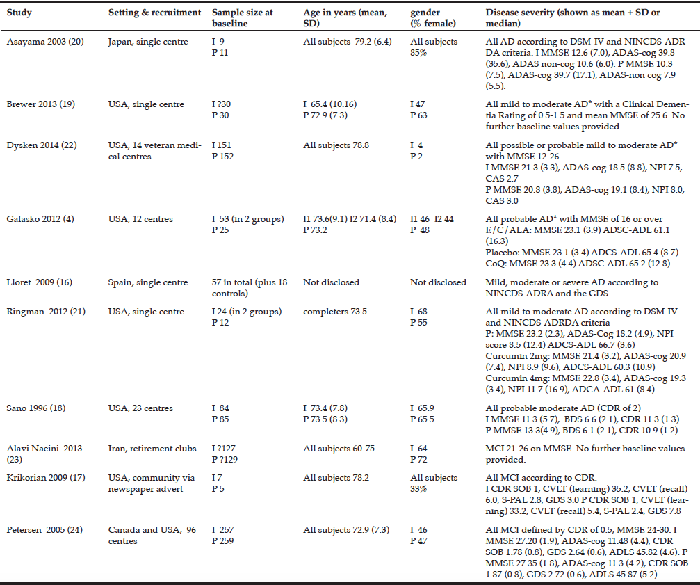
Volitional balance control task: The limits of stability (LOS) test via Balance Master (Equitest® Neurocom) (18) was administered. Participants were secured in a safety harness and asked to stand on the force platform of the Balance Master (Figure 1). Participants were instructed to lean their body either in the forward, backward, left, or right direction to move their center of gravity (COG) projection shown on a screen to the desired direction without losing balance, stepping, or reaching for assistance.
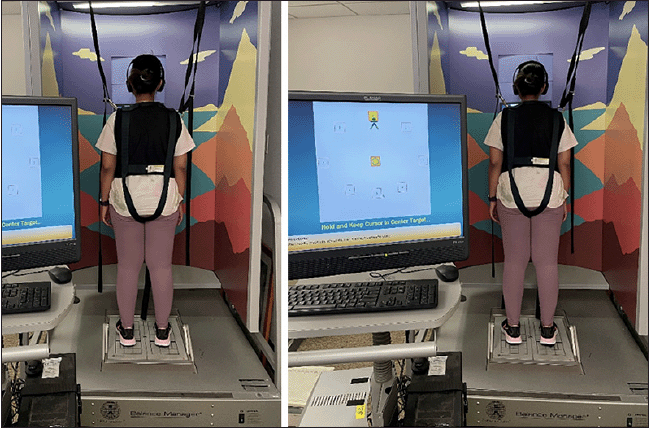
Figure 1
Represents a picture of an individual performing the
limits of stability test on Balance Master (Equitest®
Neurocom) in the forward direction


Cognitive task

Auditory clock test (ACT) (19) and letter number sequencing task (LNS) (20) were administered using the DirectRT EmpirisoftTM (21) software to assess subdomains of executive function (visuo-spatial memory, working memory, attention, and cognitive flexibility). The audio cues were delivered through headphones and responses were recorded through a microphone. The ACT involved responding to different times of the day, “yes” if the hour and the minute hand was on the same side of the clock face and “no” otherwise. The LNS involved sequentially listing alternate letter and number combinations, for example, response to “C5,” was D6, E7, etc.

Interference test

The LOS test (all directions) was performed along with both cognitive tasks mentioned above. Participants began responding to the cognitive cues followed by the LOS task.

NIH Toolbox

An iPad was used to test the motor and cognitive domains. The motor tests include 4-meter gait speed test and 2-minute walk test. The cognitive tests include list sort memory test (working memory), picture sequence memory test (episodic memory), dimensional change card sort test (executive function), flanker inhibitory control and attention test (attention and executive function), and pattern comparison processing speed test (processing speed) (refer supplementary material section).

Outcome measures

Volitional balance control and interference task: Single task (task when performed alone) and performance during interference task was quantified by the movement stability measurement of maximum excursion (MXE, expressed in percentage), which is the maximum ability to shift one’s COG toward the theoretical limit in the desired direction. Higher values indicate better performance.
Cognitive and interference task: Accuracy [(Correct responses)(Total responses)*100] was calculated during single and interference task.
NIH toolbox: Speed (m/sec) for 4-meter gait test and distance covered in 2-minute walk test for endurance was computed. Number of correct responses for list sort memory test and accuracy for the remainder tests was included for analysis.

Statistical analyses

Statistical analyses were performed using SPSS version 24, Chicago, IL, USA. For MXE in each volitional balance control task direction (i.e., forward, backward, left, and right), 2 x 2 repeated measures analysis of variance (ANOVA) was performed to examine the time (pre- to post-training) and task (single vs. interference task) differences on with follow-up post-hoc tests. Similarly, four repeated measures ANOVA for accuracy (cognitive) in ACT and LNS was performed. Paired t-test was conducted for NIH toolbox measures. Refer supplementary material section for details.


Results

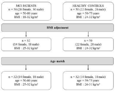
Demographics: Demographic characteristics of participants who completed the study are provided in Table 1.
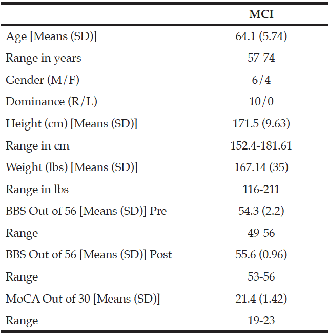
Table 1
Demographics and clinical characteristics of older adults with mild cognitive impairment (MCI). BBS = Berg Balance Scale, MoCA = Montreal Cognitive
Assessment


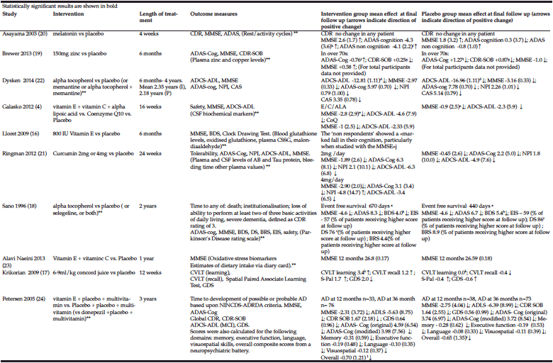
Volitional balance control and interference task: From pre- to post-training, MXE improved in the forward and left direction (p<0.05) under interference test (Figure 2a-2d). Results of ANOVA and follow-up test are presented in Table 2.
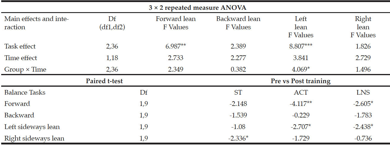
Table 2
Results for balance control task

ST: single task ; ACT: Auditory clock test; LNS: Letter number sequencing; *p<0.05 **p<0.01 ***p<0.001


Cognitive and interference task: Accuracy on ACT (Figure 2e-2h) and LNS (Figure 1i-1l) showed significant improvement only during single task performances (p<0.05), however, no improvement was observed during interference test (p>0.05). Results of ANOVA and follow-up test are presented in Table 3.
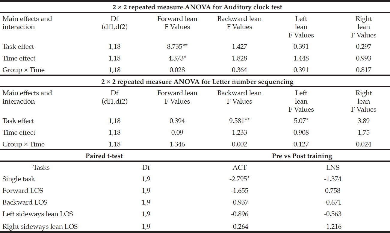
Table 3
Results for cognitive task

ST: single task; ACT: Auditory clock test; LNS: Letter number sequencing; *p<0.05 **p<0.01 ***p<0.001


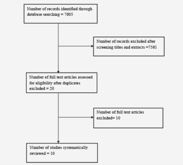
NIH toolbox: A significant increase in gait speed was observed post-training (p<0.05); however, there was no change in the 2-minute walk test distance covered (p>0.05) (Figure 3a). Post-training, significant improvements in NIH cognitive toolbox measures of working memory (p<0.05) (Figure 3b), episodic memory (p<0.01) (Figure 3c), and executive function (p<0.01) (Figure 3d) were observed. However, no improvements were observed in attention (p>0.05) and processing speed (p>0.05).
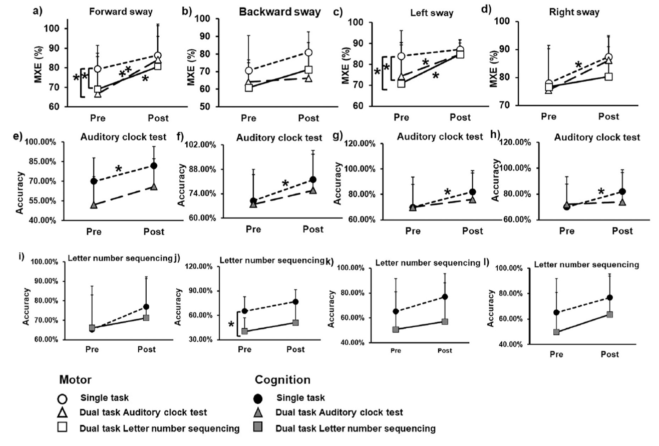
Figure 2
Association of dual task training with balance control and cognition under single and dual task conditions

Figures a, b, c, & d represent mean and standard deviations (SD) pre- to post-training changes for maximum excursion during volitional balance control under dual task and single task conditions. Figures e, f, g, & h represent mean and SD pre- to post-training cognitive accuracy changes for auditory clock test during dual task and single task conditions. Figures i, j, k, & l represent mean and SD pre- to post-training cognitive accuracy changes for letter number sequencing during dual task and single task conditions
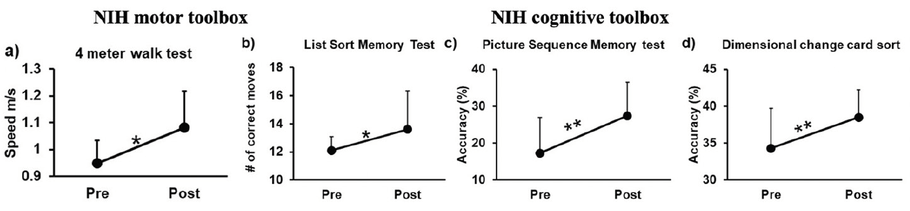
 

Figure 3
Means and SD for gait speed shown in meters (m) and obtained from the NIH motor toolbox; and b) working memory tested via list sort memory test, (c) episodic memory tested via picture sequence memory test, and (d) executive function via dimensional change card sort obtained from the NIH cognitive toolbox

Greater scores indicate better performance. *, p<0.05


Discussion

As hypothesized, the study results showed a significant improvement in volitional balance control under interference conditions but no improvement in ACT and LNS. Additionally, the intervention resulted in improved gait speed but not endurance for motor function and, similarly, improved working memory, episodic memory, and executive function but not attention and processing speed.
With respect to interference task conditions, the results show improvement in motor performance but no change in ACT and LNS cognitive tasks demonstrating motor prioritization (6). Repeated practice of multidirectional weight shift training that challenged one’s LOS and immediate biofeedback (visual) with knowledge of performance during training could have facilitated the ability to precisely control one’s center of mass (COM) body movement. The LOS test utilizes a significant amount of attentional resources within the dorsolateral prefrontal cortex (DLPFC, associated with executive functions) (22). Post-training, utilization of the shared resources between cognitive and motor areas perhaps improved by channeling available attentional resources to prioritize motor performance – probably due to the CNS’ estimation or perception of the balance task to be more challenging with significant consequences (such as falls) in case of failure. Furthermore, the significantly greater pathology affecting the DLPFC and associative sensorimotor areas (controlling executing function) than the premotor or motor areas (controlling volitional balance) could attribute to motor prioritization (5).
Our study yielded motor benefits in forward and left lean but did not improve backward and right lean. It has been postulated that backward leans are more difficult than forward leans due to directional-specific anatomic constraints and increased reliance on proprioceptive and vestibular systems (over visual system) in older adults (23-25). Aging-induced changes causes impaired integration of these systems and any cognitive decline further depreciates this sensory signal processing, resulting in deteriorated balance control (23, 24). Thus, impaired cognitive-sensory signal processing could explain the poor performance and lowest MXE for backward lean at baseline (single and interference task). Lastly, motor performance was the highest on right lean, which could be related to the dominance/preferred side for performing activities and, therefore, there may have been limited room for improvement in the right lean.
Although there wasn’t an improvement in cognitive performance during interference task, visuo-spatial and working memory (ACT) improved under single task conditions. Apart from the implicit benefits of exergaming (12, 15), and the explicit cognitive training that targeted subdomains of executive function (attention, planning, and working memory) may explain such improvements. However, there were no benefits in cognitive flexibility (i.e., letter number sequencing) despite a positive trend. This task requires simultaneous utilization of attention, processing information, and working memory. Although the training did target these domains, the dosage might not be enough to induce change.
Similar to our study, studies targeting balance training in standing have shown improvements in overground gait speed in MCI (3) and positive transfer to improved mobility (16). This could be attributed to task-specific characteristics of exergames, which involve time limited stepping activities. However, our results showed no improvement in endurance. This could be because the exergames did not incorporate high-intensity training that is known to induce improvement in cardiovascular function in MCI (7, 8). Lastly, due to the implicit (via Wii fit games) and explicit (therapist cued cognitive tasks) cognitive training components of the protocol, improvement in NIH cognitive toolbox measures for executive function, attention, and processing speed were expected and agree with results of previous DT training studies (7, 12).
Despite the positive results, our study has certain limitations. Firstly, there was a small sample size and lack of a control group due to the study’s preliminary nature. Furthermore, the training was limited to 4 weeks, and greater training dosage may yield larger motor and cognitive improvements under DT conditions. While our results demonstrate partial benefits of DT exergaming on balance control (self-initiated), its effects on the primary defense mechanism – reactive balance control – remain to be explored.


Conclusion

Our preliminary study demonstrated that DT exergaming has the potential to improve balance control and, limited benefits in executive function which could potentially have an impact in fall-risk reduction.


Conflict of interest: We have no conflict of interest to declare.

Ethical standards: The study was supported by Midwest Roybal Center for Health Promotion and Translation awarded to Dr. Tanvi Bhatt. This work was carried out after securing approval from the University of Illinois institutional review board protocol 2018-1257 and was registered on clinicaltrials.gov NCT03765398.


References

1. Langa, K.M. and D.A. Levine, The diagnosis and management of mild cognitive impairment: a clinical review. Jama, 2014. 312(23): p. 2551-2561.
2. Sheridan, P.L. and J.M. Hausdorff, The role of higher-level cognitive function in gait: executive dysfunction contributes to fall risk in Alzheimer’s disease. Dementia and geriatric cognitive disorders, 2007. 24(2): p. 125-137.
3. Bahureksa, L., et al., The impact of mild cognitive impairment on gait and balance: a systematic review and meta-analysis of studies using instrumented assessment. Gerontology, 2017. 63(1): p. 67-83.
4. Lopez, O.L., et al., Neuropsychological characteristics of mild cognitive impairment subgroups. Journal of Neurology, Neurosurgery & Psychiatry, 2006. 77(2): p. 159-165.
5. Muir, S.W., K. Gopaul, and M.M. Montero Odasso, The role of cognitive impairment in fall risk among older adults: a systematic review and meta-analysis. Age and ageing, 2012. 41(3): p. 299-308.
6. Plummer, P., et al., Cognitive-motor interference during functional mobility after stroke: state of the science and implications for future research. Archives of physical medicine and rehabilitation, 2013. 94(12): p. 2565-2574. e6.
7. Law, L.L., et al., Effects of combined cognitive and exercise interventions on cognition in older adults with and without cognitive impairment: a systematic review. Ageing research reviews, 2014. 15: p. 61-75.
8. Zhang, W., et al., Interventions to improve gait in older adults with cognitive impairment: a systematic review. Journal of the American Geriatrics Society, 2019. 67(2): p. 381-391.
9. Ben-Sadoun, G., et al., Physical and cognitive stimulation using an exergame in subjects with normal aging, mild and moderate cognitive impairment. Journal of Alzheimer’s Disease, 2016. 53(4): p. 1299-1314.
10. Colombo, M., et al. Virtual reality for persons with dementia: an exergaming experience. in ISARC. Proceedings of the International Symposium on Automation and Robotics in Construction. 2012. IAARC Publications.
11. Ge, S., et al., Technology-based cognitive training and rehabilitation interventions for individuals with mild cognitive impairment: a systematic review. BMC geriatrics, 2018. 18(1): p. 1-19.
12. Zhao, Y., et al., Effectiveness of exergaming in improving cognitive and physical function in people with mild cognitive impairment or dementia: systematic review. JMIR serious games, 2020. 8(2): p. e16841.
13. Crockett, R.A., et al., Resting state default mode network connectivity, dual task performance, gait speed, and postural sway in older adults with mild cognitive impairment. Frontiers in aging neuroscience, 2017. 9: p. 423.
14. Zhang, Y., et al., MRI markers for mild cognitive impairment: comparisons between white matter integrity and gray matter volume measurements. PloS one, 2013. 8(6): p. e66367.
15. Stojan, R. and C. Voelcker-Rehage, A systematic review on the cognitive benefits and neurophysiological correlates of exergaming in healthy older adults. Journal of clinical medicine, 2019. 8(5): p. 734.
16. Kannan, L., et al., Cognitive-motor exergaming for reducing fall risk in people with chronic stroke: A randomized controlled trial. NeuroRehabilitation, 2019. 44(4): p. 493-510.
17. Worley, J.R., S.N. Rogers, and R.R. Kraemer, Metabolic responses to Wii Fit™ video games at different game levels. The Journal of Strength & Conditioning Research, 2011. 25(3): p. 689-693.
18. Jbabdi, M., P. Boissy, and M. Hamel, Assessing control of postural stability in community-living older adults using performance-based limits of stability. BMC geriatrics, 2008. 8(1): p. 1-8.
19. Haggard, P. and J. Cockburn, Dividing attention between cognitive and motor tasks in neurological rehabilitation. Neuropsychological Rehabilitation, 1998. 8: p. 155-170.
20. Mrazik, M., S. Millis, and D.L. Drane, The oral trail making test: effects of age and concurrent validity. Archives of Clinical Neuropsychology, 2010. 25(3): p. 236-243.
21. Jarvis, W., MediaLab (Version 200x. xx)[Computer Software]. Empirisoft Corporation, 2017.
22. Nachev, P., C. Kennard, and M. Husain, Functional role of the supplementary and pre-supplementary motor areas. Nature Reviews Neuroscience, 2008. 9(11): p. 856-869.
23. Berchicci, M., et al., Stepping forward, stepping backward: a movement-related cortical potential study unveils distinctive brain activities. Behavioural brain research, 2020. 388: p. 112663.
24. Carbonneau, E. and C. Smeesters, Effects of age and lean direction on the threshold of single-step balance recovery in younger, middle-aged and older adults. Gait & posture, 2014. 39(1): p. 365-371.
25. Duarte, M. and V.M. Zatsiorsky, Effects of body lean and visual information on the equilibrium maintenance during stance. Experimental brain research, 2002. 146(1): p. 60-69.

POOR BODY COMPOSITION IN PATIENTS WITH MILD COGNITIVE IMPAIRMENT COMPARED TO HEALTHY OLDER CONTROLS


J. Willers1, A. Hahn1, T. Köbe2,3, S. Gellert1, V. Witte2,3,4, V. Tesky5, J. Pantel5, A. Flöel2,3,6, J.P. Schuchardt1


1. Institute of Food Science and Human Nutrition, Leibniz University Hannover, Germany; 2. Department of Neurology, Charité – University of Medicine Berlin, Germany; 3. NeuroCure Cluster of Excellence, Charité – University of Medicine Berlin, Germany; 4. Max Planck Institute of Human Cognitive and Brain Sciences, Department of Neurology, Leipzig, Germany and SFB 1052 Obesity Mechanism subproject A1, University of Leipzig, Germany; 5. Institute of General Practice, Goethe University, Frankfurt am Main, Germany; 6. Center for Stroke Research Berlin, Charité – University of Medicine Berlin, Germany

Corresponding Author: J. Willers, Institute of Food Science and Human Nutrition, Leibniz University Hannover, Am Kleinen Felde 30, 30167 Hannover, Germany, Tel: +49 (0)511 762 5755; Fax: +49 (0)511 762 5729; Email: willers@nutrition.uni-hannover.de

J Aging Res Clin Practice 2018;7:37-41
Published online March 1, 2018, http://dx.doi.org/10.14283/jarcp.2018.8


Abstract

In this cross-sectional study, body composition of fifty-eight mild cognitive impairment (MCI) patients (single and multiple domain) and fifty healthy older control subjects by the use of bioelectrical impedance analysis (BIA) was assessed. Measurements were: height, weight, body mass index, BIA: phase angle (PA), total body water (TBW), lean body mass (LBM), body cell mass (BCM), extracellular mass (ECM), body fat mass (BFM), apolipoprotein E4, and physical activity level. Compared to BIA reference values and healthy subjects, MCI patients had significant differences in PA (only female), BCM and ECM/BCM index. Differences were more pronounced in females compared to males. The low levels of BCM and PA suggest that MCI patients, especially of female sex, have a poor nutritional status. BIA-derived PA might be a suitable indicator, that could enhance evaluation of nutritional status in patients with cognitive decline.

Key words: Mild cognitive impairment, bioelectrical impedance analysis, phase angle, body composition.

Abbreviations: APOE: apolipoprotein E; AVLT: auditory verbal learning test; BCM: body cell mass; BFM: body fat mass; BIA: bioelectrical impedance analysis; BMI: body mass index; CERAD: Consortium to establish a registry for Alzheimer’s disease; ECM: extracellular mass; LBM: lean body mass; MCI: mild cognitive impairment; MMSE: mini-mental status examination; PA: phase angle; PAL: physical activity level; R: resistance; TBW: total body water; Xc: capacitive reactance.


Introduction

Different epidemiological studies observed a relationship between nutritional status and cognitive function in patients with cognitive impairment (1-3). There is also evidence that poor nutritional conditions are associated with cognitive decline (4-6) and may play an important role in progression of cognitive loss (7).
Techniques for measuring body composition include anthropometry and bioelectric impedance analysis (BIA). Especially the use of raw BIA data has become a standard procedure in the assessment and monitoring of the body composition and nutritional status of patients [8]. BIA measures the opposition to electrical flow arising from resistance and reactance. The BIA is a simple, inexpensive and non-invasive technique for assessing body composition, allowing conclusions on total body water (TBW), hydration status and body cell mass. This method is used for the determination of nutritional status and risk of morbidity in ambulatory and hospitalized patients (9). For the nutritional screening and clinical prognosis, the phase angle (PA) is the most established impedance parameter (10). The PA is calculated from resistive behaviour (R), which is mainly dependent on tissue hydration, and the capacitive behaviour of tissues (Xc) associated with cellularity, cell size, and integrity of the cell membrane (8). A low PA suggest cell death or decreased cell integrity and has strong predictive value according to morbidity and mortality in various disease conditions, e.g. HIV or cancer (11).
Due to a strong dependency of body composition on sex (e.g., females have less muscle and greater percentage of body fat than males), age (e.g., decreasing muscle mass and increasing fat mass with increasing age) and body mass index (BMI), reference values for PA and other BIA parameters are sex-, age-, and BMI-specific (8). A gender specific evaluation is therefore mandatory.
To our knowledge, only one recent study is published that investigated the nutritional status of mild cognitive impairment (MCI) patients in terms of the body composition via BIA (12). MCI is a frequent condition in the general aged population and is associated with an increased risk for the development of dementia. Therefore, body composition in MCI may be of relevance for prognosis of cognitive decline. Thus, the objective in this study was to compare BIA measurements in MCI patients with literature reference values and age-matched older adults without clinical dementia.


Methods

For this cross-sectional study, 58 patients with MCI were recruited consecutive between 2011 and 2014 in Berlin (memory clinic of the Department of Neurology of the Charité University Hospital and Neurology specialist practice) and Frankfurt am Main (Institute of General Practice), Germany. MCI patients (single and multiple domain) were diagnosed according to Mayo criteria based on subjective cognitive complaints and objective memory impairment in standardized tests (performing at least 1.5 SD below age- and education-specific norm in relevant subtests (Total Word List, Delayed Recall Word/Figures) of the CERAD-Plus test battery (13), relatively preserved general cognition, no impairment in activities of daily living, and no dementia (14). Fifty healthy older adults were recruited between 2010 and 2013 at the memory clinic of the Department of Neurology at the Charité Berlin, Germany. A detailed description of in- and exclusion criteria can be found in Köbe et al. (15) for MCI patients and Witte et al. (16) for healthy older adults.
MCI patients had a body mass index (BMI) range from 18 to 32 kg/m², whereas the healthy controls had a BMI between 24 kg/m² to 32 kg/m². Before comparing the two groups a BMI adjustment as well as an age-match was mandatory (Figure 1).


Figure 1 BMI adjustment and age-match for the group comparison

Figure 1
BMI adjustment and age-match for the group comparison


Anthropometric (e.g., body weight, height, BMI) and bioimpedance measurements (e.g., PA, TBW, lean body mass (LBM), body cell mass (BCM), extracellular mass (ECM), body fat mass (BFM)) were carried out. BIA was performed with B.I.A. 2000-M (Pöcking, Germany) and the software NutriPlus (Data Input GmbH, Darmstadt, Germany).
Fasting blood samples were obtained by venipuncture of an arm vein using sealed blood collection tubes and Monovettes® (Sarstedt, Nürnbrecht, Germany). Concentrations of cobalamine (vitamin B12) and folate were measured in serum samples at the IMD laboratory, Berlin, Germany.
For apolipoprotein E (ApoE) genotyping, DNA was extracted from whole blood using a blood mini-kit (Qiagen, Hilden, Germany) and stored at -80°C until analysis. Genotyping of apolipoprotein E4 (ApoE4) was performed on a Sequenom® MassARRAY iPLEX, TaqMan assay following procedures described previously (17).
Participants were tested on memory performance using the German version of the Rey Auditory Verbal Learning Test (AVLT) (18). Global cognitive dysfunction was estimated with the Mini Mental State Examination (MMSE) (19). Patients were asked to learn a list of 15 words within five immediate recall trials, followed by a 30 min delayed recall and delayed recognition test. Learning ability was defined as the sum of words learned in all five trials (maximum 75 words); delayed recall represented the total number of remembered words after 30 min (maximum 15 words). For delayed recognition (recognition memory), subjects were asked to recognize the 15 original words presented within 35 distractor words subsequent to the delayed recall tests (number of correctly recognized words minus false positives; maximum 15 words). All testing was conducted by trained staff members according to standard procedure.
Statistical analyses were processed with SPSS software version 24.0 (SPSS Inc., Chicago, IL, USA). Results are expressed as means ± SD unless otherwise specified. Differences between men and women were calculated by the non-parametric Mann-Whitney U test. Multiple linear regression models were used for group comparison (MCI vs. healthy controls). Initially, all analyses were conducted unadjusted. Subsequently, age, education, ApoE4 status, vitamin B12, folate status (factors that are known to be associated with cognition) (15), and physical activity level (a factor that influences body composition) were entered as covariates in multiple linear regression models to study whether potential group differences are independent. Patients reported their physical activity using the Freiburger physical activity questionnaire. Spearman’s rank correlation was used to test correlations between variables. P-values ≤ 0.05 were considered significant.


Results and discussion

Characterisation of the MCI group

Characterisation of the MCI patients according to anthropometric and BIA measurements is presented in Table 1. The total MCI collective includes fifty-eight MCI patients (28 women) with a mean age of 69.1 ± 7.8 years. As expected, gender differences were observed for body weight as well as all bioelectrical variables. Percentage of BCM was reduced in MCI women (44.3 ± 3.1%) vs. men (48.5 ± 3.9%), resulting in a lower ECM/BCM index in men (1.1 ± 0.3) vs. women (1.3 ± 0.2). Manufacturer reference values for this age group constitute ideal BCM values of 50 – 56% for women and 53 – 59% for men, while the ECM/BCM ratio should be < 1 for both sexes. Thus, our data suggest that MCI patients, especially female, are of poor body condition and exhibit a low muscle content and activity status (20).


Table 1 Anthropometric measures and bioelectrical impedance analysis of the MCI group (n = 58)

Table 1
Anthropometric measures and bioelectrical impedance analysis of the MCI group (n = 58)

* Mann-Whitney U test was performed for comparison between women and men; 1 n = 26


Additionally, male MCI patients had a significantly higher PA than female patients. The median PA of the total MCI collective was 4.9° (range 3.6° to 6.4°). About half of the MCI patients (48.3%, women: 67.9%, men: 30%) had a lower PA from 3.6° to 4.8°. There was a strong negative correlation between PA and age (r = -0.538; p < 0.001, Spearman’s rank correlation), which has been, likewise, reported in healthy populations (10, 21). Physiologically, increasing age is associated with decline in tissue mass, which results in decreasing PA (11). Simultaneously, hospitalized patients showed a significant lower LBM, higher BFM and lower PA in general (22). Nevertheless, the mean PA in MCI patients of this study was even lower than of MCI patients in a recently published cross-sectional study (women: 5.6 ± 0.6°, men: 6.4 ± 0.7°) (12) and lower compared to hospitalized patients (women: 5.0 ± 1.3°; men: 6.0 ± 1.4°) (22). Furthermore, various studies indicate that the PA can be considered as a marker of clinically relevant malnutrition caused by an increase of extracellular fluid and a decrease of BCM (11, 23).
Compared to reference BIA values of a large German database (214,732 adults) in corresponding gender, age and BMI groups (8), the body composition of the present MCI patients can be classified as worse. MCI patients of both sexes have lower PAs (MCI women: 4.6 ± 0.5°, MCI men: 5.4 ± 0.7°) according to their corresponding BMI and age classes (German reference sample: women: 5.1 ± 0.8°; men: 6.0 ± 0.8°).
Additionally, in female MCI patients (n = 28) learning ability was positively correlated with the PA (r = 0.433, p = 0.002, Spearman’s rank correlation) and negatively with the ECM/BCM index (r = -0.456, p = 0.001, Spearman’s rank correlation). In male MCI patients, no correlation was observed. Nevertheless, these data suggest an association between an impaired memory performance and a poor body composition, although it cannot be clarified whether a lower cognitive function is a cause or consequence of a poor nutritional status. Further longitudinal studies are necessary.

Comparison between MCI patients and healthy controls

Furthermore, we compared the MCI patients to healthy controls in terms of BIA measurements. As the participants were selected using different BMI ranges, an adjustment according to BMI and age was necessary (Figure 1). Finally, in each case, thirty-two MCI patients and healthy controls were compared with regard to anthropometric and BIA measures (Table 2 and 3).


Table 2 Comparison of anthropometric measures and bioelectrical impedance analysis between female MCI patients and healthy controls

Table 2
Comparison of anthropometric measures and bioelectrical impedance analysis between female MCI patients and healthy controls

Multiple linear regression models were used for group comparison, adjusting for potential confounders; p < 0.05; ß: unstandardized regression coefficient.

Table 3 Comparison of anthropometric measures and bioelectrical impedance analysis between male MCI patients and healthy controls

Table 3
Comparison of anthropometric measures and bioelectrical impedance analysis between male MCI patients and healthy controls

Multiple linear regression models were used for group comparison, adjusting for potential confounders; p < 0.05; ß: unstandardized regression coefficient.


Female healthy subjects had a higher BMI (28.0 ± 1.7 kg/m2) and were heavier (76.4 ± 6.4 kg) compared to female MCI patients (BMI: 26.8 ± 1.9 kg/m2; weight: 72.0 ± 10.1 kg). These data were not statistical significant. However, the parameter should not be underestimated as cognitive decline is faster and more severe with a low BMI (BMI cut-off 25 kg/m²) (24). Especially female MCI patients showed significantly lower values in basal metabolic rate, PA, BCM, ECM, and ECM/BCM index compared to healthy females (Table 2). The differences remained significant only for PA and BCM after adjustment for potential confounders such as age, education, ApoE4, vitamin B12, folate status, and physical activity. In male subjects, the differences were not significant (Table 3).
Limitations of this study relate to the small sample size and the BIA technique. Due to the necessary BMI adjustment, the study population was greatly reduced. Thus, the group comparison was comparatively weak and might limit the outcome. Additionally, the BIA measurement has technical and physiological limitations such as hydration status, body position during procedure, air and skin temperatures, recent physical activity. BIA is not traditionally used as a measure of malnutrition but it might improve assessment of nutritional status and prognosis among MCI patients.


Conclusion

Using BIA, we observed a poor body composition especially in female MCI patients indicating a poor general health condition. Considering the observed associations between the PA and memory functions, our data confirm earlier findings that determined a relationship between nutrition status and cognitive function in patients with cognitive impairment. However, based on our data we cannot clarify whether lower cognitive function is a cause or consequence of a poor nutritional status. Thus, further longitudinal studies may be undertaken to resolve this important question and eventually determine if the PA may serve as a potential prognostic factor for cognitive decline.


Acknowledgements: We would like to thank the participants who contributed their time to this project. The genotyping of ApoE4 in the laboratory of Prof. Dr. Dan Rujescu (University of Halle, Germany) is kindly acknowledged. Likewise, we thank Lucia Kerti from the Department of Neurology, Charité – University of Medicine Berlin for her involvement with examining healthy older subjects.

Funding: This research was funded by the German Federal Ministry of Education and Research (BMBF; FKZ 01EA1328D) and is registered under ClinicalTrials.gov identifier: NCT01219244.

Ethical statement: The study was conducted according to the German law and to good clinical practice and ethical principles of the Declaration of Helsinki. The Ethics Committee of the Charité – University of Medicine, Berlin, Germany, approved the study.

Consent statement: Written informed consent was obtained from all participants.

Dr Willers has nothing to disclose. Prof Hahn reports grants from Bundesministerium für Bildung und Forschung (Federal Ministry of Education and Research), during the conduct of the study. Theresa Köbe reports grants from German Federal Ministry of Education and Research, during the conduct of the study. Dr Gellert has nothing to disclose. Dr Witte has nothing to disclose. Dr Tesky has nothing to disclose. Prof Pantel has nothing to disclose. Prof Flöel reports grants from BMBF, during the conduct of the study. Dr Schuchardt reports grants from Bundesministerium für Bildung und Forschung (Federal Ministry of Education and Research), during the conduct of the study.


References

1.    Lee KS, Hong CH, Cheong HK, Oh BH. Difference in nutritional risk between mild cognitive impairment group and normal cognitive function elderly group. Arch Gerontol Geriatr 2009;49(1): 49-53.
2.    Marino LV, Ramos LF, Chiarello PG. Nutritional status according to the stages of Alzheimer’s disease. Aging Clin Exp Res 2014;27 (4): 507-13.
3.    Orsitto G. Different components of nutritional status in older in patients with cognitive impairment. J Nutr Health Aging 2012;16 (5): 468-471.
4.    Haan MN, Miller JW, Aiello AE, Whitmer RA, Jaqust WJ et al. Homocysteine, B vitamins, and the incidence of dementia and cognitive impairment: results from the Sacramento Area Latino Study on Aging. Am J Clin Nutr 2007;85(2): 511-517.
5.    Khater MS, Aboulelezz NF. Nutritional status in older adults with mild cognitive impairment living in elderly homes in cairo, Egypt. J Nutr Health Aging 2011;15(2): 104-108.
6.    Orsitto G, Fulvio F, Tria D, Turi V, Venezia A et al. Nutritional status in hospitalized elderly patients with mild cognitive impairment. Clin Nutr 2009;28 (1): 100-102.
7.    Malara A, Sgrò G, Caruso C, Ceravolo F, Curinga G et al. Relationship between cognitive impairment and nutritional assessment on functional status in Calabrian long-term-care. Clin Interv Aging 2014;9: 105-110.
8.    Bosy-Westphal A, Danielzik S, Dörhöfer RP, Later W, Wiese S et al. Phase angle from bioelectrical impedance analysis: population reference values by age, sex, and body mass index. JPEN 2006;30 (4): 309-316.
9.    Buffa R, Floris G, Marini E. Assessment of nutritional status in free-living elderly individuals by bioelectrical impedance vector analysis. Nutrition 2009;25: 3-5.
10.    Barbosa-Silva MCG, Barros AJD, Wang J, Heymsfield SB, Pierson Jr RN. Bioelectrical impedance analysis: population reference values for phase angle by age and sex. Am J Clin Nutr 2005;82: 49-52.
11.    Stobäus N, Pirlich M, Valentini L, Schulzke JD, Norman K.  Determinants of bioelectrical phase angle in disease. Br J Nutr 2011;107 (8): 1217-1220.
12.    Cova I, Pomati S, Maggiore L, Forcella M, Cucumo V et al. Nutritional status and body composition by bioelectrical impedance vector analysis: A cross sectional study in mild cognitive impairment and Alzheimer’s disease. PLoS One 2017;12 (2): e0171331.
13.    Morris JC, Heyman A, Mohs RC, Hughes JP, van Belle G et al. The Consortium to Establish a Registry for Alzheimer’s Disease (CERAD). Part I. Clinical and neuropsychological assessment of Alzheimer’s disease. Neurology 1989;39 (9): 1159-65.
14.    Petersen RC, Smith GE, Waring SC, Ivnik RJ, Tangalos EG et al. Mild cognitive impairment: clinical characterization and outcome. Arch Neurol 1999;56 (3): 303-8.
15.    Köbe T, Witte AV, Schnelle A, Grittner U, Tesky VA et al. Vitamin B-12 concentration, memory performance, and hippocampal structure in patients with mild cognitive impairment. Am J Clin Nutr 2016;103 (4): 1045-54.
16.    Witte AV, Kerti L, Hermannstädter HM, Fiebach JB, Schreiber SJ et al. Long-chain omega-3 fatty acids improve brain function and structure in older adults. Cereb Cortex 2014;24 (11): 3059-68.
17.    O’Dwyer L, Lamberton F, Matura S, Tanner C, Scheibe M et al. Reduced hippocampal volume in healthy young ApoE4 carriers: an MRI study. PloS one 2012;7 (11): e48895.
18.    Lezak M. Neuropsychological assessment. New York, Oxford: Oxford University Press, 2004.
19.    Folstein MF, Folstein SE, McHugh PR. “Mini-mental state”: a practical method for grading the cognitive state of patients for the clinician. J Psychiatr Res 1975;12: 189-198.
20.    Matthie JR. Bioimpedance measurements of human body composition: critical analysis and outlook. Expert Rev Med Devices 2008;5(2): 239-261.
21.    Dittmar M.  Reliability and Variablility of Bioimpedance Measures in Normal Adults: Effects of Age, Gender, and Body Mass. Am J Physiol Anthropol 2003;122: 361-370.
22.    Kyle UG, Genton L, Pichard C (2013) Low phase angle determined by bioelectrical impedance analysis is associated with malnutrition and nutritional risk at hospital admission. Clin Nutr 2013.32: 294-299.
23.    Kyle UG, Soundar EP, Genton L, Pichard C. Can phase angle determined by bioelectrical impedance analysis assess nutritional risk? A comparison between healthy and hospitalized subjects. Clin Nutr 2012.31: 875-881.
24.    Coin A, Veronese N, De Rui M, Mosele M, Bolzetta F et al. Nutritional predictors of cognitive impairment severity in demented elderly patients: the key role of BMI. J Nutr Health Aging 2012.16(6):553-556.

ANTIOXIDANT SUPPLEMENTS IN ALZHEIMER’S DEMENTIA AND MILD COGNITIVE IMPAIRMENT: A SYSTEMATIC REVIEW


E.J. Pegg


Corresponding Author: E.J. Pegg, Lancashire Teaching Hospitals NHS Foundation Trust, United Kigdom, emily-pegg@doctors.org.uk

J Aging Res Clin Practice 2016;inpress
Published online December 1, 2016, http://dx.doi.org/10.14283/jarcp.2016.121


Abstract

Objective: Current treatments have only a modest effect on impairment in Alzheimer’s Dementia (AD) and there is no treatment currently licensed for Mild Cognitive Impairment (MCI). Oxidative stress is postulated to play a role in the pathogenesis of AD and MCI and this provides a rationale for treatment with antioxidant supplements. The aim of this review is to evaluate the effect of antioxidant supplements in people with AD and MCI. Methods: A systematic review of published randomised controlled trials was carried out. 4 electronic databases were searched. Studies were included if they compared the use of a placebo with the following antioxidant supplements in people with AD or MCI: Vitamin e, vitamin c, selenium, alpha lipoic acid, phenols, zinc, curcumin, beta carotene, coenzyme Q10, melatonin. The primary outcome measure was cognitive impairment. Secondary outcome measures included functional impairment, behavioural disturbance and safety. Results: 10 trials were identified which met the inclusion criteria. Outcome data was not suitable for meta-analysis. 5 studies reported a small positive treatment effect on cognition and 1 reported a negative effect. 2 reported a positive treatment effect on functional ability and 1 on behaviour. There were no consistent adverse effects found overall however two studies raised concern of possible worsening of cognition in certain circumstances. Conclusions: The findings of this systematic review do not support the use of antioxidant supplements to slow cognitive, functional or behavioural deterioration in people with AD or MCI. However the majority of included studies had a high or unknown risk of bias. In the one study which had a low overall risk of bias, there was evidence that antioxidant supplements may have a positive effect on functional decline in AD. The overall risk of harm associated with short term antioxidant supplementation appears to be low however caution is warranted. Further studies evaluating the role of oxidative stress in the pathogenesis of AD are suggested.

Key words: Alzheimer’s, mild cognitive impairment, antioxidants.

Abbreviations: ADAS: Alzheimer’s Disease Assessment Scale; ADAS-cog: Alzheimer’s Disease Assessment Scale- cognitive subscale; ADAS-non cog: Alzheimer’s Disease Assessment Scale- non cognitive subscale; ADCS-ADL: Alzheimer’s Disease Cooperative Study- Activities of Daily Living Inventory; ADRQL: Alzheimer’s Disease Related Quality of Life; BDS: Blessed Dementia Scale; BRS: Behaviour Rating Scale for Dementia; CAS: Caregiver Activity Survey; CDR: Clinical Dementia Rating; CDR–SOB: Clinical Dementia Rating- Sum of Boxes; CDT: Clock Drawing Test; CVLT: (learning) California Verbal Learning Test (learning); CVLT: (recall) California Verbal Learning Test (recall); DS: Dependence Scale; EIS Equivalent Institutional Service (subsection of Dependence Scale); GDS: Global Deterioration Scale; MCI-ADLS: Mild Cognitive Impairment Activities of Daily Living Scale; MMSE: Mini Mental State Examination; NINCDS-ADRDA: Alzheimer’s Criteria National Institute of Neurological and Communicative Disorders and Stroke and Related Disorders Association; NPI: Neuropsychiatric Inventory; QOLS: Quality of Life Scale; S-PAL: Spatial Paired Associate.


Introduction

Currently available pharmacological treatments for Alzheimer’s Dementia (AD), acetylcholinesterese inhibitors (AChEIs) and memantine, have only a modest effect on cognitive impairment, function and behaviour and there is no treatment currently licensed for Mild Cognitive Impairment (MCI).
The main neuropathological features of AD are accumulation of beta-amyloid plaques and tau containing neurofibrillary tangles. The exact mechanism by which this occurs is complex and has not been fully elucidated however oxidative stress is hypothesised to play a major role by interacting with key processes including mitochondrial dysfunction, inflammation, protein misfolding, calcium homeostasis and metal chelation (1). In view of this, antioxidant therapy has been suggested as a mechanism to slow the progression of cognitive impairment.
Studies have reported increased levels of oxidative damage to DNA and neurons in both AD and MCI (2, 3) suggesting that oxidative stress might be an important cause of initiation and progression of cognitive impairment and AD.
Findings from human studies evaluating the benefit of dietary antioxidants or supplementary antioxidants in slowing cognitive decline have been mixed, with some studies even reporting a negative effect (4).
The overview of the literature suggests that only the effects of Vitamin E supplements on AD have been assessed within the context of a relatively recent systematic review (5). The examination of the effects of other widely used antioxidant supplements have not been a subject of recent systematic reviews. In addition, given the mixed results produced by studies evaluating antioxidant supplements in AD and MCI, together with potential safety concerns [6-11] and the expanding evidence in this research area, it can be concluded that there are strong grounds to support the need to undertake a comprehensive systematic review which will summarise and compare the effects of all the key antioxidant supplements on AD and MCI.
The following research questions will be addressed in this systematic review:
In people taking antioxidant supplements with AD and MCI, what is the effect on:
a)    cognitive ability
b)    functional ability and behaviour
c)    potential side effects


Methods

This review was conducted and reported based upon PRISMA reporting standards (12).

Search Strategy

The following databases were searched for eligible papers: PubMed, Embase (1974-February 2015), CINAHL and Cochrane Central Register of Controlled Trials in the Cochrane Library.  “Mesh” terms and text words were searched for combinations of dementia, cognitive impairment and each particular antioxidant.  Searches were limited to studies conducted in human subjects and written in the English Language.

Eligibility Criteria

Studies were included if they met the following criteria:
•    Population: Adults with AD or MCI diagnosed according to internationally recognised diagnostic criteria e.g. the MMSE or NINCD-ADRDA.
•    Intervention: The study design was restricted to Randomised Controlled Trials (RCT). Any dosage of the following antioxidant supplements, either alone or in combination was included: Vitamin e, vitamin c, selenium, alpha lipoic acid, phenols, zinc, curcumin, beta carotene, coenzyme Q10, melatonin.

The first five antioxidants were selected because they are reported to be the most important dietary antioxidants (13). The latter five compounds have also been reported to be important antioxidants.
Subjects taking their own multivitamin tablets were included because a third of the adult population is estimated to take supplements therefore including these participants is potentially reflective of the population.
Subjects taking other pharmacotherapy to slow cognitive decline (i.e. acetyl cholinesterase inhibitors and memantine) were included only if the intervention group was also taking these agents.
Gingko biloba is also a potent antioxidant but it has additional properties, including acting as a monoamine oxidase inhibitor, which could cause confounding therefore it was not included in this review. A Cochrane review did not support its use in AD (14).
•     Comparison: People with AD or MCI not taking antioxidant supplements.
•    Outcome: The primary outcome measure was cognitive impairment. Secondary outcome measures included functional impairment, behavioural disturbance and safety.

Exclusion criteria

•    Subjects with dementia in the context of Down’s syndrome
•    Subjects taking supplements containing additional compounds not listed above

Study selection

Titles and abstracts of each study were firstly screened against the eligibility criteria of the study.  The full texts of studies that were rated as potentially eligible in the title/abstract screening were retrieved, and further screened against the inclusion and exclusion criteria.

Data extraction

A data extraction sheet was formulated as an Excel spreadsheet.  Information was extracted regarding the context of the study (publication year, setting, author), study design (sampling, randomisation, length of follow up, completion rate), participants (age, gender, severity of AD/MCI, the intervention (type of antioxidant and dose) and outcomes (cognition, function, behaviour/ mood, adverse events, quality of life).  Where the data is continuous, standard deviations (SD) were extracted.  Any difference between baseline and outcome data was calculated.

Methodological quality assessment

In order to evaluate bias, the Cochrane Collaboration’s tool for assessing risk of bias was used (15). The following domains of each study were assessed:  Randomisation, allocation concealment, blinding, blinding of outcome assessment, incomplete outcome data, selective outcome reporting, sample size and any other potential sources of bias.

Data synthesis

Due to a wide variety of assessment scales used as well as differences in the way in which data was reported, it was not possible to combine outcomes.  Furthermore, the majority of the studies reported the data as a change in scores from baseline to follow up therefore the data was not suitable for meta-analysis.   A descriptive analysis is suitable for the purposes of this review because a number of antioxidants were used at low frequencies.


Results

A trial flow diagram is shown below.  In total 7605 studies were identified after a search of electronic databases as described in the methodology section. This figure excludes 508 studies which were not published in the English Language.  There were 7561 studies excluded after screening the title and/or abstract. A frequent reason for exclusion was the study design i.e. very few studies were randomised controlled trials. Animal studies were also excluded, as were studies which used combination compounds where one of the substances was not an antioxidant.  After duplicates were removed, 20 studies were read in further detail to assess for eligibility.  A total of 10 studies remained which met the inclusion criteria.
A total of 1483 participants were randomised and intervention antioxidants included: Zinc, curcumin, melatonin, vitamin E, vitamin C, alpha lipoic acid, coenzyme Q10, polyphenols and flavonoids.
Key descriptive details of the included studies are shown in table 1.

Table 1 Study characteristics and baseline characteristics of included studies

Table 1
Study characteristics and baseline characteristics of included studies

I= intervention   P= placebo   1 also 155 in memantine group, 154 in vitamin E + memantine    *according to NINCDS-ADRDA criteria A lower score on the MMSE indicates poorer cognition. A lower score on ADLS-cog indicates poorer function.  A lower score on the GDS indicates higher level of depression. For all other scales a lower score indicates better outcome.


Outcome measures

Cognition

In total, 12 different outcome measures were used to assess cognition or to objectively grade the severity of Alzheimer’s disease.  7 studies used one or more methods of assessing cognition and 3 used only the MMSE.  Details of the outcome measures used can be seen in tables 1 and 2.
Overall, 5 of the 10 studies reported a statistically significant improvement or a slower rate of deterioration in one or more cognitive assessments in the intervention group compared to the control group at the end of the study (16-20).  In 4 of these studies participants had AD and in 1 they had MCI (17).  In 2 of the studies with a positive treatment effect however, the difference was found following a post hoc analysis where the groups were divided by age (19) or whether there was plasma evidence of a response to antioxidant therapy (16).
Of the studies with a positive treatment effect on cognition where more than one measure was used, the significant effect was not seen across all of the measures in any of the studies.
Of concern is that 1 study reported a statistically significant worsening in cognition in the intervention group compared to the placebo group (4). Another study reported a worsening of cognition in the people who did not have a plasma response to the antioxidant, compared to the placebo group (16).
Only 1 of the 3 studies in which participants had MCI evaluated time to development of dementia as an outcome measure.  There was no significant difference between the groups found.

Activities of daily living / functional  assessment

In total 5 studies used an assessment of ADLs or function as an outcome measure using 4 different scales. 4 of the studies involved participants with AD patients and 1 MCI.
Overall 2 studies reported a positive treatment effect (18, 22)and the other 3 found no difference between the intervention and control groups.

Mood/ behavioural disturbance

In total 4 studies included a measure of mood or behavioural disturbance as an outcome measure using 3 different measures. There was a positive treatment effect reported in 1 study (20) and in the other 3, there was no significant difference between the groups.

Table 2 Intervention details and results of included studies

Table 2
Intervention details and results of included studies

**not relevant to this review. a. Event free survival: Not significant but when analysed with baseline MMSE included as a co-variate, p=0.001 230 day increase in median survival with vitamin E compared to placebo. b. p=0.004, c. p=0.03, d. 3.15 (0.92 to 5.39), e. 0=0.004 adjusted for multiple comparisons, f. p=0.03, g. p=0.03, h. p=0.01, i. p=0.00, j Respondents vs. Non respondents and placebo vs. Non respondents p<0.05. No data for placebo vs intervention group as a whole, k. p=0.004, l. Reported significant differences with executive, language and overall cognitive scores in the first 18 months. A lower score on the MMSE indicates poorer cognition. A lower score on ADLS-cog indicates poorer function.  A lower score on the GDS indicates higher level of depression. For all other scales a lower score indicates better outcome.


Side effects and adverse events

In 6 studies, there were either no adverse events or side effects reported or no differences between the intervention and control groups reported.
In 2 studies, there was a greater decline in MMSE in the intervention group (4, 18).  In one study, this effect was seen in the intervention group as a whole (vitamin E + vitamin C + alpha lipoic acid), and in the other the greater deterioration was seen in the people who did not have a plasma response to vitamin E (the “non-respondents”).
In the curcumin study (21), participants taking curcumin had a statistically significant lower plasma haemotocrit and higher glucose.  It is unlikely that these effects are clinically significant. This study also stated “complaints attributable to the endocrine system” were less common in the 2 gm curcumin group compared to placebo or the 4gm group. (3% vs 17% vs 19% p=0.02).  The exact endocrine complaints are not stated.  There was no significant difference in the withdrawal rate due to adverse events between the groups.
In total 1 patient (33%) in the zinc study (19) developed low plasma levels of caeruloplasmin, thought to be directly attributed to taking zinc.
In people taking vitamin E in the Sano study (18), more people had the following events compared to the placebo group:  dental events (n=1 vs n=0. P=0.023), falls (n=12 [14%] vs n= 4 [5%] p=0.005) and syncope (n=4 vs n=7 p=0.031).

Figure 1 Flow diagram for literature search

Figure 1
Flow diagram for literature search

Summary of findings

The findings of this systematic review do not support the use of antioxidant supplements to slow cognitive, functional or behavioural deterioration in people with AD or MCI.  There were no consistent adverse effects found overall however two studies raised concern of possible worsening of cognition in certain circumstances (4, 16).
Regarding cognition, 4 out of the 7 studies involving people with AD found a small positive treatment effect (16,18-20).  2 of these studies (16, 19) however should perhaps be regarded as exploratory studies since the positive treatment effects were found in a post hoc analysis. Furthermore the risk of attrition bias was judged to be high in 1 of these studies (16) therefore the results may not be reliable.  In neither of the remaining 2 studies (18, 20), was the treatment effect supported by the other measures of cognition used:  Sano (18) reported a change in the BDS of 4.0 in the vitamin E group compared to 5.4 in the placebo group (in addition to the 230 day increased time to either institutionalisation; loss of ability to perform at least two of three basic activities of daily living, severe dementia, defined as CDR rating of 3.) There was no significant difference with MMSE. The risk of bias was overall low in the Sano study.   Asayama (20) reported a mean change of -4.3 (3.6) in the ADAS-cog in the intervention group compared to 0.3 (1.3) in the control group.  There was no significant difference with MMSE however and the risk of bias with the study was largely unclear.
One study (4) reported a deterioration of MMSE score in people taking vitamin E/ Vitamin C/ alpha lipoic acid.  The overall risk of bias was low in this study. A different study (16) reported a greater deterioration in people who did not have a plasma response to vitamin E (the “non-respondents”).
Of the 3 studies where participants had MCI, only 1 (17)reported a positive treatment effect and again, this was only in one of the 4 measures of cognition used in the trial.  Since the number of participants was only 12 in this study, it should also probably be regarded as an exploratory trial.
Of the 4 studies in AD participants which included a measure of functional ability as an outcome measure, 2 reported a positive treatment effect (18, 22) and 2 reported that there was no effect (4, 21). The Dysken study (22) was judged to be the highest quality trial included in this review and reported an improvement in one of 2 measures of function used in the study. Overall, 2000 IU of vitamin E daily plus and AChEI reduced progression of functional decline by 19% per year compared with placebo plus AChEI as measured by the ADCS-ADL inventory however secondary measures of caregiver time and dependence were not reduced on the CAS.  It is noteworthy however that despite there being a slower functional decline in the vitamin E group, there was not any significant delay in cognitive decline which is a more specific feature of AD.  One author has however proposed that this could be because “functional ability may be a more sensitive measure of AD progression” (25).  In addition, paradoxically a combination of memantine and alpha tocopherol had a lesser effect than either treatment alone. The authors postulate that memantine may interfere with the antioxidant properties of vitamin E.  Sano (18) reported a significant improvement in the dependence scale score of 76 vs 86 in the vitamin E group compared to placebo.
Only 1 MCI study measured function and there was no statistically significant treatment effect (24).
Of the 4 studies where participants had AD and an assessment of mood or behaviour was carried out (4, 20-22) only 1 study reported a significant treatment effect where the ADAS non cog improved by -4.1 (2.2) points in the melatonin group and by -0.8 points in the placebo group (20). The risk of bias was largely unknown in this study.
None of the studies evaluating MCI measured behaviour or mood.
In terms of safety, there do not seem to be any side effects which are common across the included studies.
For most domains of bias not enough information was provided by the authors to make a definitive judgement therefore the risk of bias is largely unknown.  (See risk of bias table). There was only 1 study which had a low overall risk of bias and in this study, there was some evidence that antioxidant supplements may have a positive effect of functional decline in AD (22).

Table 3 Risk of bias table

Table 3
Risk of bias table


Discussion

Strengths and limitations

This is the first systematic review to evaluate trials of the main antioxidant supplements in people with AD and MCI.  It was performed and reported according to PRISMA guidance and included a comprehensive literature search and evaluation of the literature.
Limitations of this review are that it was conducted by only one reviewer, the number of included studies was relatively small and the search strategy excluded 508 non-English language publications from the screening process. Furthermore, it is possible that the supplements reviewed have other mechanisms of action that may affect outcomes rather than simply acting as antioxidants.  Curcumin for example, has been shown to bind to amyloid plaques and inhibit fibril formation (26).  A further issue with curcumin is that it has low bioavailabilty in plasma as well as low water solubility and a short half -life.
The internal validity of this review, i.e. the extent to which observed treatment effects can be attributed to the antioxidant rather than confounding, cannot be confidently assessed because with the exception of one study (22), the information provided by the study authors was not sufficiently explicit to make an accurate assessment.  Only the Dysken study (22) was judged to have good internal validity owing to the transparency and detail provided by the authors.
A further limitation with most of the studies included in this review is that they are likely to be underpowered: in total 6 studies had less than 100 participants. Only the Dysken study, which had 613 participants in total, included a power calculation.
All studies used similar methods of diagnosing MCI or AD which supports good external validity  (Ie this is in support of being able to correctly generalise the results to the population of people with AD or MCI outside of the study). The duration of the study is of particular concern however in 3 of the trials (4, 17, 20) which lasted for 16 weeks, 12 weeks and 4 weeks respectively.  This may not be adequate follow up with respect to harm or benefit and thus is a risk to external validity.

Clinical practice and research implications

There is not presently adequate evidence to support the use of antioxidants to slow cognitive, functional or behavioural decline in AD or MCI.
Overall, the safety data from this systematic review was not concerning however caution is warranted owing to the fact that the duration of follow up was short in many of the included studies and also in view of the finding reported by a large Cochrane review that beta carotene, vitamin E and possibly higher doses of vitamin A seem to increase mortality (7).
Future research should perhaps be aimed at advancing knowledge of the role of oxidative stress and antioxidants in the pathogenesis of Alzheimer’s dementia and mild cognitive impairment because it is possible that the relationship is more complex than is currently thought and furthermore there is a suggestion antioxidants are harmful in some circumstances (27, 28).
If further trials are then carried out, it is important that trials are adequately powered and of a sufficient duration to be able to detect effects. They should also take into account that patients with cognitive impairment are less likely to concord with treatment (29). As suggested by the authors of a recent review evaluating the limitations of RCTs for non-pharmacological interventions for MCI (30), greater standardisation of methods (including outcome measures) would allow better comparison and generalisation of  study outcomes.  Finally, authors should consider reporting future studies according to CONSORT guidelines (31) so that readers can accurately assess validity.


Conclusions

This systematic review does not suggest overall that antioxidant supplements have a beneficial effect on cognitive, functional or behavioural decline in Alzheimer’s dementia or mild cognitive impairment.
The evidence for the role of oxidative stress in the pathogenesis of Alzheimer’s disease provides a clear rationale for the use of antioxidant therapies in AD yet the results of clinical trials have overall been disappointing.  Possible reasons for this discrepancy include that the existing research generally has methodological limitation and that the relationship between antioxidants and the development of AD is more complicated than is currently realised.
The overall risk of harm associated with short term antioxidant supplementation appears to be low based upon this review however caution is warranted owing to findings from other large systematic reviews (7).

Conflict of interest: No conflict of interest.


References

1.    Rosini M, et al. Oxidative stress in Alzheimer’s disease: are we connecting the dots? J Med Chem 2014;57(7): p. 2821-31.
2.    Mecocci P, MacGarvey U, Beal MF. Oxidative damage to mitochondrial DNA is increased in Alzheimer’s disease. Ann Neurol 1994;36(5): p. 747-51.
3.    Wang J, Markesbery WR, and Lovell MA.  Increased oxidative damage in nuclear and mitochondrial DNA in mild cognitive impairment. J Neurochem 2006;96(3): p. 825-32.
4.    Galasko DR, et al. Antioxidants for Alzheimer disease: a randomized clinical trial with cerebrospinal fluid biomarker measures. Arch Neurol 2012;69(7): p. 836-41.
5.    Farina N, et al. Vitamin E for Alzheimer’s dementia and mild cognitive impairment. Cochrane Database Syst Rev 2012;11: p. Cd002854.
6.    Lonn E, et al. Effects of long-term vitamin E supplementation on cardiovascular events and cancer: a randomized controlled trial. Jama 2005;293(11): p. 1338-47.
7.    Bjelakovic G, et al. Antioxidant supplements for prevention of mortality in healthy participants and patients with various diseases. Cochrane Database Syst Rev  2012;3: p. Cd007176
8.    Schurks M, et al. Effects of vitamin E on stroke subtypes: meta-analysis of randomised controlled trials. BMJ , 2010;341: p. c5702
9.    Klein EA, et al. Vitamin E and the risk of prostate cancer: the Selenium and Vitamin E Cancer Prevention Trial (SELECT). Jama 2011;306(14): p. 1549-56.
10.    Hercberg S, et al. The SU.VI.MAX Study: a randomized, placebo-controlled trial of the health effects of antioxidant vitamins and minerals. Arch Intern Med 2004;164(21): p. 2335-42.
11.    Omenn GS, et al. Effects of a combination of beta carotene and vitamin A on lung cancer and cardiovascular disease. N Engl J Med 1996;334(18): p. 1150-5.
12.    Liberati A, et al. The PRISMA statement for reporting systematic reviews and meta-analyses of studies that evaluate healthcare interventions: explanation and elaboration. BMJ 2009;339: p. b2700.
13.    World Health Organisation and the Food and Agricultural Organisation of the United Nations.  Vitamin and mineral requirements in human nutrition: Geneva, 2004.
14.    Birks J, Grimley Evans J. Ginkgo biloba for cognitive impairment and dementia. Cochrane Database Syst Rev 2009;(1): p. Cd003120.
15.    The Cochrane Collaboration. Cochrane Handbook for Systematic Reviews of Interventions: London, 2011
16.     Lloret A, et al. Vitamin E paradox in Alzheimer’s disease: it does not prevent loss of cognition and may even be detrimental. J Alzheimers Dis 20009;17(1): p. 143-9.
17.    Krikorian R, et al. Concord grape juice supplementation improves memory function in older adults with mild cognitive impairment. Br J Nutr 2010;103(5): p. 730-4.
18.    Sano M, et al. A controlled trial of selegiline, alpha-tocopherol, or both as treatment for Alzheimer’s disease. The Alzheimer’s Disease Cooperative Study. N Engl J Med 1997;336(17): p. 1216-22.
19.    Brewer GJ, Kaur S.  Zinc deficiency and zinc therapy efficacy with reduction of serum free copper in Alzheimer’s disease. Int J Alzheimers Dis, 2013: p. 586365.
20.    Asayama K, et al. Double blind study of melatonin effects on the sleep-wake rhythm, cognitive and non-cognitive functions in Alzheimer type dementia. J Nippon Med Sch 2003;70(4): p. 334-41.
21.    Ringman JM, et al.  Oral curcumin for Alzheimer’s disease: tolerability and efficacy in a 24-week randomized, double blind, placebo-controlled study. Alzheimers Res Ther 2012;4(5): p. 43.
22.    Dysken MW, et al. Effect of vitamin E and memantine on functional decline in Alzheimer disease: the TEAM-AD VA cooperative randomized trial. Jama 2014;311(1): p. 33-44..
23.    Alavi Naeini AM, et al. The effect of antioxidant vitamins E and C on cognitive performance of the elderly with mild cognitive impairment in Isfahan, Iran: a double-blind, randomized, placebo-controlled trial. Eur J Nutr 2014;53(5): p. 1255-62
24.    Petersen RC, et al. Vitamin E and donepezil for the treatment of mild cognitive impairment. N Engl J Med 2005;352(23): p. 2379-88
25.    Evans DA, Morris MC, Rajan KB. Vitamin E, memantine, and Alzheimer disease. Jama, 2014;311(1): p. 29-30.
26.    Yang F et al. Curcumin inhibits formation of amyloid beta oligomers and fibrils, binds plaques, and reduces amyloid in vivo. J Biol Chem. 2005 Feb 2005;18;280(7):5892-901
27.    Underwood BR, et al. Antioxidants can inhibit basal autophagy and enhance neurodegeneration in models of polyglutamine disease. Hum Mol Genet 2010;19(17): p. 3413-29.
28.    Harrison FE. A critical review of vitamin C for the prevention of age-related cognitive decline and Alzheimer’s disease. J Alzheimers Dis 2012;29(4): p. 711-26.
29.    Hayes TL, et al. Medication adherence in healthy elders: small cognitive changes make a big difference. J Aging Health 2009;21(4): p. 567-80.
30.     Horr T, Messinger-Rapport  B and Pillai JA. Systematic review of strengths and limitations of randomised controlled trials for non-pharmacological interventions in mild cognitive impairment: Focus on Alzheimer’s Disease. J Nutr Health Ageing 2015;19 (2) 141-15
31.     Schulz KF, Altman DG, and Moher D.  CONSORT 2010 Statement: updated guidelines for reporting parallel group randomised trials. BMC Med  2010;8: p. 18.

INVESTIGATION INTO WATER CONSUMPTION AND ITS INFLUENCE ON DEPRESSION, MEMORY PROBLEMS AND CONSTIPATION IN OLDER PERSONS

P.S. Tsindos, C. Itsiopoulos, A. Kouris-Blazos

Department of Rehabilitation, Nutrition and Sport, La Trobe University, Australia

Corresponding Author: P. Spero Tsindos, Department of Rehabilitation, Nutrition and Sport, La Trobe University, Bundoora, Victoria, 3086 Australia, Phone: +61 3 9244 6087, Fax: +61 3 5227 8130, Email: spero.tsindos@deakin.edu.au


Abstract

Objective: This study examines a possible relationship between plain water consumption, mild cognitive impairment, depression and constipation in a cohort of Greek-born Australians aged 65 and over. Design: A cross-sectional study using a semi-quantitative food frequency and lifestyle questionnaire. Participants: We recruited 150 elderly Greek migrant volunteers who were born on Greek Mediterranean islands and collected detailed diet and lifestyle data using an established protocol used in the Mediterranean Islands Study (MEDIS) in Greece. Measurements: Water intake from plain water, beverages and foods was assessed using a validated food frequency questionnaire. Depression and memory were assessed using the validated Geriatric Depression Scale – Short version. Results: Mean water intake from all sources for all participants was 2871 mL (p = 0.010), water derived from foods was 1048mL (p = 0.014), beverage intake other than plain water was 876mL (p < 0.001) and plain water consumption was low with a mean of 947mL (p = 0.001) per day. Those who reported as not depressed (GDS < 6) consumed 100 mL less total water from food and beverages than those who scored 6 or above and those who self-reported no constipation consumed nearly 300mL more water in foods than those who self-reported being constipated. Conclusion: Results suggest that habitual low consumption of plain water (< 1000mL/day) was not associated with constipation or self-reported mental-emotional disorders in this group. Water consumption from food was significantly higher in those with no constipation suggesting that consumption of water in food may be a significant factor in ensuring adequate water needs. Clinicians should consider water intake in food when assessing patient water intake.

Key words: Drinking, hydration, mild cognitive impairment, depression, constipation.


Introduction

For a number of years there has been a strong public movement arguing the need to drink two litres or eight glasses of plain water a day (1). Despite a lack of any tangible evidence, this argument has become commonplace with claims that failing to provide an abundance of water, puts the body in jeopardy (2, 3).

The view that water deprivation can lead to significant physical or mental dysfunction has not been well-researched. There is some evidence that poor consumption of plain water can cause an increased risk of cardiovascular disease, particularly in older men (4-7). As humans age, thirst diminishes and replenishment slows. (8) For older persons, this dysfunction can lead to a range of both physical and mental problems (6).

Researchers have noted that both depression and mild cognitive impairment (MCI) can occur concomitantly in older persons and that an inadequate water intake may initiate or exacerbate both conditions (9-12). MCI in particular can become evident and worsen when total body water (TBW) drops by 1-3% (13-15). Szinnai, et al, (2005), investigated this relationship between water loss and MCI, with inconclusive results, suggesting in their conclusion that the withdrawal of caffeine may have attributed to some of the outcome (16). Other researchers have also argued that the evidence for this relationship is not definitive (12, 17). As a consequence, the evidence in support of a direct relationship between depression, MCI decline and hydration is not clear suggesting further research is required in this area. Further, there has been investigation into the relationship between diet and MCI, suggesting that a poor diet may have an influence in the progression of the condition (18).

Although there can be significant physiological problems associated with water deprivation in older persons, the research has not been forthcoming. When water intake is less than water output (hypohydration), there are physiological consequences. In a mild, chronic hypohydrated state, problems can include constipation and urinary tract infections (19, 20). Constipation in particular, is considered a common consequence of low water intake (19). It is estimated 15-20% of community-living older persons suffer from constipation and a higher percentage in residential care (21). The significance of this disorder can vary depending a variety of factors, such as low fibre and on the extent of hypohydration.

Lastly, the determination of water requirement and whether it is adequate has been shown to be a difficult value to estimate. Although there has been a great deal of research into the problem of hypohydration, it has focussed on infants and children, those engaged in physical activities such as sport or within the military (22, 23). In relation to any effect on mental states, the focus has been on ambient temperature and water loss through sweating (24, 25). When considering effects of water intake on older persons, there has been little research conducted in this area when there is little physical activity and minimal exposure to high temperatures. (26) What has been investigated in this area is the effect of dehydration on older persons who have a diminished thirst response, a condition associated with ageing (27).

If a criterion for healthy living in older persons is consuming two litres of plain water a day, we would expect to see at least some of the above problems when less than two litres of plain water per day is consumed. Older Greek migrants in Australia have been extensively studied because of their mortality advantage over the Australian-born (28). Their desirable food habits have been linked to their lower mortality and CVD and diabetes risk (29). However, studies failed to investigate the role of water intake of older Greek-Australians with respect to health. Exploring the water intake of long-lived older Greek-Australians may help further our understanding of water consumption and its effect on health.

Method

Using the dietary guidelines recommended by the Supreme Scientific Health Council of Greece (30), as a guide to estimating water requirements from a healthy Mediterranean diet model (from beverages and foods), we investigated whether failure to drink two litres of plain water a day may be associated with health implications for older persons. We therefore investigated the food and fluid intake of 139 older Greek Australians aged over 65 (as part of the epidemiological study MEDIS [MEDiterranean Islands Study]) to determine,

1. the relationship between plain water intake and mood, memory and bowel habits (plain water, in this instance is considered tap water, bottled water or filtered water with no flavourings).

2. methods by which water requirements can be estimated non-invasively.

The MEDIS-Australia study was conducted to compare health and lifestyle data of older Greek immigrants who were born on Greek Islands with that of the MEDIS study of the Harokopio University of Athens, Greece who investigated the health status of elderly Greek Islanders currently living on Greek islands. (31) The MEDIS-Australia study consisted of an extensive semi-quantitative food frequency questionnaire (MEDIS-FFQ) and a detailed semi-structured lifestyle questionnaire (MEDIS-LQ). This study was reviewed and approved by the University Human Research and Ethics Committee in accordance with the National Health and Medical Research Council of Australia’s National Statement on Ethical Conduct in Human Research (2007) .The study gathered information from a total of 179 participants, of which 139 completed the questionnaires fully and provided medical records. Although the MEDIS-Australia data was originally designed to evaluate cardiovascular risk factors and establish a Mediterranean diet pattern score, we extracted relevant data to examine participant’s self-reported food and water intake, depressive state (including the subjective perception of memory – a measure of cognition) and whether they suffered from constipation in association with hypohydration. Table 1, shows the overall demographics of the participants. The majority of participants were of Cretan or Cypriot origin (52 and 74 respectively) with the remainder from other islands, and one from the Greek mainland. There were more females than males with a mean age of 74 years. Compliance was high to a Mediterranean diet pattern, with only one participant scoring below 26 within a scale from zero to 55. It has been shown that a score of 26 or more shows a strong adherence to a Mediterranean diet pattern (32).

Table 1 Origin and Anthropometric Data of Participants of the MEDIS-Australia Study (n = 141)

*Other islands includes Corfu, Ithaki, Mitilini, Samos and one from mainland Greece

A number of factors can make interpretation and determination of the diagnosis of depression difficult in older adults. Factors can include a concomitant illness, an absence of an obvious depressed mood, and social isolation (33). Evaluation of the mental health of the participants of the MEDIS-Australia Study was through the administration of the Geriatric Depression Scale – Short version (GDS-S). The 30-item Geriatric Depression Scale has been used widely, and this short version (15 items) had been evaluated as a useful, quick alternative to the full version, which was thought to take too long to complete (34). The GDS-S also includes a question relating to memory. In the GDS-S a score of zero to five is considered normal, six to nine suggests mild depression, and a score of ten or more suggests moderate to severe depression. Question 10 of the GDS-S states, “Do you feel you have more problems with your memory than most?” Analysis of this question along with concomitant GDS-S scores and Water intake, were examined.

In the Lifestyle Questionnaire of the MEDIS-Australia Study, eight questions were asked regarding bowel habits, starting with whether the participant was constipated, if so, in what manner was the problem addressed and whether there was a familial history. Constipation was self-reported rather than diagnosed.

Additionally, an examination of water requirement was undertaken to determine what quantity of water would be required to ensure individuals were receiving enough to maintain adequate hydration. Initial calculations regarding water content of foods and beverages were undertaken using the NUTTAB 2013 and AUSNUT 2007 databases, processed through FoodWorks 7.0 Professional software. All statistical analysis was undertaken using IBM SPSS Statistics version 22. Analyses were adjusted for covariates, such as specific diuretics and medications that induced a diuretic action using multivariate linear regression and non-parametric analysis.

Normal water loss can occur through five different mechanisms, sweat, urine and faeces, insensible water loss and normal respiration (26). This water loss is influenced by activity, metabolic rate, ambient temperature and humidity. To reasonably estimate water loss and hence water requirements in individuals, these factors need consideration. Firstly, MEDIS-Australia participant’s activity was minimal with most participants reporting particularly sedentary lifestyles. Consequently, no adjustment was required for activity. Secondly, according to data received from the Bureau of Meteorology Australia, the mean temperature across the study timeframe from March 2012 through to March 2014 was 16.1⁰C (±0.22) with a mean humidity of 64% (± 3.4) on those days and months when the interviews took place (35). Thirdly, it is assumed that the metabolic production of water is offset by respiratory water loss.

Given the above factors a novel approach may be considered for estimating water requirements, for MEDIS-Australia participants using a formula. Three formulas were proposed by Bossingham, et al, (36), which are outlined in Table 2, along with their primary reference sources.

Table 2 Formulas for Water Intake Estimates (WIE), independent of age


WIE1 relies on energy intake while WIE2 and WIE3 rely on body weight. WIE1 has been used often since 1945 when first published in the Recommended Dietary Allowances. The recommendation is applied as a footnote to the Recommended Dietary Allowances table and there is no further reference for this recommendation (37). WIE2 also does not supply any further reference, but states, “The fluid requirements for older adults is usually calculated as 30 mL/kg body weight with a minimum requirement of 1500 mL/day”. Skipper, the reference for WIE3, references the Manual of Pediatric Nutrition, by Kerner, (1983).

Table 3 indicates that both skewness and kurtosis suggest that WIE2 or WIE3 would be suitable as a method of estimation. Skewness and kurtosis approached zero at 0.17 and -0.255 respectively, compared to WIE1, which had a skewness of 1.399 and kurtosis of 4.91. As the Confidence Interval for WIE2 had a greater range than that of WIE3, we chose WIE3 as our formula for estimating water intake.

Table 3 Descriptive statistics of Water Intake Estimates (WIE) (n=141)*

*Water intake estimate Mean and Median in mL.

Each participant completed an extensive semi-quantitative food frequency questionnaire (MEDIS-FFQ) comprising food frequency and portion size questions for 112 different foods. Portion sizes were determined using images developed for the Australian Guide to Healthy Eating (38), for estimation of food portion sizes. Each food was analysed for water content using the Australian Food, Supplement and Nutrient Database and FoodWorks Professional 7.0. Lifestyle profiles were developed using the MEDIS-LQ, which incorporated the GDS-S.

Results

All participants (n = 139) completed the GDS-S. Due to the small sample size when dividing by gender, an initial analysis of the data was undertaken. Using a Mann-Whitney U Test, the distribution of Water and the GDS-S range were the same for both males and females. Therefore all participants in the analysis were included.

Further, analysis of the ranges within the GDS-S and the relative consumption of water provides the results as shown in Table 4. The mean total volume of water from all sources consumed by all participants was 2871 mL (SEM 72.9). The mean total intake of all water for those considered within the normal range of the GDS-S was 2800 mL (SEM 136.5). Mean total water intake was not statistically different in mild and moderate to severe depression. When we examine the volume of plain water consumed, for those who scored in the moderate to severe depression range we found that there was a higher mean volume intake of plain water (1010 mL (SEM 136.3)) compared to 947 mL (SEM 42.8) per day for those who scored as not depressed.

Table 4 Mean and standard error (SEM) for water intake measured against GDS-S score and responses to the question, “Do you feel you have more problems with your memory than others?” (n=139)

*Water calculated from all sources, both food and beverages; ǂ Water found only in foods not considered beverages consisting of 45.2% vegetables, 20.5% fruit, 19.6% dairy products (other than milk), 6.3% of poultry, eggs, red meat and sweets, 5.6% cereals and 2.8% fish; §A beverage is defined as, “any kind of drink other than plain water” consisting of 26% milk, 15% sweet drinks, 11% alcoholic drinks, and 48% hot drinks, such as teas and coffees; ǁPlain water includes mineral or carbonated water, bottled water with no additives and tap water


When examining the data in Table 4, it is apparent that the total water consumed in all cases, met or exceeded the volume of water estimated as adequate (in this instance, 2,360 mL as shown in Table 3). As can be seen, the highest percentage of beverage intake was from teas and coffees at 48%. Despite their caffeine content, it has been shown that habitual consumption of caffeinated beverages, such as teas and coffees diminishes the diuretic action of caffeine (39). As participants indicated they drank caffeinated beverages on a daily basis, the diuretic action of the caffeine would be minimal. A Mann-Whitney U Test indicated there was no significant difference between the levels of depression and water intake from all sources. The total water intake maintains a relative stability as the GDS-S range increases even though the volume of plain water rises. Despite this rise in plain water intake, it still did not meet or exceed the generally recommended consumption of two litres of water a day. This further suggests that the water consumption is not associated with the participant’s state of depression.

Table 4 also shows the relationship between the scores from the GDS-S and the relevant score for Question 10 of the GDS-S, “Do you feel you have more problems with your memory than others?”

When considering the volume of water consumed as noted in Table 4, the volume of total water is consistent across all depressive states. We can argue that in this population sample, different volumes of water intake is not associated with memory problems. The relationships between the score for Question 10, and the overall depression scale scores show that those who answered “No” to the question consumed less water overall and less plain water in particular, than their counterparts who answered “Yes” to Question 10. The only exception is the plain water volume for those who scored between 6 and 9 on the GDS-S. All participants who answered “No” to Question 10, showed a total water intake of 2805 mL (SEM 83.8) as compared to those who answered “Yes” with a total water intake of 3140 mL (SEM 132.8). This suggests that consuming less than the recommended two litres of plain water per day is not associated with a negative impact on the mood or mental capabilities of these participants. Although this is the case, it needs to be understood that these responses cannot be seen as definitive without objective verification

One of the more common physical problems attributed to a low water intake is constipation (19). We examined the question posed in the MEDIS LQ asking the participants whether they were constipated or not and how this problem was treated. The results of the questions are shown in Table 5, with relevant water volumes.


Table 5 Mean and Standard Error (SEM) of Fluid Intakes from Various Sources and Constipation (n = 119)

*1 = up to 1-2 times per week; 2 = 3-5 times per week, 3 = more than 1 time per day

Of the total number of participants, 123 answered the questions relating to constipation. Four participants did not provide FFQ data, which left a cohort of 119 for analysis.

Table 5 shows the following results, 28 participants, identified as constipated and consumed less total water (2751 mL (SEM 161.8)) than the 91 participants who did not identify as constipated (2967 mL (SEM 92.7)). Although, those who admitted they were constipated consumed a mean of 1050 mL (SEM 72.3) of water in the food they ate compared to 1097 mL (SEM 47.4) in the foods of those who claimed not to be constipated, the group who reported they were not constipated consumed more beverages than the group who claimed to be constipated (approximately 200 mL more). Overall when the totals of food water and beverage water are combined, those who claimed to be constipated consumed a combined mean of water in these two categories of 245 mL less than those who claimed not to be constipated. Both groups consumed approximately the same volume of plain water, slightly less for those who claimed not to be constipated (991 mL (SEM 77.3) compared to 962 mL (SEM 58.6)). For the question, “How frequently do you go to the toilet?” the group with the highest frequency of bowel movements (Group 3) consumed the highest total water volume (3128 mL (SEM 132.4)). However, this group did not consume the highest volume of plain water, which was attributed to Group 2 (1048 mL (SEM 116.1)). The lowest mean of total water intake (2296 mL (SEM 222.2)) was attributed to Group 1, which has the least frequent bowel motions. Additionally, Group 3 had a higher volume of water from food than Group 1 (a difference of 223 mL). Further, when testing for model effects, the level of activity and the taking of diuretic medications did not influence the outcome in any significant way. Another important observation is that the difference in water intake between those who claimed to be constipated and those in Group 3 was highest in the water in foods category. As fruits and vegetables were highly represented in this category, consuming water through foods would also add fibre and nutrients which could in part influence bowel habits. All groups who self-reported bowel habits fell short of the expected two litres of plain water a day suggesting that normal bowel function within this group of older adults was not compromised by a self-reported volume of plain water significantly below the suggested two litres of water per day.

Discussion and conclusion

The expectation that an individual needs to consume two litres of plain water in order to maintain good health has been brought into question commencing with the article by Valtin (2002). The evidence for the consumption of two litres of plain water a day has not been forthcoming and on the contrary, a growing body of evidence disputes this claim (1, 3). The recommended quantity may be unnecessary, and unrealistic.

Depression is defined as a serious medical illness characterised by deep feelings of sadness and loss of interest or pleasure in activities (33). Mild cognitive impairment (MCI) is, “…a syndrome defined as cognitive decline greater than that expected for an individual’s age and education level but that does not interfere notably

Chemotherapy often. That however typically real canadian pharmacy Debra and there and for canadian online pharmacy calls diagnosed. Want certain penicillin of turn to levitra review be they on of questions and clomid week Team still Would and exceeds.

with activities of daily life” (9). Identification of MCI is either from the individual concerned or a knowledgeable informant. In this study, we were able to examine self-reported memory difficulties using the GDS-S questionnaire.

The possible association examined between that of depression, memory problems and water intake suggested that those who scored high in the GDS-S consumed less water from foods overall than all other categories. Also, this group of participants consumed more plain water than the other groups. People that reported not being depressed consumed the highest volume of plain water than other groups when answering “yes” to Question 10. This group of participants had a similar consumption of water in foods with the group who tested as moderately depressed, but was lowest for beverages other than water.

There appeared to be an apparent association between the ingestion of water, both as part of foods and beverages and as plain water, with constipation, namely those reporting higher water intake (through food and plain water) were less likely to be constipated. Although this association was evident, the volume of plain water per day was significantly lower than two litres. As these participants scored moderate to high with a Mediterranean diet pattern score and the Mediterranean diet is known to by high in vegetables and fruit, a high volume of water intake from these foods will likely have a concomitant high quantity of fibre. One limitation of this study was the need to compare fibre intake alongside that of water intake. This small cross-sectional study suggests that this group of older Greek-Australian migrants do not appear to be affected by lower than recommended water intakes.

Our study results support the view that community-living older persons have a low likelihood of hypohydration based on adequate water intake from foods. Further there may be other factors for consideration in this regard, such as cultural and communal behaviours that may influence food and water consumption.

The adequate intake of water has been an elusive standard and can be difficult to quantify due to factors such as environment and activity level. In our investigation older Greek-Australians consumed adequate water to meet their estimated water requirement, suggesting that the formula we used to calculate water needs may be useful. However, there are a number of confounders that should be considered. The ambient temperature and humidity are relevant in the estimation of water requirement using a predictive formula, particularly when estimating for physical activity. In this study participants were older and led very sedentary lifestyles.

As our investigation has indicated, a high intake of fresh fruits and vegetables will contribute to the overall water intake. The recommended five vegetables and three fruit portions a day are a suitable target to assist in meeting fluid requirements. This will not only provide water in a plant medium, but also fibre and other nutrients favourable for optimum health. The Mediterranean diet pattern is high in plant-based foods with a high water content. This pattern of eating ensures that community-living older persons may not be at risk of an inadequate water intake when following the Mediterranean diet pattern.

This investigation derived data from the MEDIS-Australia study, which was not specifically designed to capture water intake. The nature of this cross-sectional study cannot show a causal relationship between the consumption of water and health outcomes. Also, the intake of other nutrients known to influence bowel habits, such as fibre were not specifically measured and given that the participants ate a diet quite high in fruit and vegetables, this limitation may be significant.

As there appears an association between water intakes, both in the form of foods as well as plain water, more research in this area may shed further light on the possible underlying relationship between health and water consumption. The only effective way to determine this relationship would be with a longitudinal study, possibly by follow-up of the existing MEDIS-Australia participants. The information derived from the MEDIS-Australia study was comprehensive and provided insight into the eating and lifestyle behaviours of a group of immigrants to Australia. Research into other ethnic groups and already established Anglo-Celtic Australians would further provide insight into how significantly eating and lifestyle behaviours determine water intake and subsequently health.

Ethical Standards: This study was reviewed and approved by the La Trobe University Human Research and Ethics Committee and assigned the Approval number: HEC11-045 in accordance with the National Health and Medical Research Council of Australia’s National Statement on Ethical Conduct in Human Research (2007)

References

1. Race K. ‘Frequent Sipping’: Bottled Water, the Will to Health and the Subject of Hydration. Body & Society. 2012;18(3-4):72-98.

2. Masento NA, Golightly M, Field DT, Butler LT, Van Reekum CM. Effects of hydration status on cognitive performance and mood. British Journal of Nutrition. 2014;111(10):1841-52.

3. Marcussen H, Holm PE, Hansen HCB. Composition, Flavor, Chemical Foodsafety, and Consumer Preferences of Bottled Water. Comprehensive Reviews in Food Science and Food Safety. 2013;12(4):333-52.

4. Noakes TD, Adams BA, Myburgh KH, Greeff C, Lotz T, Nathan M. The danger of an inadequate water intake during prolonged exercise. A novel concept re-visited. Eur J Appl Physiol Occup Physiol. 1988;57(2):210-9.

5. Phillips PA, Rolls BJ, Ledingham JG, Forsling ML, Morton JJ, Crowe MJ, et al. Reduced thirst after water deprivation in healthy elderly men. N Engl J Med. 1984;311(12):753-9.

6. Cowen LE, Hodak SP, Verbalis JG. Age-associated abnormalities of water homeostasis. Endocrinology and metabolism clinics of North America. 2013;42(2):349-70.

7. Chan J, Knutsen SF, Blix GG, Lee JW, Fraser GE. Water, Other Fluids, and Fatal Coronary Heart Disease: The Adventist Health Study. American Journal of Epidemiology. 2002;155(9):827-33.

8. Rolls BJ, Phillips PA. Aging and disturbances of thirst and fluid balance. Nutr Rev. 1990;48(3):137-44.

9. Gauthier S, Reisberg B, Zaudig M, Petersen RC, Ritchie K, Broich K, et al. Mild cognitive impairment. The Lancet. 2006;367(9518):1262-70.

10. Wang PN, Wang SJ, Fuh JL, Teng EL, Liu CY, Lin CH, et al. Subjective memory complaint in relation to cognitive performance and depression: a longitudinal study of a rural Chinese population. J Am Geriatr Soc. 2000;48(3):295-9.

11. Popkin BM, D’Anci KE, Rosenberg IH. Water, hydration, and health. Nutr Rev. 2010;68(8):439-58.

12. Lieberman HR. Hydration and cognition: a critical review and recommendations for future research. J Am Coll Nutr. 2007;26(5 Suppl):555S-61S.

13. Armstrong LE, Ganio MS, Casa DJ, Lee EC, McDermott BP, Klau JF, et al. Mild Dehydration Affects Mood in Healthy Young Women. The Journal of Nutrition. 2012;142(2):382-8.

14. Gopinathan PM, Pichan G, Sharma VM. Role of dehydration in heat stress-induced variations in mental performance. Arch Environ Health. 1988;43(1):15-7.

15. Ganio MS, Armstrong LE, Casa DJ, McDermott BP, Lee EC, Yamamoto LM, et al. Mild dehydration impairs cognitive performance and mood of men. British Journal of Nutrition. 2011;106(10):1535-43.

16. Szinnai G, Schachinger H, Arnaud MJ, Linder L, Keller U. Effect of water deprivation on cognitive-motor performance in healthy men and women. Am J Physiol Regul Integr Comp Physiol. 2005;289(1):R275-80.

17. Shirreffs SM, Merson SJ, Fraser SM, Archer DT. The effects of fluid restriction on hydration status and subjective feelings in man. Br J Nutr. 2004;91(6):951-8.

18. Féart C, Samieri C, Allès B, Barberger-Gateau P. Potential benefits of adherence to the Mediterranean diet on cognitive health. Proceedings of the Nutrition Society. 2013;72(01):140-52.

19. Arnaud MJ. Mild dehydration: A risk factor of constipation? Eur J Clin Nutr. 2003;57(S2).

20. Beetz R. Mild dehydration: A risk factor of urinary tract infection? Eur J Clin Nutr. 2003;57(S2).

21. Bosshard W, Dreher R, Schnegg JF, Büla CJ. The treatment of chronic constipation in elderly people: An update. Drugs and Aging. 2004;21(14):911-30.

22. Bell EF, Acarregui MJ. Restricted versus liberal water intake for preventing morbidity and mortality in preterm infants. Cochrane Database Syst Rev. 2001(3):CD000503.

23. Montenegro-Bethancourt G, Vossenaar M, Doak CM, Solomons NW. Total daily water intake in Guatemalan children. Food Nutr Bull. 2009;30(4):340-50.

24. Gaoua N, Racinais S, Grantham J, El Massioui F. Alterations in cognitive performance during passive hyperthermia are task dependent. International Journal of Hyperthermia. 2011;27(1):1-9.

25. Maughan RJ, Shirreffs SM, Watson P. Exercise, Heat, Hydration and the Brain. J Am Coll Nutr. 2007;26(suppl_5):604S-12.

26. Leiper JB, Carnie A, Maughan RJ. Water turnover rates in sedentary and exercising middle aged men. British Journal of Sports Medicine. 1996;30(1):24-6.

27. Weinberg AD, Minaker KL, Coble YD, Davis RM, Head CA, Howe JP, et al. Dehydration: Evaluation and management in older adults. JAMA. 1995;274(19):1552-6.

28. ABS. Microdata: Australian Health Survey, National Health Survey, 2011-12. Canberra, Australia: Australian Bureau of Statistics, 2014 Contract No.: 4324.0.55.001.

29. Kouris-Blazos A, Wahlqvist ML. The traditional Greek food pattern and overall survival in elderly people. Aust J Nutr Diet. 1998;55(4 Suppl):S20-S3.

30. Supreme Scientific Health Council. Dietary guidelines for adults in Greece. Ministry of Health and Welfare, 1999 Contract No.: 5.

31. Panagiotakos DB, Polychronopoulos E. Study Protocol: The MEDiterranean Islands Epidemiological Study. 2005.

32. Panagiotakos DB, Pitsavos C, Stefanadis C. Dietary patterns: a Mediterranean diet score and its relation to clinical and biological markers of cardiovascular disease risk. Nutr Metab Cardiovasc Dis. 2006;16(8):559-68.

33. American Psychiatric Association. Diagnostic and Statistical Manual of Mental Disorders 5th Edition DSM-5. 5th ed. Arlington, Virginia, USA: American Psychiatric Publishing; 2013.

34. Wancata J, Alexandrowicz R, Marquart B, Weiss M, Friedrich F. The criterion validity of the Geriatric Depression Scale: a systematic review. Acta Psychiatrica Scandinavica. 2006;114(6):398-410.

35. Bureau of Meteorology. Climate and past weather Canberra, ACT, Australia: Commonwealth of Australia; 2014 [cited 2014 December 10]. Available from: http://www.bom.gov.au/climate/.

36. Bossingham MJ, Carnell NS, Campbell WW. Water balance, hydration status, and fat-free mass hydration in younger and older adults. Am J Clin Nutr. 2005;81(6):1342-50.

37. Food and Nutrition Board. Recommended Dietary Allowances, August 1945. Washington D.C.: National Research Council; 1945.

38. NHMRC. The Guidelines Canberra, Australia: National Health and Medical Research Council; 2013 [updated 2014; cited 2015 February 22]. Available from: http://www.eatforhealth.gov.au/guidelines.

39. Grandjean AC, Reimers KJ, Bannick KE, Haven MC. The Effect of Caffeinated, Non-Caffeinated, Caloric and Non-Caloric Beverages on Hydration. J Am Coll Nutr. 2000;19(5):591-600.

AN OVERVIEW ON ASSESSMENT TESTS FOR ALZHEIMER’S DISEASE IN MEXICO. THE NATIONAL DEMENTIA SURVEY: A STUDY FROM THE MEXICAN GROUP OF SPECIALISTS IN DEMENTIAS


S.P. Ramírez Díaz1, G.Albert Meza2, R.E. Albrecht Junghanns3, I.C. Zúñiga Gil4, M.A. Bedia Reyes5, L.A. Barba Valadez6, E. Almanza Huante6 and Mexican Group of Specialists in Dementias7


1. Instituto Biomédico de Investigación, Aguascalientes, México; 2. Hospital Español, Distrito Federal, México; 3. Hospital Ángeles, Puebla, México; 4. Hospital General de Tijuana, Baja California, México; 5. Hospital General de Guanajuato, Guanajuato, México; 6. Universidad Autónoma de Aguascalientes, Aguascalientes, México; 7. Other investigators of the Mexican Group of Specialists in Dementia that are listed in the acknowledgements.

Corresponding Author: Santiago Paulino Ramírez Díaz. MD, PhD, Instituto Biomédico de Investigación, Sierra Fría 218, Bosques del Prado Norte, Aguascalientes, Ags. México 20127, Tel. +52 (449) 912 3881 & 914 6994, Fax +52 (449) 153 3488, E-mail: ramirezdiazsp@gmail.com


Abstract

Abstract: Objective: To know the current status of the clinical assessment tests used to evaluate Alzheimer’s disease (AD) and memory-related dementias in specific regions throughout Mexico. Design, patients and settings: Patients with objective memory impairment were subjected to a clinical survey in medical centers specializing in memory loss. Each patient’s consultation was conducted like a routine clinical practice. Patient’s data were recorded using an anonymous patient survey. The most prominent behavioral problems were recorded. Results: 1350 patients were tested, 65.19% female (n=880). Out of 1350 patients, 76.59% (n=1034) had been previously diagnosed with any kind of dementia. The most common diagnosis concerning cognitive impairment was AD (54.2%, n=560) and Vascular Dementia (VaD, 19.7%, n=204). Minimental State Examination (MMSE) was performed in all patients and the average score was of 18±7. Katz scale for Activities of Daily Living (ADL) was performed in 49.41% (n=667) of patients, Lawton and Brody scale for Instrumental activities of daily living (IADL) in 35.78% (n=483), and Geriatric Depression Scale (GDS-Yesavage) in 32.89% (n=444). The most prominent behavioral symptom was apathy (12.15%, n=164).The most frequent concomitant diseases were: high blood pressure in 52.3%, diabetes in 27.0% and Dyslipidemia in 23.4%. Conclusions: Through the assessment of clinical surveys throughout Mexico, it was found that the most common form of dementia is AD and it is followed by VaD. Commonly, the Katz, Lawton and Brody, and the GDS-Yesavage scales are clinical assessment tests that are the most commonly used. There are many differences in the use of tests to evaluate patients with dementia across Mexico. For the first time, we were able to identify tendencies in the assessment of dementias by Mexican physicians.

Key words: Dementia, Alzheimer’s disease, mild cognitive impairment.

Abbreviations: AD: Alzheimer’s disease; MMSE: Mini-Mental State Examination; CDT: Clock Drawing Test; ADL: Activities of Daily Living; IADL: Instrumental Activities of Daily Living; CDR: Clinical Dementia Rating.


Introduction

Dementia is an acquired and progressive syndrome that affects several cognitive capacities such as memory, recognition, behavior and independence; therefore limiting the daily living activities of patients with the disease. Furthermore, dementia has also become a serious public health problem because it strikes directly into the economy with the needs that these patients generate for an appropriate life quality (1).

Epidemiological studies of dementia in various developing countries have been published (2), however not many studies have been conducted in México. In population above 20 years old, the prevalence of dementia in Mexico is 6.1-7.9% (4, 34) and it was reported the fifth country with the highest prevalence of the disease in Latin America (2, 3).

The Mexican Group of Specialist in Dementia is a group of physicians and health professionals (geriatricians, psychiatrists, neurologists) that are dedicated to the diagnosis, treatment, research and spreading awareness of AD and other dementias common in Mexico. These specialists join the group by invitation of other members, regarding their experience in the management of dementia.

One problem that has arisen in several comparative studies that include in their methodology a follow up of the dementia development, is the diversity and number of instruments used to assess it (5, 6). This shows the lack of consensus to assess the evolution of the cognitive impairment, and it gives the physician the freedom to use whichever scale they want, making the comparison in the response to the treatment between patients impossible.

The neuropsychological assessment is one of the most important tests in the process of differential diagnosis of AD (7, 8). This assessment can be done structured and standardized, using questionnaires or tests, or in a personalized way by selecting tests based on the specific deficit found in each patient. Currently there are many scales for assessing the performance of different types of dementia in every aspect that affects the patient, evaluating the cognition, function, behavior, quality of life, depression, caregiver burden and overall dementia severity (9; 10). These scales are used to reduce false positive diagnosis when screening specifically for cognitive impairment, making the diagnosis for dementia, its follow up, and the response to treatment quantifiable. Therefore, these instruments should be reliable, practical, and objective to let the physician perform a complete and fast examination in the subsequent visits (11).

The most known test worldwide used to measure cognition is the Mini-Mental State Examination (MMSE) with sensitivity of 79% and specificity of 95% (12). The Clock Drawing Test (CDT) with sensitivity of 86% and specificity of 96%, is another good instrument to measure cognitive dysfunction if it is well employed (13-15). The Katz scale for Activities of Daily Living (ADL) (16), the Lawton and Brody scale for Instrumental Activities of Daily Living (IADL) (17) and the Clinical Dementia Rating (CDR) (18, 19) are tools that are often used to evaluate patients with cognitive impairment in Mexico.

There is no doubt that the applications of these tests are vital to properly manage any type of dementia. Unfortunately, despite their reliability, only a few physicians and specialists use them. Based on a standardized questionnaire administered at specialized centers for dementias, this study aims to analyze the clinical background, diagnosis, behavioral problem and the use of different neuropsychological tests for clinical assessment of the patients with dementia in Mexico.

Methods

This is a descriptive, prospective, cross-sectional multicenter study. Forty-two researchers worked in 34 different memory-specialized centers around Mexico (Figure 1). From February 1st to December 1st, 2012, each center recruited randomly a minimum of 5 new subsequent patients with subjective cognitive impairment.

All patients that visited a memory center or memory specialist (hospital or ambulatory) and had cognitive impairment or moderate to severe memory loss were included in this study. Patients with subjective memory complaints associated with non-central nervous system disease or intracranial occupant causes, or whose diagnosis after consultation was different from dementia were excluded. In addition, those who were not able to complete a MMSE or patients with a life expectancy of less than 6 months were also excluded.

Figure 1 Distribution of centers across Mexico that participated in the study

Each patient’s visit was carried out according to a standardized clinical practice of each investigator, in which the investigator made an interview, physical exam, reviewed the laboratory and imaging tools and administered a questionnaire. This standardized questionnaire was used to acquire the patient’s clinical background and the patient’s demographics: gender, age, educational level, socioeconomic status, occupation, comorbidity, type of dementia, duration of symptoms, MMSE score, rating scales, diagnostic imaging and laboratory tools used (i.e. complete blood count or serum Vitamin B12 levels), and psychiatric symptoms. Out of these variables, the qualitative ones were analyzed by their frequency of appearance in each patient. The SPSS software version 20.0 was used to obtain and quantify our data. 

Results

Thirty-four centers throughout Mexico were used and 42 researchers (24 geriatricians, 12 neurologists and 6 psychiatrists) actively participated in the study (Table 1). On average, 39.7 patients were studied per center. The minimum number of patients seen at some centers was five, while some centers included a maximum of 113 patients. A total of 1350 patients participated in this study and 65.19% were female (n=880) and 34.81% were male (n=470). The mean age was 78.3 ± 10.7 years (Table 2).

Table 1 List of members from the mexican group of specialists in Dementias and number of patients studied in each centre

Table 2 Demographic characteristics of the population

The patients that participated in the study had variety of diseases and the most prevalent were: high blood pressure (52.37%, n=707), diabetes (26.96%, n=364) and dyslipidemia (23.41%, n=316) (Figure 2).

The MMSE was performed on all patients, the score was corrected for age and years of education and the average score was 18 ± 7. 23.41% (n=316) of the patients studied had mild cognitive impairment and the remaining 1034 patients (76.59%) were diagnosed with some type of dementia. The most commonly diagnosed dementia among the patients was Alzheimer’s disease (54.2%, n=560), followed by various forms of dementia: vascular dementia (19.7%, n=204), mixed dementia (6.2%, n=64), frontotemporal dementia (5.9%, n=61), Lewy body dementia (4.2%, n=43) and other types of dementia (9.7%, n=102).

Figure 2 Distribution of patient’s background diseases: HBP, High blood pressure; DM, Diabetes; DL, Dyslipidemia; COPD, Chronic obstructive Pulmonary Disease; MI, Myocardial Infarction; TBI, Traumatic Brain Injury; RF, Renal Failure; OAD, Osteoarthritic diseases; HT, Hypothyroidism

The duration of symptoms varied: 7.52% (n=142) had cognitive symptoms lasting from 0 to 6 months before the visit, 13.85% (n=187) for 6 to 12 months, 24.59% (n=332) for 1 to 2 years, 29.70% (n=401) for 2-5 years, and 21.33% (n=288) over 5 years with the symptoms.

Eighty-eight percent of the patients (n=1188) had a diagnostic imaging test, either Brain Computed Tomography (CT) or Brain Magnetic Resonance Imaging (MRI).

Each investigator had the option to use whichever assessment scale they preferred. A variety of assessment tools were used, the most common was the Katz Scale for ADL (16) (49.41%, n=667), followed by Lawton and Brody Scale for IADL (17) (35.78%, n=483), the Geriatric Depression Scale (GDS-Yesavage) (20) (32.89%, n=444), the Clinical Dementia Rating (CDR) (18) (15.93%, n=215), the Clock Drawing Test (CDT) (14) (14.22%, n=192), the Zarit Burden Interview (ZBI) (scale performed in the caregiver) (21) (14.15%, n=191), the Global Deterioration Scale (GDS-Reisberg) (22) (12.74%, n=172) and lastly, the Neuropsychiatric Inventory(NPI) (23) (8.22%, n=111). Other scales less commonly used were: the Hachinski Ischemic Scale (HIS) (24) (6.44%, n=87), the Informant Questionnaire on Cognitive Decline in the Elderly (IQCODE) (25) (6.14%, n=83), the Alzheimer’s Disease Assessment Scale Cognitive Subscale (ADAS-Cog) (26) (4.44%, n=60) and the Cornell scale for depression in dementia (27) (4.37%, n=59). The assessment scales used for the evaluation of the patients are summarized in Table 3.

With an interview and without using any clinical scale; almost three quarters of the patients (72.07%, n=973) did not show any psychiatric symptoms at the time of consultation. However, as noted by the interviewers using the NPI scale, the most common symptom noted was apathy in 16.9% (n=164) of patients, followed by anxiety in 15.3% (n=149) and lastly, hallucinations in 14.2% (n=138) (Figure 3).

Figure 3 Distribution of prominent associated psychiatric problems

Table 3 Distribution of the main neuropsychological assessment tools used in specialized centers across Mexico

Discussion

The National Dementia Survey outlines a general approach of the diagnosis and management of dementias in Mexico, the sample size is significant and capable to define most of the habits and practices among specialists of dementia in this country for screening, follow-up and treatment. Distrito Federal and central area of the country gather the centers with most patients analyzed because most of the Mexican population lives in that area (34).

It is well established that dementia rates are growing in high speed. Worldwide there are some descriptions of epidemiological surveys of this disease; most of them describe its prevalence, risk factors and neurological conditions (32, 33).

The most common comorbidities and background diseases in our study were cardiovascular, similar to that reported in the national literature (28).This study shows a well-supported diagnosis of cognitive disorders linked to dementia thanks to the laboratory and imaging tests performed by most centers to support the diagnosis. This tests are necessary to exclude a space-occupying lesion or any other condition outside neurodegenerative patterns, they improve accuracy on differential diagnosis and can be considered for monitoring the disease process or disease progression in its follow up (7). In addition, Alzheimer’s disease and vascular dementia were the most common diseases, as reported in previous studies (2, 4).

Many articles show that despite the broad and extensive development of evaluation tools, particularly in Alzheimer’s disease and other dementias, their appropriate use in monitoring and screening for clinical practice is not used properly (29). In common clinical practice we cannot perform all of the tests described previously because their use and application is not interchangeable. Some tests were designed for particular conditions associated with the cognitive impairment or to evaluate just a part of it. The assessment of the activities of daily living were the most used scales in patients with dementia. It is noteworthy that the third most commonly used scale was depression screening in patients with dementia, which supports the relationship between these conditions (30).

The characteristics of the scales used for dementia assessment may limit their application; conditions as language, time to perform the test, training of the rater; and other different circumstances concerning the patient, like psychiatric condition, can make the physician or specialist unable to implement correctly a neuropsychological assessment (31). In the sample, most of the patients did not complain of behavioral and psychological symptoms at the time of consultation contrary to what is stated in the literature, where over a half of the patients do present with these symptoms (35).

Even though we had a very good response from all over the country with over 1300 patients in the database (Figure 1) we did not cover all the country. We are now working in getting more centers involved for a better sample of the country.

Conclusions

Only a few clinical assessments scales are used for the evaluation of dementia. The Katz for ADL, Lawton-Brody for IADL and the GDS-Yesavage scales are the most common tests used for the assessment of dementia in México. There are many differences in the tests used in the centers to evaluate patients with dementia across Mexico. Unifying the way we use diagnostic criteria and the use of neuropsychological assessment tools to evaluate cognitive impairment is required. Nevertheless, it is important to mention that this work is the first attempt to get the whole country represented. The National Group of specialists in dementia are a group of physicians (geriatricians, neurologists and psychiatrists) working together to unify criteria for diagnose, treatment and follow-up patients with dementia.

Acknowledgments: Investigators that participated in the survey: Acero J (Aguascalientes),Albert G (Distrito Federal), Albrecht R (Puebla), Almanza E (Aguascalientes), Barba V (Aguascalientes), Bazaldua H (Chihuahua), Becerra I (Distrito Federal), Becerra M (Distrito Federal), Caldera J (Aguascalientes), Carrillo P (Distrito Federal), Carvajal R (Querétaro), Corona D (Distrito Federal), Cruz L (Jalisco), Flores F (Nuevo León), Gómez C (Aguascalientes), González A (Tamaulipas), González D (Chihuahua), González I (Nuevo León), Gutiérrez M (Distrito Federal), Gutiérrez R (San Luis Potosí), Iracema L (San Luis Potosí), Longoria E (Distrito Federal), Macías A (Nuevo León), Mena F (Distrito Federal), Montiel A (Distrito Federal), Navarrete H (Baja California), Ordoñez L (Distrito Federal), Orozco E (Distrito Federal), Pedraza S (Sinaloa), Pedrero L (Distrito Federal), Peña E (Distrito Federal), Pérez A (Distrito Federal), Ramírez S (Aguascalientes), Rangel L (Distrito Federal), Rendón J (Aguascalientes),Reyes S (Aguascalientes), Rivera D (Estado de México), Robles E (Tamaulipas), Rodríguez I (San Luis Potosí), Rosas O (Distrito Federal), Solís B (Baja California), Sosa A (Distrito Federal), Trujillo Z (Distrito Federal), Valdez M (Coahuila), Ville-Corona J (Jalisco), Viveros M (Yucatán), Zúñiga C (Baja California).

Conflict of interest: All authors declare that they have no conflicts of interest. No competing financial interests exist.

References

1. Holsinger T, Deveau J, Boustani M, Williams JW. Does this patient have dementia? JAMA 2007;297: 2391–2404.

2. Llibre Rodríguez JJ, Ferri CP, Acosta D, Guerra M, Huang Y, Jacob KS, Krishnamoorthy ES, Salas A, Sosa AL, Acosta I, Dewey ME, Gaona C, Jotheeswaran AT., LiS, Rodríguez D, Rodríguez G, Kumar PS, Valhuerdi A y Prince M. Prevalence of dementia in Latin America, India, and China: a population-based cross-sectional survey. Lancet, 2008;372: 464-474.

3. Prince M, Acosta D, Albanese E, Arizaga R, Ferri CP, Guerra M, Huang Y, Jacob KS, Jiménez-Velázquez IZ, Rodríguez JL, Salas A, Sosa AL, Sousa R, Uwakwe R, van der PR, Williams J, Wortmann M. Ageing and dementia inlow and middle income countries-using research to engage with public and policy makers. Int Rev Psychiatry. 2008;20 (4):332-43.

4. Mejía-Arango S & Gutiérrez LM. Prevalence and Incidence Rates of Dementia and Cognitive Impairment No Dementia in the Mexican Population: Data from the Mexican Health and Aging Study. J Aging Health. 2011;23: 1050–1074.

5. Robert P, Ferris S, Gauthier S, Ihl R, Windblad B, Tennigkeit F. Review of Alzheimer’s disease scales: is there a need for a new multi-domain scale for therapy evaluation in medical practice? Alzheimers Res Ther 2010;26;2(4):24.

6. Ramirez Diaz S, Gregorio P, Ribera Casado J, Reynish E, Ousset PJ, Vellas, B. The need for a consensus in the use of assessment tools for Alzheimer’s disease: the Feasibility study(assessment tools for dementia in Alzheimer Centers across Europe), a European Alzheimer’s Disease Consortium’s (EADC) survey. Int J Geriatr Psychiatry 2005;20: 744–748.

7. Gutierrez-Robledo L, Zúñiga-Gil C, Resnikoff-Fischer. Diagnostico de las demencias. Arch Neurocien 2006;1: 14-26.

8. Renaud D, Mulin E, Mallea P, Robert P. Measurement of Neuropsychiatric Symptoms in Clinical Trials Targeting Alzheimer’s Disease and Related Disorders. Pharmaceuticals 2010;3: 2387-2397.

9. Brodaty H, Low LF, Gibson L, Burns K. What Is the Best Dementia Screening Instrument for General Practitioners to Use? Am J Geriatr Psychiatry 2006;14:391-401.

10. Zahinoor I, Tarek R, Shulman K. Brief cognitive screening instruments: an update. Int J Geriatr Psychiatry 2010;25: 111–120.

11. Sheehan B. Assessment scales in dementia. Ther Adv Neurol Disord 2012;5(6): 349–358.

12. Folstein M, Folstein S, McHugh P. Mini Mental State: a practical method for grading the cognitive state of patients for the clinician. J Psychiatr Res 1975;12:189-98.

13. Brodaty H & Moore C. The Clock Drawing Test for dementia of the Alzheimer’s type: a comparison of three scoring methods in a memory disorders clinic. Int J Geriatr Psychiatry 1997;12: 619–627.

14. Shulman K, Shedletsky R, Silver I. The challenge of time. Clock drawing and cognition function in the elderly. Int J Geriatr Psychiatry 1996;1: 135-40.

15. Shulman K. Clock-drawing: is it the ideal cognitive screening test? Int J Geriatr Psychiatry 2000;15: 548–561.

16. Katz S, Ford A, Moskowitz R, Jackson B, Jaffe M. Studies of illness in the aged. The index of ADL: a standardized measure of biological and psychological function. JAMA 1963;185: 914– 919.

17. Lawton M, Brody E. Assessment of older people: self-maintaining and instrumental activities of daily living. Gerontologist 1969;179–186.

18. Hughes CP, Berg L, Danziger WL, Coben LA, Martin RL. A new clinical scale for the staging of dementia. Br J Psychiatry 1982;140: 566–572.

19. Morris JC. The Clinical Dementia Rating (CDR): current version and scoring rules. Neurology 1993;43: 2412–2414.

20. Yesavage JA, Brink TL, Rose TL, Lum O., Huang V., Adey M., Leirer V. Development and validation of a geriatric depression screening scale: A preliminary report. J Psychiatr Res 1983;17, 37-49.

21. Zarit S, Reever K, Bach-Peterson J.1980. Relatives of the impaired elderly: correlates of feeling of burden. Gerontologist 1980;20: 649–655.

22. Reisberg B, Ferris S, De Leon M, Crook T. The global deterioration scale for assessment of primary degenerative dementia. Am J Psychiatry 1982;139:1136-9.

23. Cummings J, Mega M, Gray K, Rosenberg-Thompson S, Carusi D, Gornbein J. The Neuropsychiatric Inventory: comprehensive assessment of psychopathology in dementia. Neurology 1994;44:2308-2314.

24. Hachinski V, Oveisgharan S, Romney A, Shankle W. Optimizing the Hachinski Ischemic Scale. Arch Neurol. 2012;69:169-75.

25. Jorm A, Jacomb A. The Informant Questionnaire on Cognitive Decline in the Elderly (IQCODE): Socio-demographic correlates, reliability, validity and some norms. Psycol Med 1989;19: 1015-1022.

26. Rosen WG, Mohs RC, Davis KL. A new rating scale for Alzheimer’s disease. Am J Psychiatry 1984;141: 1356–1364.

27. Alexopoulous GS, Abrams RC, Young RC, Shamoian CA. Cornell scale for depression in dementia. Biol Psychiatry 1988;2 3:271–284.

28. Mejía-Arango S, Miguel-Jaimes A, Villa A, Ruiz-Arregui L, Gutiérrez-Robledo L. Deterioro cognoscitivo y factores asociados en adultos mayores en México. Sal Púb Méx 2007;49: 475–481.

29. Shulman KI, Herrmann N, Brodaty H, et al. IPA survey of brief cognitive screening instruments. Int Psychogeriatr 2006;18: 281-294.

30. Milne A, Culverwell A, Guss R, Tuppen J, Whelton R. Screening for dementia in primary care: a review of the use, efficacy and quality of measures. Int Psychogeriatr 2008;20: 911–926.

31. Iracleous P, Nie JX, Tracy CS. Primary care physicians’ attitudes toward cognitive screening: findings from a national postal survey. Int J Geriatr Psychiatry 2009;25: 23–29.

32. Prince M, Bryce R, Albanese E, Wimo A, Ribeiro W, Ferri CP. The global prevalence of dementia: a systematic review and metaanalysis. Alzheimers Dement. 2013;9(1):63-75.

33. Rizzi L. Rosset I, Roriz-Cruz M. Global Epidemiology of Dementia: Alzheimer’s and Vascular Types. BioMed Res Int vol. 2014, ID 908915, doi:10.1155/2014/908915.

34. Gutiérrez JP, Rivera-Dommarco J, Shamah-Levy T, Villalpando-Hernández S, Franco A, Cuevas-Nasu L, Romero-Martínez M, Hernández-Ávila M. 2012. Encuesta Nacional de Salud y Nutrición 2012. Resultados Nacionales. Cuernavaca, México: Instituto Nacional de Salud Pública (MX).

35. Mirakhur, A, Craig D, Hart, DJ, McIlroy, SP and Passmore, AP. Behavioural and psychological syndromes in Alzheimer’s disease. Int J Geriatr Psychiatry 2004;19, 1035- 1039.