jarlife journal
Sample text

AND option

OR option

ORAL N-ACETYLCYSTEINE LOWERS PLASMA HOMOCYSTEINE IN ADULTS ON A BACKGROUND OF ANABOLIC RESISTANCE TRAINING


W. Hildebrandt1,2, H. Krakowski-Roosen2,3, H. Renk2,4, A. Künkele2,5, R. Sauer2,6, D. Tichy7, L. Edler7, R. Kinscherf1


1. Department of Medical Cell Biology, Institute of Anatomy and Cell Biology, Philipps-University of Marburg, Robert-Koch-Str. 8, 35032 Marburg, Germany;
2. Former Department of Immunochemistry, Deutsches Krebsforschungszentrum, Im Neuenheimer Feld 280, D-69120 Heidelberg, Germany; 3. Applied Sport Sciences, University of Applied Sciences Hamm-Lippstadt, Marker Allee 76-78, 59063 Hamm, Germany; 4. University Children’s Hospital Tübingen, Department of Paediatric Cardiology, Pulmology and Intensive Care Medicine, Hoppe-Seyler Str. 1, 72076 Tübingen, Germany; 5. Department of Pediatric Oncology and Hematology, Charité – University Hospital Berlin, Augustenburger Platz 1, 13353 Berlin, Germany; 6. Department of Neurology, General Hospital Fürth, Jakob-Henle-Straße 1, 90766 Fürth, Germany; 7. Division of Biostatistics, Deutsches Krebsforschungszentrum, Im Neuenheimer Feld 280, 69120 Heidelberg, Germany

Corresponding Author: Prof. Dr. med. Wulf Hildebrandt, Department of Medical Cell Biology, Institute for Anatomy and Cell Biology, University of Marburg, Robert-Koch-Straße 8, 35032 Marburg, Tel. +49-6421-28-64042, Fax: +49-6421-28 -68983, e-mail: Wulf.Hildebrandt@staff.uni-marburg.de

J Aging Res Clin Practice 2019;8:44-48
Published online May 27, 2019, http://dx.doi.org/10.14283/jarcp.2019.8


Abstract

Lowering high plasma levels of homocysteine (tHcy) by folate/vitamin-B-supplementation only unsufficiently protects against cardiovascular diseases and dementia. To enhance therapeutic options, we evaluated whether the significant tHcy-lowering effect of oral N-acetylcysteine (NAC) in sedentary adults (-11.71%  [12]) is still detectable on a background of anabolic resistance training (RT) which moderately decreases tHcy itself. Reanalysing a previous randomized controlled double-blinded clinical trial, we compared the effect of oral NAC (8 weeks 1.8 g/d, n=9) to that of placebo (n=8) on postabsorptive tHcy in healthy middle-aged subjects (tHcy 11.82±0.69 µM) undergoing 8 weeks of supervised progressive RT. NAC (+RT) led to a significantly greater reduction of tHcy (-13.97±5.81%) than placebo (+RT) (-3.85±4.81%) as confirmed by ANOVA (P<0.05) adjusting for methionine plasma levels and gain in strength. This add-on effect of NAC (~-10%) suggests that combining cysteine supplementation with RT may offer a novel (additional) option to lower tHcy in an aging population.

Key words: Aging, exercise, thiol, cysteine, prevention.


Introduction

Elevated total plasma levels of homocysteine (tHcy) has long been considered to be a pro- oxidative/-inflammatory risk factor of endothelial dysfunction, atherosclerosis and related cardiovascular endpoints (1-3). However, available large-scale trials on tHcy-lowering (~-25%) through folate/B-vitamins supplementation have shown the cardiovascular benefit to be limited to stroke (1). Presently, in line with its age-related increase and its role in oxidative stress (4), tHcy is emerging as a factor of age-related neuronal degeneration though the benefit of folate/ B-vitamins remains to be proven (5). Moreover, tHcy is implicated in the age-related decline of skeletal muscle mass and function which critically limit mobility and life-span (6-8).
Given the insufficient prevention through folate/B-vitamins, alternative/additional options for tHcy-lowering (ideally via different mechanisms) are needed: Resistance training (RT), strongly suggested for maintenance of skeletal muscle mass (8), has been shown to moderately lower tHcy by ~5-6% possibly via methionine incorporation into myofibrillar proteins (9, 10).
As another option, the thiol compound N-acetylcysteine (NAC) is considered to lower tHcy by increasing renal tHcy clearance via thiol-exchange at (albumin) disulfide-binding sites (11, 12). Upon intravenous bolus application, NAC acutely lowers tHcy by up to -50% (2). More relevant to primary prevention, several weeks of oral 1.8 g/d NAC lead to a tHcy decrease by -11.7% which is associated with reductions in blood pressure (12). While this NAC effect on tHcy was demonstrated in healthy sedentary adults, it remains to be proven on a background of RT, because RT lowers tHcy itself.
We therefore explored unpublished data of a randomized, double-blind, placebo-controlled trial on the effect of 8 weeks 1.8 g/d NAC orally taken during an 8-weeks-program of anabolic RT in healthy adults. The tHcy reduction attributable to NAC was quantified and compared with the outcome of a previous trial on an identical dose of oral NAC dose in sedentary subjects (12).


Methods

Subjects

Seventeen healthy normotensive middle-aged adults were recruited to the randomized placebo-controlled trial on a background of progressive RT (Table 1). Calculation of sample size (n=8-9 per treatment arm) was based on published tHcy-lowering effects of NAC (12) or RT (10) alone. The study was approved by the Ethical Committee the University of Heidelberg (L-157/2003-2, 11.11.2003) and complied with the Declaration of Helsinki (1996). No registration in an ICMJE-approved public trials registries had been required for this study completed before June 2004. Main exclusion criteria were: tHcy>30 µM, NAC or vitamin supplementation, NAC intolerance, cardiovascular, renal, metabolic or any other disease. Body composition was analysed by measurement of electrical impedance and reactance using the TVI-10 body composition analyzer (FM Service GmbH, Leverkusen, Germany).

Trial medication and supplementation

1.8 g per day NAC (Fluimucil, Zambon, Bresso, Italy) or placebo (Lactose) were taken orally over 8 weeks as 3×3 200 mg capsules (white, size 2, blinded with regard to the characteristic NAC smell). To exclude nutritional or endogeneous limitations in creatine availability during RT (10), 1 g/d oral creatine (DSM Fine Chemicals Austria GmbH, Linz, Austria) was supplemented throughout.

Blood parameters

Antecubital venous blood samples were drawn between 8:00 and 10:00 a.m. after >12 h overnight fast and >48 h abstinence from RT for fluormetric determination of tHcy by high-performance-liquid-chromatography (HPLC; Abbott Laboratory, Wiesbaden, Germany). The acid-soluble plasma thiol concentration was measured photometrically and the acid-soluble plasma concentrations of cystine (cysteine-disulfide) and of methionine determined by HPLC (Amino Acid Analyzer LC 3000, Eppendorf, Hamburg, Germany) as described (12, 13).

Training intervention

The 8-week-protocol of progressive concentric isokinetic RT of the knee extensors and flexors comprised 16 professionally supervised sessions (60 min, 2/week) using the Multi-Joint-System Isomed-2000™ (D+R Ferstl, Hemau, Germany). The subjects’ knee extensor peak torque (PT) was assessed under isokinetic (80° range of motion (ROM), angular velocity (AV) 60° s-1) and isometric (flexion angle of 40°, 3 maximal voluntary contractions covering 7 s) conditions at each session. The isokinetic training consisted of three sets of 12 flexion-extension-cycles at (progressively adjusted) 75% of the individual isokinetic PT at a ROM of 80° and AV of 60° s-1, was performed by the left and the right leg separately and guided by visual monitor feed-back of a preset individual torque trace.

Statistics

Descriptive statistical analyses report means (±standard error of the mean (S.E.M.)) of the quantitative characteristics collected on pre- and post-treatment as well as their intra-individual absolute and percentage changes (Table 1). Differences between the two arms, i.e. NAC+RT and placebo+RT, were tested for tHcy as the primary endpoint as well as for the secondary endpoints by the Student’s two-sample unpaired t-test (Table 1). Differences in tHcy changes between the NAC+RT and placebo+RT arms were also assessed using an analysis of variance (ANOVA) to adjust for covariate effects, in particular i) the plasma level of methionine as a major source of tHcy and ii) the RT-related gain in isometric PT. Furthermore, a multivariate analysis of variance (MANOVA) was applied to test for the interaction ‘time’ by ‘medication’ (NAC versus placebo) as described [12]. In addition, the paired t-test or the Wilcoxon test when the t-test was inadequate was applied to each treatment arm, to detect significant differences between pre-and post-treatment values. P-values were reported as statistically significant when P<0.05. The SPSS-software (version 22.0 SPSS Inc., Chicago, IL, USA) was used throughout.

Results

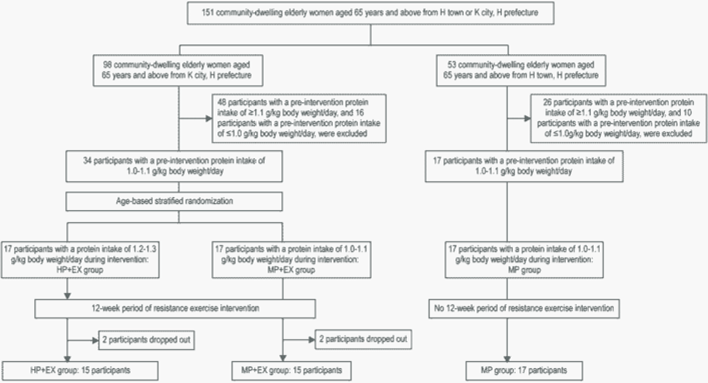
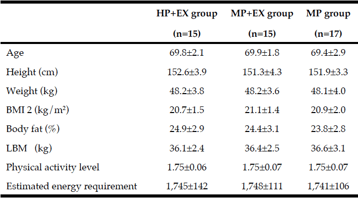
Baseline anthropometric data, muscle function, as well as plasma amino acids were comparable between the NAC and placebo treatment arms (Table 1). Mean baseline tHcy of the total study population was 11.83±0.70 µM, i.e. slightly above the values (9.53±0.35 µM) of our previous trial on NAC in 82 sedentary subjects [12] which had been ca 8 years younger (43.5±3.5 vs. 51.7±2.1 years). Eight weeks of RT yielded substantial and significant increases in isometric and isokinetic PT in both treatment arms, differing neither in absolute nor in percentage terms of strength gain (Table 1). The concomitant small increases in body weight, BMI and BCM (at stable body fat) were also not different between the NAC and placebo arm (though significant within the NAC arm). As a main finding, tHcy significantly decreased with 8 weeks of NAC treatment (-13.97±5.81%, p=0.046 by paired t-test) but not with placebo (-3.85±4.81%) (Table 1; Fig. 1, right panel). For comparison, in our previous placebo-controlled trial in sedentary male subjects (Fig. 1, left panel,[12]) tHcy significantly decreased with NAC (-11.71±3.04%, P<0.001) but not with placebo (4.09±3.59%, P>0.05). ANOVA with adjustment for plasma methionine levels and gain in isometric PT detected a significant difference between the NAC and placebo effect on tHcy (P=0.048; see § in Fig. 1 and Table 1). This result was further scrutinized and confirmed by MANOVA (P=0.048, factor ‘time’ by ‘medication’) when adjusting for the same covariates. The increase in plasma thiol was found to be non-significantly higher with NAC (0.95±1.13 µM, 34.73±30.71%) than with placebo (0.24±0.52 µM, 6.41±10.73%). A similar trend was observed for plasma cystine (cysteine-disulfide). Methionine was significantly increased with NAC only (P=0.02) (Table 1).
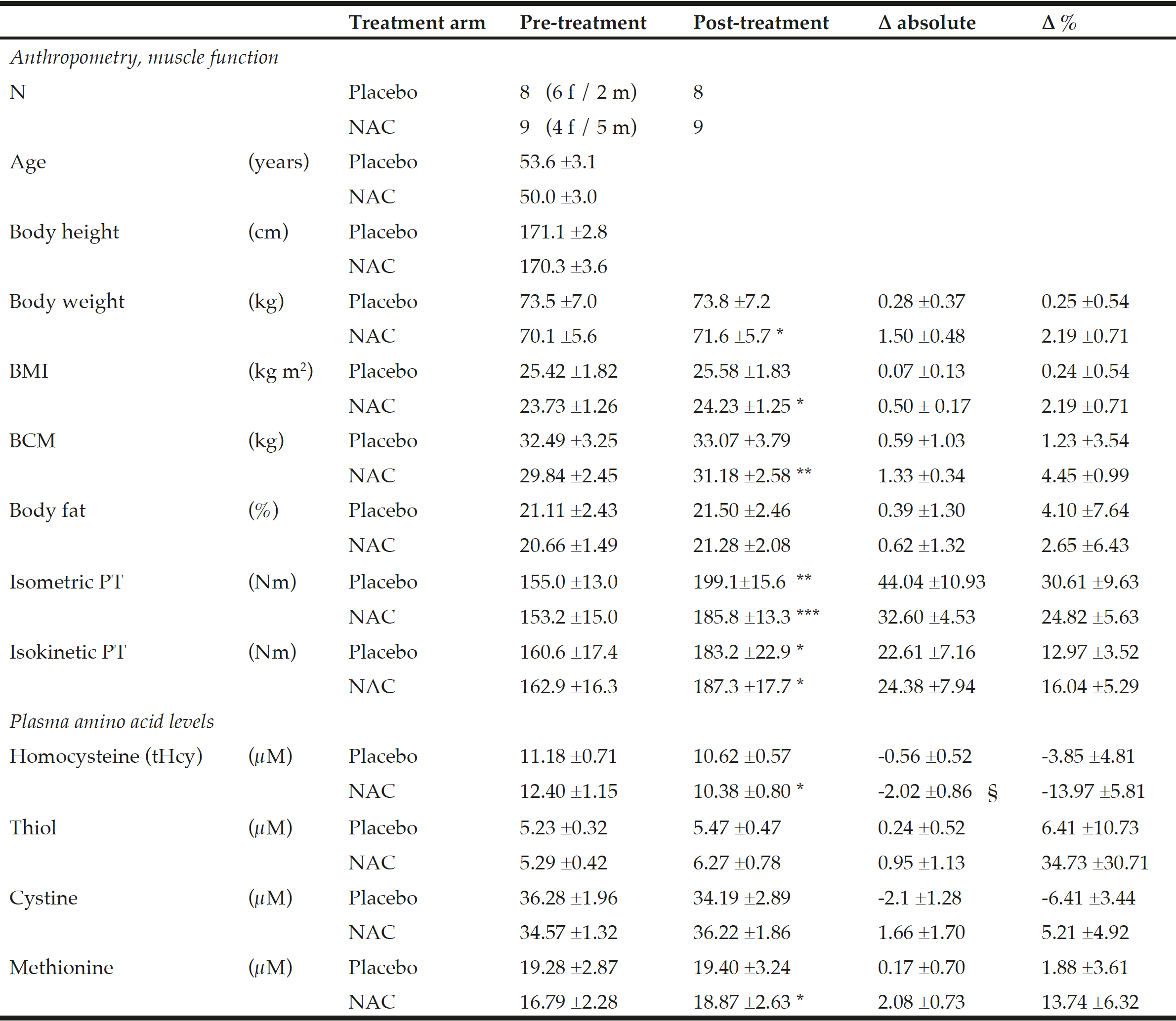
Table 1 Anthropometry, muscle function and amino acid plasma levels before and after NAC and placebo treatment during ongoing resistance training

Table 1
Anthropometry, muscle function and amino acid plasma levels before and after NAC and placebo treatment during ongoing resistance training

Data show the mean ±standard error of the mean (S.E.M.); BMI = body mass index; BCM = body cell mass;  PT = peak torque of right knee extensor. * for P<0.05, ** for P<0.01 and *** for P<0.001 by paired t-test or the Wilcoxon test for post- vs pre-treatment values separately for the NAC or the placebo arm. A significant effect of NAC vs. placebo on the primary endpoint tHcy was assessed by  ANOVA comparing pre-to-post changes between the two treatment arms with adjustments for methionine and pre-to-post gain in isometric PT as covariates (see §, P=0.048).
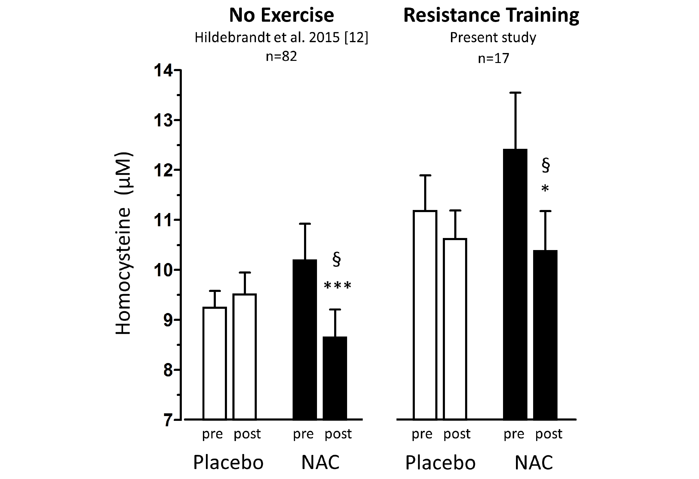
 

Figure 1 Total homocysteine plasma levels (tHcy) before and after 1.8g/d oral NAC or placebo treatment of non-exercising subjects (previous study (12), left panel,n=82) and of subjects undergoing anabolic resistance training (present study, right panel, n=17)

Figure 1
Total homocysteine plasma levels (tHcy) before and after 1.8g/d oral NAC or placebo treatment of non-exercising subjects (previous study (12), left panel,n=82) and of subjects undergoing anabolic resistance training (present study, right panel, n=17)

Percentage homocysteine changes with placebo and NAC amounted to +4.13±3.61% and -11.71±3.04 % (without training, left panel) and to -3.85±4.81% and -13.97±5.81% (with resistance training, right panel), respectively. According to (M)ANOVA with adjustments for confounders the effect of NAC on tHcy was significantly different from that of placebo in both studies: § P=0.001, without training, left panel; § P=0.048, with resistance training, right panel. For details see ‘Statistics’ within ‘Methods’ section.  Data represent means±S.E.M.; * for p>0.05 and *** for p<0.001 by Student’s t-test for paired observation.  


Discussion

Though of limited size, this randomized double-blind clinical trial showed for the first time (generated the hypothesis), that a dose of 1.8 g NAC /d for 8 weeks, orally taken on a background of effectively anabolic RT, significantly lowered tHcy by -13.97%, while a non-significant decrease of -3.85% tHcy occured with placebo (RT alone). This resulting ‘add-on’ effect of NAC of around -10% tHcy reduction is well in line with our recent findings in non-training males (-11.71±3.04%) taking an identical oral NAC dose (12). Notably, two covariates were presently identified to significantly impact the detected NAC effect on tHcy (warranting consideration as confounders in previous and future trials): The RT-related (likely anabolic) gain in isometric PT and the plasma methionine level as a main and nutritionally variable source of tHcy (3). Likely due to the limited muscle mass involved (lower limb vs. whole body) the presently observed tHcy-lowering of -3.85% with RT (plus placebo) remained slightly below the published range of -5 to -6% (9, 10) despite creatine supplementation. Importantly however, NAC did not compromise the outcome of RT suggested as an anti-aging intervention. Of note, the present postabsorptive measurements (12 h after the last NAC dose) may not reflect the transient NAC-induced increase in plasma thiol (mainly cysteine) levels (13) which likely is associated with a large transient tHcy decrease of up to -45% (2, 11). The potential of NAC to acutely decrease tHcy – largely underestimated under postabsorptive conditions – might offer an option to attenuate predictable diurnal/nutritional tHcy peaks by well-timed and adjusted NAC intake.
As NAC was tolerated well and without adverse effect on the outcome of RT, a combination of oral NAC with RT warrants further evaluation as an anti-aging intervention against tHcy-related degeneration limiting functional capacity. Indeed, in elderly subjects (>75 years), the combination of 1.8 g/d NAC with RT was previously shown by us to significantly enhance functional capacity while decreasing plasma TNFα levels (14). Moreover, NAC is able to improve both, the ventilatory and the erythropoietin response to hypoxia (13) beside several other vital functions which clearly decline with age and respond to thiol redox signals – in line with a non-radical oxidative stress theory of aging (15).


Acknowledgments: We gratefully acknowledge the expert laboratory assistance of Ute Winter and Helge Lips.

Conflict of Interest: The authors declare that they have no conflict of interest.

Ethical Standards: The authors declare that the experiment (clinical trial) complied with the current law of the country (Germany) where they were performed. The study was approved approved by the Ethical Committee the University of Heidelberg (L-157/2003-2, 11.11.2003).


References

1.    Martí-Carvajal AJ, Solà I, Lathyris D. Homocysteine-lowering interventions for preventing cardiovascular events. Cochrane Database Syst Rev. 2017 Aug 17;8:CD006612.doi: 10. 1002/14651858.CD006612.put;
2.    Scholze A, Rinder C, Beige J, Riezler R, Zidek W, Tepel M. Acetylcysteine reduces plasma homocysteine concentration and improves pulse pressure and endothelial function in patients with end-stage renal failure. Circulation 2004;109:369-374.
3.    Kanani PM, Sinkey CA, Browning RL, Allaman M, Knapp HR, Haynes WG. Role of oxidant stress in endothelial dysfunction produced by experimental hyperhomocys-t(e)inemia in humans. Circulation 1999;100:1161-1168.
4.    Ventura E, Durant R, Jaussent A, Picot MC, Morena M, Badiou S, et al. Homocysteine and inflammation as main determinants of oxidative stress in the elderly. Free Radic Biol Med 2009;46:737-744.
5.    Clarke R, Bennett D, Parish S, Lewington S, Skeaff M, Eussen SJ, et al. B-Vitamin Treatment Trialists’ Collaboration. Effects of homocysteine lowering with B vitamins on cognitive aging: meta-analysis of 11 trials with cognitive data on 22,000 individuals. Am J Clin Nutr 2014;100:657-666. doi: 10.3945/ajcn.113.076349. Epub 2014 Jun 25.
6.    Veeranki S, Winchester LJ, Tyagi SC. Hyperhomocysteinemia associated skeletal muscle weakness involves mitochondrial dysfunction and epigenetic modifications. Biochim Biophys Acta 2015;1852:732-741. doi: 10.1016/j.bbadis.2015.01.008. Epub 2015 Jan 20.
7.    Guralnik JM, Ferrucci L, Simonsick EM, Salive ME, Wallace RB. Lower-extremity function in persons over the age of 70 years as a predictor of subsequent disability. N Engl J Med 1995;332:556-561.
8.    Liu CJ, Latham NK. Progressive resistance strength training for improving physical function in older adults. Cochrane Database Syst Rev. 2009; 8(3):CD002759. doi: 10.1002/14651858.CD002759.
9.    Silva Ade S, da Mota MP. Effects of physical activity and training programs on plasma homocysteine levels: a systematic review. Amino Acids 2014;46:1795-1804.
10.    Steenge GR, Verhoef P, Greenhaff PL. The effect of creatine and resistance training on plasma homocysteine concentration in healthy volunteers. Arch Intern Med 2001;161:1455-1456.
11.    Ventura P, Panini R, Abbati G, Marchetti G, Savioli G. Urinary and plasma homocysteine and cysteine levels during prolonged oral N-acetylcysteine therapy. Pharmacology 2003; 68:105-114.
12.    Hildebrandt W, Sauer R, Bonaterra G, Dugi KA, Edler L, Kinscherf  R. Oral N-acetylcysteine reduces plasma homocysteine concentrations regardless of lipid or smoking status. Am J Clin Nutr  2015;102:1014-1024.
13.    Hildebrandt W, Alexander S, Bartsch P, Dröge W. Effect of N-acetylcysteine  on the hypoxic ventilatory response and erythropoietin production: linkage between plasma thiol redox state and O2-Chemosensitivity. Blood 2002;99:1552-1555.
14.    Hauer K, Hildebrandt W, Sehl Y, Edler L, Oster P, Dröge W. Improvement in muscular performance and decrease in tumor necrosis factor level in old age after antioxidant treatment. J Mol Med 2003; 81:118-125.
15.    Go YM, Jones DP. Redox theory of aging: implications for health and disease. Clin Sci (Lond) 2017; 31:1669-1688. doi: 10.1042/CS20160897. Print 2017 Jul 15.

EFFECT OF INCREASED DAILY INTAKE OF PROTEIN, COMBINED WITH A PROGRAM OF RESISTANCE EXERCISES, ON THE MUSCLE MASS AND PHYSICAL FUNCTION OF COMMUNITY-DWELLING ELDERLY WOMEN


H. Mori1, Y. Tokuda2


1. Diabetes Therapeutics and Research Center, University of Tokushima, , Tokushima, Tokushima, Japan; 2. Faculty of Health Science Department, Hyogo University, Hiraoka, Kakogawa, Hyogo, Japan

Corresponding Author: Dr. Hiroyasu Mori, Diabetes Therapeutics and Research Center, University of Tokushima, 3-18-15 Kuramoto, Tokushima, Tokushima 770-8503, Japan, Phone number: +81 886337587, Fax Number: +81 886337589, Email: hiroyam31@gmail.com


J Aging Res Clin Practice 2016;inpress
Published online December 1, 2016, http://dx.doi.org/10.14283/jarcp.2016.124


Abstract

Abstract: Background: In elderly women, significant loss of muscle mass due to declining levels of estrogen secretion is a health concern. Increasing the recommended dietary allowance of protein intake has been included as a general health guideline to prevent age-related sarcopenia. Objectives: To investigate effects of light-to-moderate resistance training combined with increased protein intake on the muscle mass, strength, and physical function of community-dwelling elderly women. Design: The 12-week training program combined weight-bearing and resistance band exercises, performed 3 times per week. Setting: Hyogo Prefecture, in either City K or Town H. Practical Intervention: Women were randomly allocated to three groups: exercise with protein intake adjusted to the recommended daily allowance (RDA) of 1.0–1.1 g/kg body weight/day (MP+EX group); exercise with protein intake adjusted above the RDA level at 1.2–1.3 g/kg body weight/day (HP+EX group); and a control group receiving classroom-based session on nutrition management, with protein intake adjusted to the RDA level (MP group). Measurements: Body weight and physical composition were measured by multi-frequency bioelectrical impedance analysis. Results: Exercise prevented decreases in muscle mass and strength and in performance of physical function tasks (p<0.05). Increasing dietary intake of protein above RDA level increased muscle mass (p<0.01), walking speed (p<0.01) and knee extensor strength (p<0.05). Conclusion: Adjusting protein intake to 1.2–1.3 g/kg body weight/day, in combination with light-to-moderate resistance training, can improve body composition and physical function in elderly women. The result of this study could be effective in lowering the incidence of age-related sarcopenia.

Key words: Community-dwelling elderly, exercise, muscle mass, protein intake, sarcopenia.


Introduction

Aging is associated with a decrease in skeletal muscle mass and strength which leads to limitations in physical activities, increased risk for falls and overall lowering of the quality of life of elderly people (1–7). A specific health concern is the significant loss of muscle mass in elderly women due to declining levels of estrogen secretion (8). The resulting loss of strength is one of the primary factors of the higher incidence of falls in older women, compared to older men. Increasing the recommended dietary allowance of protein intake has been included as a general health guideline to prevent age-related sarcopenia (9-11).
The Japanese recommendations for dietary reference intakes (DRIs) indicate a daily protein intake of 1.0–1.1 g/kg body weight/day for people aged >65 years (12). These DRIs do not include protein intake levels necessary for elderly individuals performing resistance exercises for the purpose of increasing skeletal muscle mass and physical function, knowledge required to optimize outcomes of exercise (13).
According to the Long-Term Care Prevention Service, which aims to increase musculoskeletal system function in the elderly, few elderly individuals practice high-intensity resistance exercises using large-scale training equipment. Moderate-intensity resistance exercises performed using body weight and resistance bands provide an easy way for elderly people to exercise for the purposes of increasing skeletal muscle mass and physical function to prevent sarcopenia. Accordingly, the aim of our study was to evaluate the effectiveness of adjusting total protein intake to levels of 1.2–1.3 g/kg body weight/day, a level exceeding the DRIs, on skeletal muscle mass and physical function in elderly women performing moderate intensity resistance exercises that used their own body weight and resistance bands, thereby preventing sarcopenia.


Methods

Participants

Participants were 151 Japanese women, ≥65 years old, living in Hyogo Prefecture, in either City K or Town H.
With the aim of using the DRI level of protein intake (1.0–1.1 g/kg/day12) as a baseline reference, a dietary survey was used to screen the daily protein intake of prospective participants. Based on this dietary survey, 26 participants were excluded as their total daily protein intake was <1.0 g/kg/day, and 74 participants for a daily protein intake >1.1 g/kg/day. Remaining participants were randomly allocated to three groups of 17 participants: an exercise intervention group with protein intake adjusted to the DRI of 1.0–1.1 g/kg/day (MP+EX group); an exercise intervention group with protein intake adjusted above the DRI at 1.2–1.3 g/kg/day (HP+EX group); and a control group who participated in classroom-based session on nutrition management, with their protein intake adjusted to the DRI of 1.0–1.1 g/kg/day (MP group). A stratified randomization strategy was used to achieve a comparable age distribution across groups. Over the study period, 4 participants withdrew due to family circumstances or poor physical conditioning, 2 participants from the HP+EX group and 2 participants from the MP+EX group. The information from these 4 participants was excluded from analysis, with the final study group formed of 15 participants in the HP+EX group, 15 participants in the MP+EX group, and 17 participants in the MP group (Figure 1).


Figure 1 Flow chart of study participation

Figure 1
Flow chart of study participation

Using participants with a pre-intervention protein intake of 1.0-1.1g/kg body weight/day, we randomly divided the participants into a high protein intake group of 1.2-1.3g/kg body weight/day (HP+EX group) and a moderate protein intake group of the usual 1.0-1.1g/kg body weight/day (MP+EX group) during an intervention of light-to-moderate intensity resistance exercise. We also established a moderate protein intake group of the usual 1.0-1.1 g/kg body weight.

Intervention design

A 12-week program was implemented. Groups HP+EX and MP+EX completed a standardized program of resistance exercise and a nutrition management, with MP group completed a classroom-based program on nutrition.

Measurement of body weight and physical composition

Body weight and physical composition were measured by multi-frequency bioelectrical impedance analysis (In Body 430, Bio Space, Seoul, Korea). The following parameters of body composition were obtained: body mass index (BMI), body fat percentage, lean body mass (LBM), and total limb muscle mass, which was calculated separately for the upper and lower limbs, and for the trunk.

Measurement of physical functions

The following measures of physical function were evaluated: grip strength, knee extension strength, 5-m maximum walking speed, and timed up-and-go (TUG). Grip and knee extension strength were measured by hand held dynamometry (T.K.K5401; Takei Instruments, Tokyo, Japan. μ-tus F-100; ANIMA., Tokyo, Japan). Measures were taken twice, and the maximum value recorded for analysis.

Nutritional survey

A nutritional survey was conducted to document: total energy intake (TEI), total and adjusted protein intake (g/kg/day), total fat intake, and total carbohydrate intake. The survey was complemented by food weighing methods (Excel Eiyou version 5.0; Kenpakusha, Tokyo, Japan). The nutrition questionnaire and weighing were completed for 5 consecutive days before the start of the program, and daily during the 12-week intervention.

Daily activity survey

Participants recorded their daily activity for three consecutive days before the start of the program, with the information used to calculate each participant’s regular physical activity level (PAL). Again, individual interviews were used to complete missing information. The estimated energy requirement (EER) to meet each individual’s PAL was calculated as follows: EER = basal metabolic rate × body weight (kg) × PAL (12).

Exercise intervention

The exercise intervention consisted of a series of body weight resisted and resistance band exercises, performed 3 times per week for 12 weeks. Twice per week, exercises were performed under supervision of an exercise specialist, with participants completing the third session at home.

Resistance exercises

Body weight resisted exercises consisted of the following: abdominal crunches, rising and sitting from a chair; leg extensions; standing heel kicks, and calf raises. Five exercises were also performed with elastic bands (REP BAND; Magister Corporation, Chattanooga, TN): arm curls, pull-ups, leg extensions, squats, and sit-ups. Elastic bands of five difference resistance were used, with the level of resistance individually adjusted using 1 maximum repetition (1RM) testing for the upper and lower limbs prior to the intervention. The resistance load was modified in standardized fashion over the 12-week program as follows: for weeks 1-4, participants performed 2 sets of 10 repetitions each of the body weight resisted exercises and 2 sets of 20 repetitions of the resistance band exercises, at a resistance load of 50% 1RM; for weeks 5-8, participants performed 2 sets of 15 repetitions each of the body weight resisted exercises and 2 sets of 15 repetitions each of the resistance band exercises at 60% 1RM; and for weeks 9-12, participants performed 3 sets of 15 repetitions each of the body weight resisted exercises and 3 sets of 12 repetitions each of the resistance band exercises at 70% 1RM.

Nutritional intervention

Total protein intake in the HP+EX group was adjusted to a daily level of 1.2–1.3 g/kg/day. Participants’ were advised to increase their protein intake in their standard meals (breakfast, lunch, and dinner). The total protein intake for participants in the MP+EX and MP groups was adjusted to 1.0–1.1 g/kg/day.

Nutritional management

Nutritional management was provided by a nutritionist, based on the DRIs (14) and results of the nutrition survey conducted before the intervention. Nutritional management was provided according to the following schedule: once to all participants prior to the onset of the study; as part of the group nutrition guidance sessions in intervention weeks 1-2; and on an individual basis, twice per week, for intervention weeks 3-12. At these individual sessions, protein intake was verified to ensure compliance with group-specific levels. Daily activity surveys were also reviewed to ensure sufficient TEI for all participants.

Statistical analysis

Individual changes in EER and TEI, pre- and post-intervention, were evaluated using paired t-test analysis. Between-group differences in outcome variables, measured pre- and post-intervention, were evaluated using two-way analysis of variance (ANOVA). Changes in measured outcomes of muscle mass, physical function, and nutrient intake were evaluated between groups (HP+EX, MP+EX and MP) and time (pre- and post-intervention) using a repeated measures ANOVA with group as an independent factor and time as the repeated factor. For identified main effects and interactions, multiple comparisons were performed using the Tukey post hoc analysis, with specific change in pre- and post-intervention values compared using an unpaired one-way ANOVA. All statistical analyzes were performed using IBM SPSS statistical software (IBM, Tokyo, Japan), with the level of significance defined as a p value < 0.05.


Results

Baseline measures of body composition and physical function are listed in Table 1. Pre-intervention, measures were comparable between groups. Adherence to the schedule of resistance exercise of three sessions per week for 12 weeks was comparable for the two exercise groups, with a mean (SD) number of sessions completed of 34.3±1.2, out of a possible maximum of 36, for the HP+EX group (95.3%) and 34.0±1.0 for the MP+EX group (94.4%).


Table 1 Physical characteristics of subjects pre-intervention

Table 1
Physical characteristics of subjects pre-intervention

Mean value ± Standard deviation;  The results of a non-paired one-way ANOVA showed no differences in the characteristics of the 3 groups pre-intervention; HP+EX group: Protein intake of 1.2-1.3 g/kg body weight/day during the 12-week period of exercise intervention; MP+EX group: Protein intake of 1.0-1.1 g/kg body weight/day during the 12-week period of exercise intervention; MP group: Protein intake of 1.0-1.1 g/kg body weight/day for the 12-week period; BMI: Body mass index; LBM: Lean body mass


Changes in measures of muscle mass pre- and post-intervention are reported in Table 2. A significant main effect of exercise on limb muscle mass was identified (p<0.05) with the largest change identified for the HP+EX group (p<0.001 compared to both the MP+EX and MP groups). A significant group X limb interaction was identified (p<0.05), with a significantly higher increase in muscle mass for the HP+EX group, compared to the MP group, for the upper limbs (p<0.05), trunk (p<0.001) and lower limbs (p<0.001). Comparing HP+EX and MP+EX groups, the magnitude of change in muscle mass was higher for the HP+EX group only for the lower limbs (p<0.05).

Table 2 Comparison of limb muscle mass and physical functions pre-/post-intervention

Table 2
Comparison of limb muscle mass and physical functions pre-/post-intervention

Mean value ± Standard deviation; Two-way ANOVA by time (Pre- and Post-intervation period) × group (HP+EX, MP+EX, MP group)* p <0.05, ** p <0.01,*** p <0.001; One-way ANOVA and post-hoc by Tukey test, Significant difference with MP group: †p <0.05, ††p <0.01, †††p <0.001, Significant difference with MP+EX group: ‡p <0.05, ‡‡p <0.01


Effects of the intervention physical function are reported in Table 2. The ANOVA identified significant effects of exercise on knee extensor strength and 5-m maximum walking speed, as well as knee extensor strength x TUG interaction. Change in knee extensor strength was higher for the MP+EX group, compared to the MP group (p<0.01), with the highest magnitude of change obtained by the HP+EX group (p<0.001 compared to MP; p<0.05 compared to MP+EX group). Findings were similar for 5-m maximum walking speed, with greater improvement in walking speed in the MP+EX compared to MP groups (p<0.05), and largest increase for the HP+EX group (p<0.001 compared to MP; p<0.01 compared to MP+EX group). The improvement in TUG was greater for the HP+EX compared to the MP group (p<0.01).
Nutritional intake, TEI, EER, and levels of total protein intake, adjusted protein intake/kg body weight, fat, and carbohydrates are listed in Table 3. Significant changes in TEI and protein intake (TEI, p<0.05; protein, p<0.001), as well as an interaction between adjusted protein intake and body weight (p<0.001) were identified. The magnitude of change was higher for the HP+EX group compared to the MP group (TEI, p<0.05; protein intake/kg, p<0.001) and the MP+EX group (TEI, p<0.05; protein intake/kg, p<0.001).

Table 3 Comparison pre-intervention and during intervention

Table 3
Comparison pre-intervention and during intervention

Mean value ± Standard deviation, Two-way ANOVA by time (Pre- and post-intervation period) × group (HP+EX, MP+EX, MP group), * p <0.05, ** p <0.01, ***p <0.001; One-way ANOVA and post-hoc by Tukey test, significant difference with MP group: †p <0.05, †††p <0.001, Significant difference with MP+EX group:  ‡p <0.05, ‡‡‡p <0.001


Discussion

In this study, we evaluated the effectiveness of providing an adjusted protein intake of 1.2-1.3 g/kg/day during a 12-week program of light-to-moderate intensity weight-bearing and resistance band exercises on the skeletal muscle mass and physical function of elderly women. The adjusted protein intake improved selected parameters of body composition and physical function, namely lower limb muscle mass, knee extension strength, and 5-m maximum walking speed. The mean protein intake for the HP+EX group was 11.4 ± 1.8 g/day, and increased by approximately 0.22 ± 0.02 g/kg/day over the 12-week program. Fat and carbohydrate intake were comparable between groups and TEI was never below EER.
Our current results support our a priori hypothesis of the necessity of increasing protein intake to optimize outcomes of exercise on muscle mass and physical function in the elderly. In fact, total limb muscle mass increased by 0.9±0.3 kg when combining increased protein intake and exercise (HP+EX), compared to 0.2±0.4 kg for exercise alone (MP+EX). Our measured effect of exercise alone is comparable to results reported by Kim et al. (15) of a 0.29 kg increase in total limb mass for elderly Japanese women completing a 12-week program. Kim et al. (15) combined resistance exercises with amino acid supplements, reporting an increase of approximately 0.34 kg in total limb muscle mass. Using an adjusted protein intake, our program surpassed these levels with a mean increase in total limb mass of 0.9 kg, with the most significant increase noted for the lower limbs. This lower limb bias may be explained by performance of a higher number of lower limb exercises. An important difference in our study, compared to Kim et al. (15), is that we graduated the workload of our program over the 12-weeks.
An important outcome of our study was the identification of a 0.2±0.7 kg decrease in total limb muscle mass and 0.2±1.0 kg decrease in knee extension strength for participants in the MP group over the study period. This emphasizes the need to adjust protein intake, at least to the minimum DRI of 1.0–1.1 g/kg/day, in combination with a light-to-moderate program of resistance training, to maintain muscle mass, with further adjustment in protein intake yielding more significant results.
According to Chevalier et al. (16), a dietary protein intake of 0.8 g/kg/day is insufficient for increasing the skeletal muscle mass of elderly persons and intake should be increased to levels of at least 1.2 g/kg/day. Several physiological reasons contribute to the increased requirement of protein intake in the elderly. Drummond et al. (17) reported a delay and/or decrease of the signaling responses to amino acids in skeletal muscle cells with age. Volpi et al. (18) further demonstrated that a higher proportion of the amino acids absorbed in the gastrointestinal tract of elderly persons is metabolized in the small intestine and liver. Although specific physiological measurements related to protein metabolism were not included in our study, our outcomes provide evidence of insufficient muscle protein synthesis during resistance exercise with a moderate adjustment of protein intake to levels of 1.0–1.2 g/kg/day and that a higher adjustment to levels of 1.2–1.3 g/kg/day are required to enhance effectiveness of resistance training on muscle mass and strength.
Our light-to-moderate level of resistance was sufficient to maintain muscle mass and strength in the MP+EX group. Levinger et al. (19) reported a 1.1-kg increase in skeletal muscle mass of elderly participants performing a 10-week program of high-intensity resistance exercises. In their review of research evidence for resistance training in elderly individuals, Miyachi et al. (20) indicated that an exercise intensity of 80% 1RM or above is required to increase the skeletal muscle mass with a single intervention of exercise. It is important to note that the participants in our study group had a relatively low PAL of 1.75; as none of our participants engaged in regular resistance training, prescription of a high-intensity program was not possible. Future research is required to more clearly elucidate the effectiveness of supplemented protein intake and different levels of resistance training for enhancing gains in muscle mass and strength. However, we do emphasize that our program of light-to-moderate resistance was effective, with a practice rate of approximately 95%, and can be safely implemented as a home program for elderly individuals who are relatively deconditioned.
The benefits of combining a light-to-moderate resistance training program with nutritional supplementation in lowering the risk for sarcopenia in elderly individuals is supported by previous research. In their review comparing the transient response of muscles to high- and low-intensity resistance training regimes, Mallinson et al. (21) reported an increase in muscle protein synthesis (and increased muscle protein reserves) with high-intensity resistance exercise, performed at 90% 1RM, while low-intensity resistance exercises, performed at 30% 1RM, stimulated muscle protein synthesis. Therefore, light-to-moderate intensity weight-bearing and resistance band resistance exercise can have benefits for elderly women, who can easily perform these exercises.
In our study, we assumed that a training resistance of 50–70% 1RM would have little stimulus on protein synthesis in skeletal muscle. As a result, we did not include a control group of adjusted high protein intake alone, with no participation in the program of exercise. Therefore, we are unable to determine if the increase in muscle mass for the HP+EX group results from the increase in protein intake or from the combination of increased protein intake and exercise. Future research is required to clarify the pathway of muscle mass increase associated to a combination of supplemented protein intake and exercise. Another important limitation of our study is the inclusion only of women to control for known sex-specific differences in age-related muscle atrophy and sarcopenia (22, 23). Future research will need to verify effectiveness of protein supplementation in elderly men. Moreover, we estimated muscle mass from BIA measurements. There are currently no population norms of BIA measurements for elderly women, and research is needed to establish norms compared to DEXA (24). Finally, the present study is that the sample size was too small, and the conclusion was not well supported by this data. For example, SD of total limb muscle mass is 1.9 and the change by intervention was 0.9kg in HP+EX group. Required sample size to make this change as significant is 37, which is more than twice of the actual number of sample.
Our study provides evidence of the benefit of increasing protein intake as one component of a resistance training program to improve muscle mass and strength in elderly women.


Funding: This work was supported by JSPS KAKENHI Grant Number 25750353. The sponsors had no role in the design and conduct of the study; in the collection, analysis, and interpretation of data; in the preparation of the manuscript; or in the review or approval of the manuscript.

Acknowledgements: None.

Conflict of Interest: All of the authors declare that they have no conflicts of interest regarding this paper.

Ethical standards: Ethics Committee of Hyogo University (approved # 12003).


References

1.     Evans WJ. What is sarcopenia? J Gerontol A Biol Sci Med Sci 1989;50A:5–8.
2.     Janssen I, Baumgartner RN, Ross R. Skeletal muscle cutpoints associated with elevated physical disability risk in older men and women. Am J Epidemiol 2004;159:413 – 421.
3.     Roubenoff R, Hughes VA. Sarcopenia: current concepts. J Gerontol A Biol Sci Med Sci 2000;55:716–724.
4.     Robinson S, Cooper C, Aihie SA. Nutrition and sarcopenia: a review of the evidence and implications for preventive strategies. J Aging Res 2012;2012.(ID 510801). http://dx.doi.org/10.1155/2012/510801
5.     Christie J. Progressive resistance strength training for improving physical function in older adults. Int J Older People Nurs 2011;6:244 – 246.
6.     Peterson MD, Rhea MR, Sen A. Resistance exercise for muscular strength in older adults: a meta-analysis. Ageing Res Rev 2010;9:226–237.
7.     Taaffe DR. Sarcopenia: exercise as a treatment strategy. Aust Fam Physician 2006;35:130–134.
8.     Doherty TJ. Invited review: aging and sarcopenia. J Appl Physiol 2003;95:1717–1727.
9.     Beasley JM, Shikany JM, Thomson CA. The role of dietary protein intake in the prevention of sarcopenia of aging. Nutr Clin Pract 2013;28:684–690.
10.     Morley JE, Argiles JM, Evans WJ, et al. Society for Sarcopenia, Cachexia, and Wasting Disease. Nutritional recommendations for the management of sarcopenia. J Am Med Dir Assoc 2010;11:391–396.
11.     Volkert D, Sieber CC. Protein requirements in the elderly. Int J Vitam Nutr Res 2011;81:109–119.
12.     Nakade M, Imai E, Tsubota-Utsugi M. Systematic classification of evidence for dietary reference intakes for Japanese 2010 (DRIs-J 2010) in adults and future prospects of DRIs in Asian countries. Asia Pac J Clin Nutr 2013;22:474–489.
13.     Eckard T, Lopez J, Kaus A, Aden J. Home exercise program compliance of service members in the deployed environment: an observational cohort study. Mil Med 2015;180:186–191.
14.     Mori H, Niwa M. Effect of nutritional care and whey protein supplementation on the body composition and physical function in older adults after combined resistance and aerobic exercise. Jpn J Nutr Diet 2014;72:12–20. [in Japanese]
15.     Kim HK, Suzuki T, Saito K, et al. Effects of exercise and amino acid supplementation on body composition and physical function in community-dwelling elderly Japanese sarcopenic women: a randomized controlled trial. J Am Geriatr Soc 2012;60:16–23.
16.     Chevalier S, Gougeon R, Nayar K, Morais JA. Frailty amplifies the effects of aging on protein metabolism: role of protein intake. Am J Clin Nutr 2003;78:422–429.
17.     Drummond MJ, Dreyer HC, Pennings B, et al. Skeletal muscle protein anabolic response to resistance exercise and essential amino acids is delayed with aging. J Appl Physiol 2008;104:1452–1461.
18.     Volpi E, Mittendorfer B, Wolf SE, Wolfe RR. Oral amino acids stimulate muscle protein anabolism in the elderly despite higher first-pass splanchnic extraction. Am J Physiol 1999;277:513–520.
19. Levinger I, Goodman C, Hare DL, Jerums G, Seling S. The effect of resistance training on functional capacity and quality of life in individuals with high and low numbers of metabolic risk factors. Diabetes Care 2007;30:2205–2210.
20.     Miyachi M, Ando D, Oida Y, et al. Possible treatments for sarcopenia: systematic review on the effect of exercise intervention. Jpn J Geriatr 2011;48:51–54. [in Japanese]
21.     Mallinson JE, Murton AJ. Mechanisms responsible for disuse muscle atrophy: potential role of protein provision and exercise as countermeasures. Nutrition 2012;29:22–28.
22.     Sanada K, Miyachi M, Tanimoto M, et al. A cross-sectional study of sarcopenia in Japanese men and women: reference values and association with cardiovascular risk factors. Eur J Appl Physiol 2010;10:57–65.
23.     Basaria S, Coviello AD, Travison TG. Adverse events associated with testosterone administration. N Engl J Med 2010;8:109–122.
24.     Buckinx F, Reginster JY, Dardenne N, et al. Concordance between muscle mass assessed by bioelectrical impedance analysis and by dual energy X-ray absorptiometry: a cross-sectional study. BMC Musculoskelet Disord 2015;16:60.

PLASMA LEVELS OF INTERLEUKIN-6 AND SOLUBLE TUMOR NECROSIS FACTOR RECEPTOR ARE ASSOCIATED WITH MUSCLE PERFORMANCE IN PRE-FRAIL COMMUNITY-DWELLING OLDER WOMEN?


L. Paccini Lustosa1, L. Souza Máximo Pereira1, P.Parreira Batista2, D.A. Gomes Pereira1, J.M. Domingues Dias1, A. Netto Parentoni3


1. PhD, Prof. Physiotherapy Department, Universidade Federal de Minas Gerais, Belo Horizonte, MG, Brazil; 2. Post-graduate program in Rehabilitation Sciences, Universidade Federal de Minas Gerais, Belo Horizonte, MG, Brazil; 3. PhD, Prof. Physiotherapy Department, Universidade Federal dos Vales do Jequitinhonha e Mucuri, Diamantina, MG, Brazil.

Corresponding Author: Lygia Paccini Lustosa, Av. Prof. Antonio Carlos, 6627 – Pampulha – Belo Horizonte – Minas Gerais – Brazil, Cep: 31.270-901 –e-mail: lygia.paccini@gmail.com – tel.: 55 (31) 99831854/ 34094791

J Aging Res Clin Practice 2016;inpress
Published online September 1, 2016, http://dx.doi.org/10.14283/jarcp.2016.113


Abstract

Aim: Increased plasma levels of interleukin (IL)-6 and tumor necrosis factor (TNF)-α have been associated with frailty syndrome and reduced muscle strength in older. Sarcopenia influenced loss of mobility and functional independence, and contributed to frailty syndrome. Furthermore, sarcopenia mainly entails a decrease in type II muscle fibers, with consequent loss of muscle power; this could occur as a result of a lack of physical activity. Objective: To examine the correlation of muscular performance and the plasma levels of IL-6 and soluble TNF receptor (sTNFr) in pre-frail community-dwelling women. Methods: The study included 32 pre-frail women (≥ 65ys). The measurements were plasma concentrations of IL-6 and sTNFr1 (ELISA); muscle strength (isokinetics Biodex System). The muscle resistance program constituted 75% of maximum load (3 times/week, 10 weeks). Statistical analysis were made through Pearson and Spearman correlation (α = 5%). Results: There was a significant inverse correlation between sTNFr1 and muscle strength, pre- (r = −0.36, P = .04) and post-training (r = −0.37, P = .04) and, a significant positive correlation between IL-6 and muscle strength (r = 0.45, P = .01). Conclusion: The correlations found between the inflammatory mediators and the measures of muscular performance evaluated before and after training suggest that, as the muscles increase their ability to generate power, sTNFr concentrations decrease, and the levels of IL-6 increase. Muscle resistance exercises should be encouraged in pre-frail older women to induce the release of cytokines.

Key words: Frail, older women, IL-6, sTNFr, exercise.


Introduction

Sarcopenia is a term used to describe a degeneration of the musculoskeletal system, which can be related to changes in the immune and endocrine systems, among others (1, 2). Sarcopenia mainly entails a decrease in type II muscle fibers, with consequent loss of muscle power; this could occur as a result of a lack of physical activity (3-7). Moreover, sarcopenia may have a greater health impact on older women than men, as women have a longer life expectancy and greater rates of morbidity (7, 8). Schaap et al (2006) reported a positive correlation between sarcopenia and elevated plasma levels of pro-inflammatory cytokines, including interleukin-6 (IL-6), C-reactive protein, and tumor necrosis factor-alpha (TNF-α) (4). Doherty (2003) reported that sarcopenia influenced loss of mobility and functional independence, and contributed to frailty syndrome (6). Frailty syndrome has been described as a clinical, multi-factorial syndrome characterized by 3 distinct actions: deregulation of the neuroendocrine system, deregulation of the immune system, and the induction of sarcopenia (9, 10). Thus, sarcopenia may be associated with a sub-threshold state of chronic inflammation that is characteristic of older people (11).
Ferrucci et al (2002) concluded that the reduced ability to perform daily functional activities was associated with high levels of IL-6 and TNF-α, and the loss of muscular strength (12). These authors pointed to the deleterious effect of high concentrations of these cytokines in muscle tissue. Previous studies in our laboratory have demonstrated an inverse correlation between plasma IL-6 levels and muscle strength of lower limbs and hand grip in institutionalized older individuals at rest (13, 14). Pereira et al (2009) and Oliveira et al (2008) found an inverse correlation between manual and knee extensor muscle strength with plasma levels of IL-6 (15,16). Greiwe et al (2001) reported that an increase in plasma levels of TNF-α is associated with loss of muscle mass, and that concentric resistance exercises could reduce plasma expression of this cytokine in their older participants (17).
In this context, some authors have suggested that physical activity, or a program of specific resistance exercises, could reduce the plasma levels of pro-inflammatory mediators and possibly reduce the deleterious consequences of these cytokines in musculoskeletal tissue (5, 13, 17-19). The explanation behind this hypothesis relates to the fact that IL-6 can be released by muscle contraction (named myokine), independently of TNF-α, thereby inducing the release of other anti-inflammatory cytokines (IL-10 and IL-1ra) that could reduce plasma concentrations of TNF-α (5, 17-20). These assumptions are even more significant when considering the muscular and inflammatory systems of older individuals with frailty syndrome. Moreover, a recent study demonstrated an improvement in muscle strength and function after a resistance exercise program, but no changes in inflammatory mediators following the program. However, discontinuation of this program increased the plasma levels of TNF-α (21).
The objective of this study was therefore to assess the correlation between the muscle strength of knee extensors and plasma indexes of IL-6 and sTNFr1, before and after a resistance exercise program for the lower limbs in pre-frail older women.


Methods

This study was a cross-sectional analysis as part of a randomized, blind, crossover clinical trial approved by the Research Ethics Committee of Universidade Federal de Minas Gerais, decree ETIC 321/2007. The protocol for this study was registered in BioMed Central (BMC) under number ISRCTN62824599 (http://www.controlled-trials.com/ISRCTN62824599). All participants signed an informed consent form before starting the study, and were recruited from the clinics of 2 universities, through verbal invitation.  After the initial evaluation, the participants started training (3 times/week, for 10 weeks) at 75% of maximal load. The physiotherapist responsible for the intervention had no knowledge of the evaluations performed. The evaluators had no knowledge of the group to which each participant belonged (21, 22).

Sample

Thirty-two community-dwelling older women (aged 65 years and older) were selected; pre-frail criteria, according to the phenotype proposed by Fried et al (2010) were used.2 All participants answered a questionnaire aimed to characterize the sample in terms of clinical and socio-demographic aspects.
Exclusion criteria were cognitive impairment (Mini Mental State Exam, 1994) (23), orthopedic and neurological diseases that could affect test outcomes, acute inflammatory disease, cancer, and use of drugs that act on the immune system.

Measuring Instruments

The plasma levels of IL-6 and sTNFr1 were measured by enzyme-linked immunosorbent assay using high sensitivity kits (Quantikine®HS, R&D Systems, Minneapolis, USA). The samples were analyzed by a micro-plate reader set to 490 nm and corrected for wavelength at 650 nm. The blood sample analyses of plasma concentrations of IL-6 and sTNFr1 were performed on different days from the muscle tests, with at least a 48-h interval and always in the morning between 8 and 10 am. A qualified professional performed the blood collection, following the necessary standards and procedures. Five milliliters of blood was collected and centrifuged at 1,500 rpm for 15 min to separate the plasma. The plasma was properly identified and stored in a freezer at −70°C. The analyses were performed in duplicate, and the results were presented as the average of the 2 measures ± standard deviation, in pg/ml.
The muscle performance of the knee extensor muscles were measured by an isokinetic dynamometer (Biodex System 3 Pro®) at an angular velocity of 60º/s and 180º/s. At each velocity, 3 training repetitions at sub-maximal effort were used to familiarize the participants with the procedure. The isokinetic evaluation was conducted by measuring 5 and 15 repetitions at maximum effort, at angular velocities of 60º/s and 180º/s, respectively. Participants were motivated during the test by using clapping and verbal encouragement. This standardized version of the test has been used in previous studies [16]. For the analyses, the variable, i.e. work, was standardized by body weight, average power, and peak torque at the angular velocities of 60º/s and 180º/s.

Intervention

The resistance exercise program was conducted during a period of 10 weeks, with 3 sessions per week. Each session consisted of exercises performed in groups of 4−6 participants, with direct guidance and supervision by a physiotherapist. The exercises targeted the lower limbs, particularly the knee extensors, using open and closed kinetic chain exercises, and a load of 75% of the participant’s maximal load (24). The choice of exercises and program dynamic was based on previous studies (24) and is in agreement with the previously published study protocol (25).

Statistical Analysis

The sample size was calculated considering a confidence interval of 95%, an alpha (α) value of 5%, and a standard error of 20%. To test for the normality of the data, the Anderson Darling test was performed, and a Box Cox transformation for optimal lambda (λ) was done for the IL-6 variable as it was not normally distributed. The correlations between variables were made by Pearson and Spearman correlation test. The level of significance was set at α = 5%.


Results

This study included 32 pre-frail older women. All volunteers were classified as pre-frail, according to criteria described by Fried et al (1, 2) and 16 (1 in 2 cases) out of the 32 older women evaluated had 2 positive criteria. The most prevalent criteria were reduction in hand grip strength (43.8%), low caloric expenditure (43.8%), reduction in gait speed (34.4%) and reported exhaustion (25%). The clinical and demographic characteristics of each group are shown in Table 1.


Table 1 Demographic and characteristics of participants

Table 1
Demographic and characteristics of participants

SD, standard deviation; MEEM,Mini Mental State Exam


The analyses of correlation were done before and after the exercises. Before training, there was a poor but significant inverse correlation between the plasma concentration of sTNFr1 and work, which was standardized by body weight in the angular velocity of 180º/s (r = −0.36, P = .04), peak torque at 180º/s (r = −0.38, P = .03) and average power at 180º/s (r = −0.40, P = .02), showing that sTNFr1 concentrations were lower when the power, peak torque, and average power increased (Table 2). After exercises, there was a poor but significant inverse correlation between the concentration of sTNFr1 and the measures of standardized work by body weight and average power at 180º/s (r = −0.37, P = .04; r = −0.37, P = .04, respectively).
Furthermore, there was a positive significant correlation between the plasma concentration of IL-6 and the peak torque and average power at 60º/s (r = 0.45, P = .01; r = 0.44, P = .01, respectively) and at 180º/s (r = 0.46, P = .01; r = 0.37, P = .04, respectively). These results showed an increase in IL-6 associated with an increase in peak torque and muscle power, suggesting that this cytokine was released after training (Table 2).
The statistical analyses showed improvement on the muscular power and on the functional capacity after training period, but there was no difference on the inflammatory mediators (data not shown, but previously published). Likewise, after the period of 10 weeks of follow-up there was statistical difference on the sTNFr measures (data not shown, but previously published) (21).


Table 2 Correlation between sTNFr1 and IL-6 with muscular variables, pre- and post-intervention

Table 2
Correlation between sTNFr1 and IL-6 with muscular variables, pre- and post-intervention

* Significant difference; IL-6, interleukin-6; sTNFr, soluble tumor necrosis factor receptor.


Discussion

The aim of this study was to assess the correlation between the muscle strength of knee extensors and plasma indexes of IL-6 and sTNFr1 before and after a resistance exercise program for the lower limbs in pre-frail older women. The results showed that there was a significant inverse correlation between sTNFr1 and the muscle strength parameters, before and after training. Furthermore, a significant positive correlation between IL-6 and the muscle parameters was detected after training, indicating a probable anti-inflammatory effect of IL-6 released by muscular contraction after resistance exercises.
These findings are in agreement with the results of some authors who suggested using the term myokine for the cytokines that are released by muscle contraction, in particular, IL-6 (11, 19, 20). According to these authors, in response to muscle contraction, type I and II fibers induce the release of IL-6 (11, 19, 20). Thus, this cytokine would exert a local effect on the muscle, and peripheral effects via the induction and inhibition of other pro- and anti-inflammatory cytokines, thereby increasing glucose levels, which are needed for muscle contraction and fat oxidation (18, 19, 26, 27). In this context, there is evidence of an increase in IL-1ra and IL-10 and a reduction in TNF-α after physical exercise, suggesting that exercise has anti-inflammatory effects (19, 21, 28). Therefore, the significant correlations found in this study are in agreement with the literature, showing that greater muscular performance is associated with lower concentrations of sTNFr and higher concentrations of IL-6.
Another argument about these associations has been suggested, concerning the mechanism of this phenomenon. Febbraio et al (2002) and Petersen et al (2005) showed that the plasma levels of IL-6 tend to increase in response to an increase in adrenal sympathetic response induced by the β-adrenergic pathway (11, 19). Therefore, modifications in the glycogen available for muscle contraction would be sufficient to initiate greater release of IL-6, which, in turn, would alter levels of sTNFr (11, 19, 20). The present study did not intend to elucidate the physiological mechanisms that occur during the release of mediators, but the significant correlations that were found suggest that improved muscular performance is one factor that can modify the plasma concentrations of these inflammatory mediators. However, this hypothesis must be further investigated in future studies with an adequate methodology to explain such mechanisms.
Muscle resistance-training programs have been identified as a positive factor that influences the plasma levels of some cytokines, such as IL-6 and TNF-α (20). Febbraio et al (2002) and Petersen et al (2005) demonstrated that IL-6 can be released by muscle activation, independently of TNF-α, after performing strenuous exercise that involves large muscle groups (11, 19). These authors argued that the muscle could be considered an endocrine organ owing to its participation in the release of cytokines, having consequential endocrine and paracrine actions (11, 18, 20, 27). In this context, IL-6 can induce other anti-inflammatory cytokines (IL-10 and IL-1ra) and thereby inhibit the deleterious effects of TNF-α in muscular tissue (19, 27). Therefore, considering the association between muscle strength and inflammatory mediators, our findings suggest that there are modifications occurring in relation these cytokine mediators’ causing functional limitations before the clinical detection of loss of strength in older people. These modifications could be triggered and/or exacerbated by not performing physical activities. However, this phenomenon may also be exacerbated owing to the fluctuating condition and vulnerability of patients with frailty syndrome, which was the target sample of this study.
Finally, studies on the pathogenesis of sarcopenia are not yet conclusive. Several factors may be involved in the loss of muscle mass and strength that are inherent to aging and its association with the inability to perform some activities (2). One of the factors involved, which is currently being studied and may contribute to muscle loss, is the increase in fat between muscle fibers (27). Besides complicating the physiology of muscle contraction, obesity may also contribute to the increased plasma levels of inflammatory mediators (18, 27). Even though this study did not aim to verify the correlation between obesity and sarcopenia, the body mass index of the volunteers (29.3 kg/m2 ± 4.1 kg/m2) at basal levels suggests that this variable may have influenced the observed results. Since body mass index was not controlled for different stages of the study, this variable may be a limitation of the study. The observation that the participants were overweight and had a greater abdomen/hip circumference reinforces this hypothesis. However, at this time, a link between high body mass index and muscle strength is a speculative observation that should be investigated in future work.
The correlations found between the inflammatory mediators and the measures of muscular performance evaluated before and after training suggest that, as the muscles increase their ability to generate power, sTNFr concentrations decrease, and the levels of IL-6 increase. Physiotherapists and health professionals who investigate functional and muscular performance in older people must consider the silent activities of inflammatory mediators in their studies.


Acknowledgements: The authors acknowledge the Conselho Nacional de Desenvolvimento Científico e Tecnológico and, Fundação de Amparo à Pesquisa do Estado de Minas Gerais and, Pro-Reitoria de Pesquisa da Universidade Federal de Minas Gerais for supporting this work. The authors certify that they have complied with the ethical guidelines for authorship and publishing in the Journal of Cachexia, Sarcopenia and Muscle 2010, 1:7-8 (von Haeling S, Morley JE, Coats AJ and Anker SD). This study was approved by the appropriate ethics comitee (Universidade Federal de Minas Gerais, Belo Horizonte, Brazil) and has therefore been performed in accordance with the ethical standards laid down in the 1964 Declaration of Helsinki and its later amendments. Supported by the Conselho Nacional de Desenvolvimento Científico e Tecnológico and, Fundação de Amparo à Pesquisa do Estado de Minas Gerais and, Pro-Reitoria de Pesquisa da Universidade Federal de Minas Gerais.

Disclosure: No potential conflicts of interest were signed. No commercial party having a direct financial interest in the results of the research supporting this article has or will confer a benefit on the authors or on any organization with which the authors are associated.


References

1.     Fried LP, Ferrucci L, Darer J et al. Untangling the concepts of disability, frailty, and comorbidity: implications for improved targeting and care. J Gerontol Biol Sci Med Sci 2004;59:255−263.
2.    Fried LP, Tangen CN, Walston J et al. Frailty in older adults: Evidence for a phenotype. J Gerontol A Biol Sci Med Sci 2001;56:146−156.
3.    Cherniack EP, Florez HJ, Troen BR. Emerging therapies to treat frailty syndrome in the elderly. Alter Med Review 2007;12:246−258.
4.    Schaap LA, Pluijm SMF, Deeg DJH et al. Inflammatory markers and loss muscle mass (Sarcopenia) and strength. Am J Med 2006;119:526−527.
5.    Cesari M, Fielding RA, Pahor M et al. Biomarkers of sarcopenia in clinical trials – recommendations from the International Working Group on Sarcopenia. J Cachexia Sarcopenia Muscle 2012;3:181-190. DOI: 101007/s13539-012-0078-2.
6.    Doherty TJ. Physiology of aging. Invited review: aging and sarcopenia. J Appl Physiol 2003;95:1717−1727.
7.    Clarck BC, Manini TM. Sarcopenia ≠ Dynapenia. J Gerontol Med Sci 2008;63A:829−834.
8.    Janssen I. The healthcare costs of sarcopenia in the United States. J Am Geriatr Soc 2004;52:80−85.
9.    Ershler WB, Keller ET. Aged-associated increased interleukin-6 gene expression, late-life diseases, and frailty. Annu Rev Med 2000;51:245−270.
10.    Cohen HJ, Harris T, Pieper CF. Coagulation and activation of inflammatory pathways in the development of functional decline and mortality in the elderly. Am J Med 2003;114:180−187.
11.    Febbraio MA, Pedersen BK. Muscle-derived interleukin-6: mechanisms for activation and possible biological roles. Faseb J 2002;16:1335−1347.
12.    Ferrucci L, Penninx BWJH, Volpato S et al. Change in muscle strength explains accelerated decline of physical function in older women with high interleukin-6 serum levels. J Am Geriatr Soc 2002;50:1947−1954.
13.    Pedersen BK. IL-6 signaling in exercise and disease. Biochem Soc Trans 2007;35:1295−1297.
14.    Roubenoff R. Catabolism of aging: is it an inflammatory process? Curr Opin Nutr Metab Care 2003;6:295−299.
15.    Pereira LSM, Narciso FMS, Oliveira MG et al. Correlation between manual muscle strength and interleukin-6 (IL-6) plasma levels in elderly community-dwelling women. Arch Gerontol Geriatr 2009;48:313–316.
16.    Oliveira DMG, Narciso FMS, Santos MLAS et al. Muscle strength but not functional capacity is associated with plasma interleukin-6 levels of community-dwelling elderly women. Brazil J Med Biol Res 2008;41:1148−1153.
17.    Greiwe JS, Cheng B, Rubin DC et al. Resistance exercise decreases skeletal muscle tumor necrosis factor-α in frail elderly humans. Faseb J 2001;15:475−482.
18.    Plomgaard P, Keller P, Keller C et al. TNF-α, but not IL-6, stimulates plasminogen activator inhibitor-1 expression in human subcutaneous adipose tissue. J Appl Physiol 2005;95:2019−2023.
19.    Petersen AMW, Pedersen BK. The anti-inflammatory effect of exercise. J Appl Physiol 2005;98:1154–1162.
20.    Brandt C, Pedersen BK. The role of exercise-induced myokines in muscle homeostasis and the defense against chronic diseases. J Biomed & Biotech 2010; doi:10.1155/2010/520258.
21.    Lustosa LP, Pereira LSM, Coelho FM et al. Impact of an exercise program on muscular and functional performance and plasma levels of interleukin 6 and soluble receptor tumor necrosis factor in prefrail community-dwelling older women: a randomized controlled trial. Arch Phys Med Rehabil 2013;94:660−666.
22.    Lustosa LP, Silva JP, Coelho FM et al. Impact of resistance program on functional capacity and muscular strength of knee extensor in pre-frailty community-dwelling older women: a randomized crossover trial. Brazil J Phys Ther 2011;15:318−324.
23.    Bertolucci P, Brucki S, Campacci S. O mini-exame do estado mental em uma população geral. Arq Neuropsiquiatr 1994;52:1−7.
24.    Eyigor S, Karapolat H, Durmaz B. Effects of a group-based exercise program on the physical performance, muscle strength and quality of life in older women. Arch Gerontol and Geriat 2007;43:259−271.
25.    Lustosa LP, Coelho FM, Silva JP et al. The effects of a muscle resistance program on the functional capacity, knee extensor muscle strength and plasma levels of IL-6 and TNF-alpha in pre-frail elederly women: a randomized crossover clinical trial – a study protocol. Trials J 2010;11:82. doi:10.1186/1745-6215-11-82.
26.    Reuben DB, Judd-Hamilton L, Harris TB et al. The associations between physical activity and inflammatory markers in high-functioning older persons: MacArthur studies of successful aging. J Am Geriatr Soc 2003;51:1125−1130.
27.    Starkie R, Ostrowski SR, Jauffred S et al. Exercise and IL-6 infusion inhibit endotoxin-induced TNF-alfa production in humans. Faseb J 2003;17:884−886.
28.    Pereira DS, Mateo ECC, Queiroz BZ et al. TNF-α, IL6, and IL10 polymorphisms and the effect of physical exercise on inflammatory parameters and physical performance in elderly women. Age 2013;doi:10.1007/s11357-013-9515-1.