jarlife journal
Sample text

AND option

OR option

ANTIOXIDANT SUPPLEMENTS IN ALZHEIMER’S DEMENTIA AND MILD COGNITIVE IMPAIRMENT: A SYSTEMATIC REVIEW


E.J. Pegg


Corresponding Author: E.J. Pegg, Lancashire Teaching Hospitals NHS Foundation Trust, United Kigdom, emily-pegg@doctors.org.uk

J Aging Res Clin Practice 2016;inpress
Published online December 1, 2016, http://dx.doi.org/10.14283/jarcp.2016.121


Abstract

Objective: Current treatments have only a modest effect on impairment in Alzheimer’s Dementia (AD) and there is no treatment currently licensed for Mild Cognitive Impairment (MCI). Oxidative stress is postulated to play a role in the pathogenesis of AD and MCI and this provides a rationale for treatment with antioxidant supplements. The aim of this review is to evaluate the effect of antioxidant supplements in people with AD and MCI. Methods: A systematic review of published randomised controlled trials was carried out. 4 electronic databases were searched. Studies were included if they compared the use of a placebo with the following antioxidant supplements in people with AD or MCI: Vitamin e, vitamin c, selenium, alpha lipoic acid, phenols, zinc, curcumin, beta carotene, coenzyme Q10, melatonin. The primary outcome measure was cognitive impairment. Secondary outcome measures included functional impairment, behavioural disturbance and safety. Results: 10 trials were identified which met the inclusion criteria. Outcome data was not suitable for meta-analysis. 5 studies reported a small positive treatment effect on cognition and 1 reported a negative effect. 2 reported a positive treatment effect on functional ability and 1 on behaviour. There were no consistent adverse effects found overall however two studies raised concern of possible worsening of cognition in certain circumstances. Conclusions: The findings of this systematic review do not support the use of antioxidant supplements to slow cognitive, functional or behavioural deterioration in people with AD or MCI. However the majority of included studies had a high or unknown risk of bias. In the one study which had a low overall risk of bias, there was evidence that antioxidant supplements may have a positive effect on functional decline in AD. The overall risk of harm associated with short term antioxidant supplementation appears to be low however caution is warranted. Further studies evaluating the role of oxidative stress in the pathogenesis of AD are suggested.

Key words: Alzheimer’s, mild cognitive impairment, antioxidants.

Abbreviations: ADAS: Alzheimer’s Disease Assessment Scale; ADAS-cog: Alzheimer’s Disease Assessment Scale- cognitive subscale; ADAS-non cog: Alzheimer’s Disease Assessment Scale- non cognitive subscale; ADCS-ADL: Alzheimer’s Disease Cooperative Study- Activities of Daily Living Inventory; ADRQL: Alzheimer’s Disease Related Quality of Life; BDS: Blessed Dementia Scale; BRS: Behaviour Rating Scale for Dementia; CAS: Caregiver Activity Survey; CDR: Clinical Dementia Rating; CDR–SOB: Clinical Dementia Rating- Sum of Boxes; CDT: Clock Drawing Test; CVLT: (learning) California Verbal Learning Test (learning); CVLT: (recall) California Verbal Learning Test (recall); DS: Dependence Scale; EIS Equivalent Institutional Service (subsection of Dependence Scale); GDS: Global Deterioration Scale; MCI-ADLS: Mild Cognitive Impairment Activities of Daily Living Scale; MMSE: Mini Mental State Examination; NINCDS-ADRDA: Alzheimer’s Criteria National Institute of Neurological and Communicative Disorders and Stroke and Related Disorders Association; NPI: Neuropsychiatric Inventory; QOLS: Quality of Life Scale; S-PAL: Spatial Paired Associate.


Introduction

Currently available pharmacological treatments for Alzheimer’s Dementia (AD), acetylcholinesterese inhibitors (AChEIs) and memantine, have only a modest effect on cognitive impairment, function and behaviour and there is no treatment currently licensed for Mild Cognitive Impairment (MCI).
The main neuropathological features of AD are accumulation of beta-amyloid plaques and tau containing neurofibrillary tangles. The exact mechanism by which this occurs is complex and has not been fully elucidated however oxidative stress is hypothesised to play a major role by interacting with key processes including mitochondrial dysfunction, inflammation, protein misfolding, calcium homeostasis and metal chelation (1). In view of this, antioxidant therapy has been suggested as a mechanism to slow the progression of cognitive impairment.
Studies have reported increased levels of oxidative damage to DNA and neurons in both AD and MCI (2, 3) suggesting that oxidative stress might be an important cause of initiation and progression of cognitive impairment and AD.
Findings from human studies evaluating the benefit of dietary antioxidants or supplementary antioxidants in slowing cognitive decline have been mixed, with some studies even reporting a negative effect (4).
The overview of the literature suggests that only the effects of Vitamin E supplements on AD have been assessed within the context of a relatively recent systematic review (5). The examination of the effects of other widely used antioxidant supplements have not been a subject of recent systematic reviews. In addition, given the mixed results produced by studies evaluating antioxidant supplements in AD and MCI, together with potential safety concerns [6-11] and the expanding evidence in this research area, it can be concluded that there are strong grounds to support the need to undertake a comprehensive systematic review which will summarise and compare the effects of all the key antioxidant supplements on AD and MCI.
The following research questions will be addressed in this systematic review:
In people taking antioxidant supplements with AD and MCI, what is the effect on:
a)    cognitive ability
b)    functional ability and behaviour
c)    potential side effects


Methods

This review was conducted and reported based upon PRISMA reporting standards (12).

Search Strategy

The following databases were searched for eligible papers: PubMed, Embase (1974-February 2015), CINAHL and Cochrane Central Register of Controlled Trials in the Cochrane Library.  “Mesh” terms and text words were searched for combinations of dementia, cognitive impairment and each particular antioxidant.  Searches were limited to studies conducted in human subjects and written in the English Language.

Eligibility Criteria

Studies were included if they met the following criteria:
•    Population: Adults with AD or MCI diagnosed according to internationally recognised diagnostic criteria e.g. the MMSE or NINCD-ADRDA.
•    Intervention: The study design was restricted to Randomised Controlled Trials (RCT). Any dosage of the following antioxidant supplements, either alone or in combination was included: Vitamin e, vitamin c, selenium, alpha lipoic acid, phenols, zinc, curcumin, beta carotene, coenzyme Q10, melatonin.

The first five antioxidants were selected because they are reported to be the most important dietary antioxidants (13). The latter five compounds have also been reported to be important antioxidants.
Subjects taking their own multivitamin tablets were included because a third of the adult population is estimated to take supplements therefore including these participants is potentially reflective of the population.
Subjects taking other pharmacotherapy to slow cognitive decline (i.e. acetyl cholinesterase inhibitors and memantine) were included only if the intervention group was also taking these agents.
Gingko biloba is also a potent antioxidant but it has additional properties, including acting as a monoamine oxidase inhibitor, which could cause confounding therefore it was not included in this review. A Cochrane review did not support its use in AD (14).
•     Comparison: People with AD or MCI not taking antioxidant supplements.
•    Outcome: The primary outcome measure was cognitive impairment. Secondary outcome measures included functional impairment, behavioural disturbance and safety.

Exclusion criteria

•    Subjects with dementia in the context of Down’s syndrome
•    Subjects taking supplements containing additional compounds not listed above

Study selection

Titles and abstracts of each study were firstly screened against the eligibility criteria of the study.  The full texts of studies that were rated as potentially eligible in the title/abstract screening were retrieved, and further screened against the inclusion and exclusion criteria.

Data extraction

A data extraction sheet was formulated as an Excel spreadsheet.  Information was extracted regarding the context of the study (publication year, setting, author), study design (sampling, randomisation, length of follow up, completion rate), participants (age, gender, severity of AD/MCI, the intervention (type of antioxidant and dose) and outcomes (cognition, function, behaviour/ mood, adverse events, quality of life).  Where the data is continuous, standard deviations (SD) were extracted.  Any difference between baseline and outcome data was calculated.

Methodological quality assessment

In order to evaluate bias, the Cochrane Collaboration’s tool for assessing risk of bias was used (15). The following domains of each study were assessed:  Randomisation, allocation concealment, blinding, blinding of outcome assessment, incomplete outcome data, selective outcome reporting, sample size and any other potential sources of bias.

Data synthesis

Due to a wide variety of assessment scales used as well as differences in the way in which data was reported, it was not possible to combine outcomes.  Furthermore, the majority of the studies reported the data as a change in scores from baseline to follow up therefore the data was not suitable for meta-analysis.   A descriptive analysis is suitable for the purposes of this review because a number of antioxidants were used at low frequencies.


Results

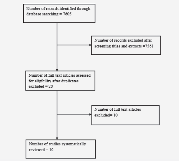
A trial flow diagram is shown below.  In total 7605 studies were identified after a search of electronic databases as described in the methodology section. This figure excludes 508 studies which were not published in the English Language.  There were 7561 studies excluded after screening the title and/or abstract. A frequent reason for exclusion was the study design i.e. very few studies were randomised controlled trials. Animal studies were also excluded, as were studies which used combination compounds where one of the substances was not an antioxidant.  After duplicates were removed, 20 studies were read in further detail to assess for eligibility.  A total of 10 studies remained which met the inclusion criteria.
A total of 1483 participants were randomised and intervention antioxidants included: Zinc, curcumin, melatonin, vitamin E, vitamin C, alpha lipoic acid, coenzyme Q10, polyphenols and flavonoids.
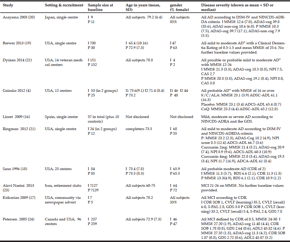
Key descriptive details of the included studies are shown in table 1.

Table 1 Study characteristics and baseline characteristics of included studies

Table 1
Study characteristics and baseline characteristics of included studies

I= intervention   P= placebo   1 also 155 in memantine group, 154 in vitamin E + memantine    *according to NINCDS-ADRDA criteria A lower score on the MMSE indicates poorer cognition. A lower score on ADLS-cog indicates poorer function.  A lower score on the GDS indicates higher level of depression. For all other scales a lower score indicates better outcome.


Outcome measures

Cognition

In total, 12 different outcome measures were used to assess cognition or to objectively grade the severity of Alzheimer’s disease.  7 studies used one or more methods of assessing cognition and 3 used only the MMSE.  Details of the outcome measures used can be seen in tables 1 and 2.
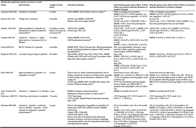
Overall, 5 of the 10 studies reported a statistically significant improvement or a slower rate of deterioration in one or more cognitive assessments in the intervention group compared to the control group at the end of the study (16-20).  In 4 of these studies participants had AD and in 1 they had MCI (17).  In 2 of the studies with a positive treatment effect however, the difference was found following a post hoc analysis where the groups were divided by age (19) or whether there was plasma evidence of a response to antioxidant therapy (16).
Of the studies with a positive treatment effect on cognition where more than one measure was used, the significant effect was not seen across all of the measures in any of the studies.
Of concern is that 1 study reported a statistically significant worsening in cognition in the intervention group compared to the placebo group (4). Another study reported a worsening of cognition in the people who did not have a plasma response to the antioxidant, compared to the placebo group (16).
Only 1 of the 3 studies in which participants had MCI evaluated time to development of dementia as an outcome measure.  There was no significant difference between the groups found.

Activities of daily living / functional  assessment

In total 5 studies used an assessment of ADLs or function as an outcome measure using 4 different scales. 4 of the studies involved participants with AD patients and 1 MCI.
Overall 2 studies reported a positive treatment effect (18, 22)and the other 3 found no difference between the intervention and control groups.

Mood/ behavioural disturbance

In total 4 studies included a measure of mood or behavioural disturbance as an outcome measure using 3 different measures. There was a positive treatment effect reported in 1 study (20) and in the other 3, there was no significant difference between the groups.

Table 2 Intervention details and results of included studies

Table 2
Intervention details and results of included studies

**not relevant to this review. a. Event free survival: Not significant but when analysed with baseline MMSE included as a co-variate, p=0.001 230 day increase in median survival with vitamin E compared to placebo. b. p=0.004, c. p=0.03, d. 3.15 (0.92 to 5.39), e. 0=0.004 adjusted for multiple comparisons, f. p=0.03, g. p=0.03, h. p=0.01, i. p=0.00, j Respondents vs. Non respondents and placebo vs. Non respondents p<0.05. No data for placebo vs intervention group as a whole, k. p=0.004, l. Reported significant differences with executive, language and overall cognitive scores in the first 18 months. A lower score on the MMSE indicates poorer cognition. A lower score on ADLS-cog indicates poorer function.  A lower score on the GDS indicates higher level of depression. For all other scales a lower score indicates better outcome.


Side effects and adverse events

In 6 studies, there were either no adverse events or side effects reported or no differences between the intervention and control groups reported.
In 2 studies, there was a greater decline in MMSE in the intervention group (4, 18).  In one study, this effect was seen in the intervention group as a whole (vitamin E + vitamin C + alpha lipoic acid), and in the other the greater deterioration was seen in the people who did not have a plasma response to vitamin E (the “non-respondents”).
In the curcumin study (21), participants taking curcumin had a statistically significant lower plasma haemotocrit and higher glucose.  It is unlikely that these effects are clinically significant. This study also stated “complaints attributable to the endocrine system” were less common in the 2 gm curcumin group compared to placebo or the 4gm group. (3% vs 17% vs 19% p=0.02).  The exact endocrine complaints are not stated.  There was no significant difference in the withdrawal rate due to adverse events between the groups.
In total 1 patient (33%) in the zinc study (19) developed low plasma levels of caeruloplasmin, thought to be directly attributed to taking zinc.
In people taking vitamin E in the Sano study (18), more people had the following events compared to the placebo group:  dental events (n=1 vs n=0. P=0.023), falls (n=12 [14%] vs n= 4 [5%] p=0.005) and syncope (n=4 vs n=7 p=0.031).

Figure 1 Flow diagram for literature search

Figure 1
Flow diagram for literature search

Summary of findings

The findings of this systematic review do not support the use of antioxidant supplements to slow cognitive, functional or behavioural deterioration in people with AD or MCI.  There were no consistent adverse effects found overall however two studies raised concern of possible worsening of cognition in certain circumstances (4, 16).
Regarding cognition, 4 out of the 7 studies involving people with AD found a small positive treatment effect (16,18-20).  2 of these studies (16, 19) however should perhaps be regarded as exploratory studies since the positive treatment effects were found in a post hoc analysis. Furthermore the risk of attrition bias was judged to be high in 1 of these studies (16) therefore the results may not be reliable.  In neither of the remaining 2 studies (18, 20), was the treatment effect supported by the other measures of cognition used:  Sano (18) reported a change in the BDS of 4.0 in the vitamin E group compared to 5.4 in the placebo group (in addition to the 230 day increased time to either institutionalisation; loss of ability to perform at least two of three basic activities of daily living, severe dementia, defined as CDR rating of 3.) There was no significant difference with MMSE. The risk of bias was overall low in the Sano study.   Asayama (20) reported a mean change of -4.3 (3.6) in the ADAS-cog in the intervention group compared to 0.3 (1.3) in the control group.  There was no significant difference with MMSE however and the risk of bias with the study was largely unclear.
One study (4) reported a deterioration of MMSE score in people taking vitamin E/ Vitamin C/ alpha lipoic acid.  The overall risk of bias was low in this study. A different study (16) reported a greater deterioration in people who did not have a plasma response to vitamin E (the “non-respondents”).
Of the 3 studies where participants had MCI, only 1 (17)reported a positive treatment effect and again, this was only in one of the 4 measures of cognition used in the trial.  Since the number of participants was only 12 in this study, it should also probably be regarded as an exploratory trial.
Of the 4 studies in AD participants which included a measure of functional ability as an outcome measure, 2 reported a positive treatment effect (18, 22) and 2 reported that there was no effect (4, 21). The Dysken study (22) was judged to be the highest quality trial included in this review and reported an improvement in one of 2 measures of function used in the study. Overall, 2000 IU of vitamin E daily plus and AChEI reduced progression of functional decline by 19% per year compared with placebo plus AChEI as measured by the ADCS-ADL inventory however secondary measures of caregiver time and dependence were not reduced on the CAS.  It is noteworthy however that despite there being a slower functional decline in the vitamin E group, there was not any significant delay in cognitive decline which is a more specific feature of AD.  One author has however proposed that this could be because “functional ability may be a more sensitive measure of AD progression” (25).  In addition, paradoxically a combination of memantine and alpha tocopherol had a lesser effect than either treatment alone. The authors postulate that memantine may interfere with the antioxidant properties of vitamin E.  Sano (18) reported a significant improvement in the dependence scale score of 76 vs 86 in the vitamin E group compared to placebo.
Only 1 MCI study measured function and there was no statistically significant treatment effect (24).
Of the 4 studies where participants had AD and an assessment of mood or behaviour was carried out (4, 20-22) only 1 study reported a significant treatment effect where the ADAS non cog improved by -4.1 (2.2) points in the melatonin group and by -0.8 points in the placebo group (20). The risk of bias was largely unknown in this study.
None of the studies evaluating MCI measured behaviour or mood.
In terms of safety, there do not seem to be any side effects which are common across the included studies.
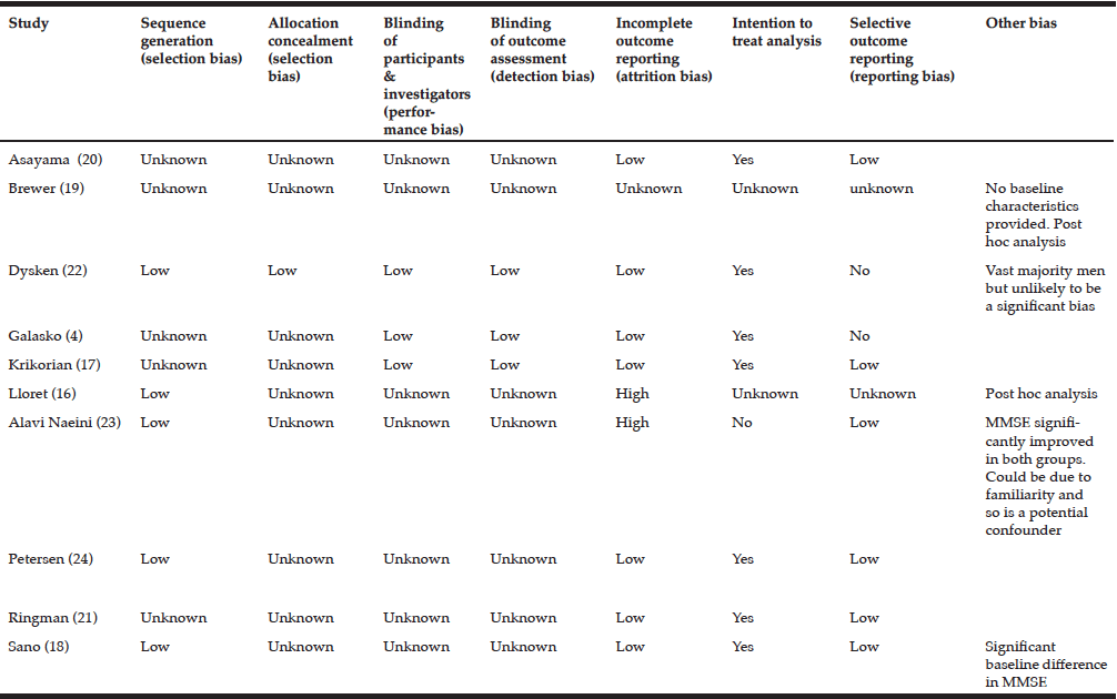
For most domains of bias not enough information was provided by the authors to make a definitive judgement therefore the risk of bias is largely unknown.  (See risk of bias table). There was only 1 study which had a low overall risk of bias and in this study, there was some evidence that antioxidant supplements may have a positive effect of functional decline in AD (22).

Table 3 Risk of bias table

Table 3
Risk of bias table


Discussion

Strengths and limitations

This is the first systematic review to evaluate trials of the main antioxidant supplements in people with AD and MCI.  It was performed and reported according to PRISMA guidance and included a comprehensive literature search and evaluation of the literature.
Limitations of this review are that it was conducted by only one reviewer, the number of included studies was relatively small and the search strategy excluded 508 non-English language publications from the screening process. Furthermore, it is possible that the supplements reviewed have other mechanisms of action that may affect outcomes rather than simply acting as antioxidants.  Curcumin for example, has been shown to bind to amyloid plaques and inhibit fibril formation (26).  A further issue with curcumin is that it has low bioavailabilty in plasma as well as low water solubility and a short half -life.
The internal validity of this review, i.e. the extent to which observed treatment effects can be attributed to the antioxidant rather than confounding, cannot be confidently assessed because with the exception of one study (22), the information provided by the study authors was not sufficiently explicit to make an accurate assessment.  Only the Dysken study (22) was judged to have good internal validity owing to the transparency and detail provided by the authors.
A further limitation with most of the studies included in this review is that they are likely to be underpowered: in total 6 studies had less than 100 participants. Only the Dysken study, which had 613 participants in total, included a power calculation.
All studies used similar methods of diagnosing MCI or AD which supports good external validity  (Ie this is in support of being able to correctly generalise the results to the population of people with AD or MCI outside of the study). The duration of the study is of particular concern however in 3 of the trials (4, 17, 20) which lasted for 16 weeks, 12 weeks and 4 weeks respectively.  This may not be adequate follow up with respect to harm or benefit and thus is a risk to external validity.

Clinical practice and research implications

There is not presently adequate evidence to support the use of antioxidants to slow cognitive, functional or behavioural decline in AD or MCI.
Overall, the safety data from this systematic review was not concerning however caution is warranted owing to the fact that the duration of follow up was short in many of the included studies and also in view of the finding reported by a large Cochrane review that beta carotene, vitamin E and possibly higher doses of vitamin A seem to increase mortality (7).
Future research should perhaps be aimed at advancing knowledge of the role of oxidative stress and antioxidants in the pathogenesis of Alzheimer’s dementia and mild cognitive impairment because it is possible that the relationship is more complex than is currently thought and furthermore there is a suggestion antioxidants are harmful in some circumstances (27, 28).
If further trials are then carried out, it is important that trials are adequately powered and of a sufficient duration to be able to detect effects. They should also take into account that patients with cognitive impairment are less likely to concord with treatment (29). As suggested by the authors of a recent review evaluating the limitations of RCTs for non-pharmacological interventions for MCI (30), greater standardisation of methods (including outcome measures) would allow better comparison and generalisation of  study outcomes.  Finally, authors should consider reporting future studies according to CONSORT guidelines (31) so that readers can accurately assess validity.


Conclusions

This systematic review does not suggest overall that antioxidant supplements have a beneficial effect on cognitive, functional or behavioural decline in Alzheimer’s dementia or mild cognitive impairment.
The evidence for the role of oxidative stress in the pathogenesis of Alzheimer’s disease provides a clear rationale for the use of antioxidant therapies in AD yet the results of clinical trials have overall been disappointing.  Possible reasons for this discrepancy include that the existing research generally has methodological limitation and that the relationship between antioxidants and the development of AD is more complicated than is currently realised.
The overall risk of harm associated with short term antioxidant supplementation appears to be low based upon this review however caution is warranted owing to findings from other large systematic reviews (7).

Conflict of interest: No conflict of interest.


References

1.    Rosini M, et al. Oxidative stress in Alzheimer’s disease: are we connecting the dots? J Med Chem 2014;57(7): p. 2821-31.
2.    Mecocci P, MacGarvey U, Beal MF. Oxidative damage to mitochondrial DNA is increased in Alzheimer’s disease. Ann Neurol 1994;36(5): p. 747-51.
3.    Wang J, Markesbery WR, and Lovell MA.  Increased oxidative damage in nuclear and mitochondrial DNA in mild cognitive impairment. J Neurochem 2006;96(3): p. 825-32.
4.    Galasko DR, et al. Antioxidants for Alzheimer disease: a randomized clinical trial with cerebrospinal fluid biomarker measures. Arch Neurol 2012;69(7): p. 836-41.
5.    Farina N, et al. Vitamin E for Alzheimer’s dementia and mild cognitive impairment. Cochrane Database Syst Rev 2012;11: p. Cd002854.
6.    Lonn E, et al. Effects of long-term vitamin E supplementation on cardiovascular events and cancer: a randomized controlled trial. Jama 2005;293(11): p. 1338-47.
7.    Bjelakovic G, et al. Antioxidant supplements for prevention of mortality in healthy participants and patients with various diseases. Cochrane Database Syst Rev  2012;3: p. Cd007176
8.    Schurks M, et al. Effects of vitamin E on stroke subtypes: meta-analysis of randomised controlled trials. BMJ , 2010;341: p. c5702
9.    Klein EA, et al. Vitamin E and the risk of prostate cancer: the Selenium and Vitamin E Cancer Prevention Trial (SELECT). Jama 2011;306(14): p. 1549-56.
10.    Hercberg S, et al. The SU.VI.MAX Study: a randomized, placebo-controlled trial of the health effects of antioxidant vitamins and minerals. Arch Intern Med 2004;164(21): p. 2335-42.
11.    Omenn GS, et al. Effects of a combination of beta carotene and vitamin A on lung cancer and cardiovascular disease. N Engl J Med 1996;334(18): p. 1150-5.
12.    Liberati A, et al. The PRISMA statement for reporting systematic reviews and meta-analyses of studies that evaluate healthcare interventions: explanation and elaboration. BMJ 2009;339: p. b2700.
13.    World Health Organisation and the Food and Agricultural Organisation of the United Nations.  Vitamin and mineral requirements in human nutrition: Geneva, 2004.
14.    Birks J, Grimley Evans J. Ginkgo biloba for cognitive impairment and dementia. Cochrane Database Syst Rev 2009;(1): p. Cd003120.
15.    The Cochrane Collaboration. Cochrane Handbook for Systematic Reviews of Interventions: London, 2011
16.     Lloret A, et al. Vitamin E paradox in Alzheimer’s disease: it does not prevent loss of cognition and may even be detrimental. J Alzheimers Dis 20009;17(1): p. 143-9.
17.    Krikorian R, et al. Concord grape juice supplementation improves memory function in older adults with mild cognitive impairment. Br J Nutr 2010;103(5): p. 730-4.
18.    Sano M, et al. A controlled trial of selegiline, alpha-tocopherol, or both as treatment for Alzheimer’s disease. The Alzheimer’s Disease Cooperative Study. N Engl J Med 1997;336(17): p. 1216-22.
19.    Brewer GJ, Kaur S.  Zinc deficiency and zinc therapy efficacy with reduction of serum free copper in Alzheimer’s disease. Int J Alzheimers Dis, 2013: p. 586365.
20.    Asayama K, et al. Double blind study of melatonin effects on the sleep-wake rhythm, cognitive and non-cognitive functions in Alzheimer type dementia. J Nippon Med Sch 2003;70(4): p. 334-41.
21.    Ringman JM, et al.  Oral curcumin for Alzheimer’s disease: tolerability and efficacy in a 24-week randomized, double blind, placebo-controlled study. Alzheimers Res Ther 2012;4(5): p. 43.
22.    Dysken MW, et al. Effect of vitamin E and memantine on functional decline in Alzheimer disease: the TEAM-AD VA cooperative randomized trial. Jama 2014;311(1): p. 33-44..
23.    Alavi Naeini AM, et al. The effect of antioxidant vitamins E and C on cognitive performance of the elderly with mild cognitive impairment in Isfahan, Iran: a double-blind, randomized, placebo-controlled trial. Eur J Nutr 2014;53(5): p. 1255-62
24.    Petersen RC, et al. Vitamin E and donepezil for the treatment of mild cognitive impairment. N Engl J Med 2005;352(23): p. 2379-88
25.    Evans DA, Morris MC, Rajan KB. Vitamin E, memantine, and Alzheimer disease. Jama, 2014;311(1): p. 29-30.
26.    Yang F et al. Curcumin inhibits formation of amyloid beta oligomers and fibrils, binds plaques, and reduces amyloid in vivo. J Biol Chem. 2005 Feb 2005;18;280(7):5892-901
27.    Underwood BR, et al. Antioxidants can inhibit basal autophagy and enhance neurodegeneration in models of polyglutamine disease. Hum Mol Genet 2010;19(17): p. 3413-29.
28.    Harrison FE. A critical review of vitamin C for the prevention of age-related cognitive decline and Alzheimer’s disease. J Alzheimers Dis 2012;29(4): p. 711-26.
29.    Hayes TL, et al. Medication adherence in healthy elders: small cognitive changes make a big difference. J Aging Health 2009;21(4): p. 567-80.
30.     Horr T, Messinger-Rapport  B and Pillai JA. Systematic review of strengths and limitations of randomised controlled trials for non-pharmacological interventions in mild cognitive impairment: Focus on Alzheimer’s Disease. J Nutr Health Ageing 2015;19 (2) 141-15
31.     Schulz KF, Altman DG, and Moher D.  CONSORT 2010 Statement: updated guidelines for reporting parallel group randomised trials. BMC Med  2010;8: p. 18.

STIMULATORY EFFECT OF ACUTE SINGLE DOSE OF DRIED WHOLE COFFEE CHERRY POWDER ON NRF2 ACTIVITY IN FRESHLY ISOLATED BLOOD CELLS. A SINGLE-BLIND, PLACEBO CONTROLLED CROSS-OVER PILOT CLINICAL STUDY


T. Reyes-Izquierdo1, B. Nemzer2, R. Argumedo1, M. Cervantes1, Z. Pietrzkowski1


1. Futureceuticals Inc.; 16259 Laguna Canyon Rd, Ste 150, Irvine, CA USA 92618; 2. Futureceuticals Inc.; 2692 N. State Rt. 1-17., Momence, IL, USA 60954

Corresponding Author: Tania Reyes-Izquierdo, 16259 Laguna Canyon Rd Ste 150, Irvine, CA, 92618 USA, Phone +1 949 502 4496, Fax +1 949 502 4987, Email: treyes@futureceuticals.com

J Aging Res Clin Practice 2016;5(3):120-127
Published online August 11, 2016, http://dx.doi.org/10.14283/jarcp.2016.109


Abstract

Background: NF-E2-related factor 2 (Nrf2) is a transcription factor that participates in the regulation of antioxidant expression during increased oxidant stress. Several phytochemicals and food products have shown to trigger Nrf2 activity. In this pilot placebo-controlled study the effects of a single dose of dried whole coffee cherry powder (“WCCP”) on Nrf2 levels were tested. Objectives: To characterize a blend of WCCP and evaluate the potential effects on mTOR and Nrf2 in healthy subjects. Design: In this cross over study, subjects were given placebo or a single dose of 1000mg WCCP on day 1 and 2. Blood was collected for four time points. Participants served as their own controls. Setting: After supplementation, blood samples were processed for mTOR and Nrf2 analysis. Blood ATP, glucose and lactate were also measured. Participants: Ten healthy subjects, ages ranging from 22 to 35 years and BMI ranging from 24.1 to 30 kg/m² were selected to participate. Results: One 1000 mg dose of WCCP resulted in an average 44% increase of NRf2 levels 180 minutes after ingestion (p=0.03 ). Phosphorylated mTOR (Ser 2448) was reduced at 180 minutes after supplement; when compared to placebo. Correlation (“Corr”) analyses revealed that increases in Nrf2 appear to be associated with mTOR reduction. Blood glucose and extracellular ATP levels were not changed. Conclusions: WCCP increased Nrf2 3 hours after ingestion. Additional testing is required to verify the potency of WCCP on Nrf2, as well as any potential correlation between mTOR (S2448) reduction and increased levels of Nrf2 after supplementation.

Key words: Nrf2 activation, mTOR, dried whole coffee cherry powder, antioxidants.


Introduction

Nuclear factor NF-E2-related factor 2 (Nrf2) is a key transcription factor in the regulation of antioxidant expression during increased oxidant stress (1, 2). This key cell-defense gene regulates the expression of cyto-protective proteins that detoxify harmful cellular compounds, neutralizes reactive oxygen species, directly or indirectly modulates the inflammatory response and immune system, and assists in the repair or removal of damaged macromolecules (2-5). Increased Nrf2 expression has previously been associated with improvements in neurodegenerative, autoimmune, diabetic and renal disease in animal and in vitro models (6).
Activation of Nrf2 and phosphoinositide 3-kinase (PI3K), serine-threonine kinase (AKT), and the mammalian target of rapamycin (mTOR) signaling pathways, (also known as PI3K)/AKT/mTOR signaling pathways, respectively), have been observed in certain human cancers and it has been postulated that said activations may be central to tumor development and progression (7). Increased expression of Nrf2 and decreased expression of mTOR has been associated with diminished frequency of tumor development and with smaller tumors in animal models (7). More interestingly, inhibition of PI3K)/AKT/mTOR has been associated with extended life spans in insects, invertebrates, and mammals (8-10). Collectively, these pathways present an attractive target for human longevity investigations.
Polyphenols have been suggested as potential therapeutic compounds that may have a positive effect on several pathological conditions such as neurodegenerative diseases, diabetes, certain cancers and cardiovascular diseases (11, 12). Dietary polyphenols have been reported to induce the expression of enzymes involved in cellular antioxidant defenses (13).
Several phytochemicals and food-derived products have similarly been shown to trigger Nrf2 activity (2, 14). Green coffee beans have been reported to contain large amounts of polyphenolic antioxidants, such as chlorogenic, caffeic, ferulic, and n-coumarinic acids (15).
Here we have evaluated the potential effects of the novel antioxidant activities inherent in dried whole cherries from the coffee plant on mTOR and Nrf2 expression in healthy humans as a preliminary study on potential for coffee cherry to support human health.


Materials and Methods

Materials

5-O-caffeoylquinic acid, (-)-epicatechin, procyanidin dimer B2, quercetin-3-glucoside and rutin were obtained from AASC Ltd (Southampton, UK). Methanol and acetonitrile were obtained from Rathburn Chemicals (Walkburn, Scotland). Formic acid was obtained from Fisher Scientific (Loughborough, UK). Dried, ground whole coffee cherry powder samples, commercially marketed as ”CoffeeBerry® Brand whole coffee fruit” was obtained from FutureCeuticals, Inc. (Momence, IL USA). Primary caffeine standard was obtained from USP (Rockville, MD). Perchloric acid (HPLC grade) and acetonitrile (HPLC grade) were purchased from Sigma-Aldrich Corp. (St. Louis, MO, USA). Primary Sorbent Amine (PSA) was obtained from Supelco Inc. (Bellefonte, PA, USA).
Dulbecco’s phosphate buffered saline (PBS) and water were purchased from Sigma-Aldrich Corp. Co. (St Louis, MO, USA). Low protein binding microtubes were obtained from Eppendorf (Hauppauge, NY, USA) and RC DC Protein Assay Kit II was from Bio-Rad (Hercules, CA, USA). ATP-luciferase assays were obtained from EMD Millipore (Billerica, MA, USA). Heparin capillary blood collection tubes were obtained from Safe-T-Fill® (Ram Scientific Inc. Yonkers, NY). Accutrend® Lactate Point of Care and BM-Lactate Strips® were from Roche (Mannheim, Germany). Accu-Chek® Compact Plus glucometer and Accu-Chek® test strips were from Roche Diagnostics (Indianapolis, IN, USA). TransAM® Nrf2 detection assay was from Active Motif (Carlsbad, CA, USA). Phospho m-TOR (Ser 2448), phospho m-TOR (Ser 2481) and total m-TOR were from Cell Signaling Technologies (Danvers, MA, USA).

Chemistry Analyses

Chlorogenic acids, procyanidins, flavanols and flavonols of WCCP were characterized by LC-MS (n) and quantified by UV absorbance (16, 17). Analysis was carried out on a Thermo Acella HPLC system comprising of an auto-sampler with sampler cooler maintained at 6ºC, an Accela photodiode array (PDA) detector (Thermo Fisher Scientific, San Jose, CA, USA) scanning from 200-600 nm. Samples (5 or 10μl) were injected onto a 150 x 3.0mm C18 Accucore (Thermo Fisher Scientific, Waltham, MA, USA) maintained at 40ºC and eluted with a 5-10-50% gradient of 1.0% formic acid and acetonitrile at 700 μL/min over 0-10-20 minutes. After passing through the absorbance detector, the eluant was split, and 200 uL/min was directed to the electrospray interface of an ExactiveTM Orbitrap mass spectrometer (Thermo Fisher Scientific, Waltham, MA USA). Samples were run in negative ionization mode, and the scan range was from 150 to 1200 amu with resolution set to 60,000.Peak identifications were based on co-chromatography with authentic standards, when available, as well as absorbance spectra and published MS2 mass spectra data.
Quantification of hydroxycinnamic compounds was obtained by comparison to an authentic standard of 5-O-caffeoylquinic acid, range 5 to 750 ng at 325 nm, and caffeine at 275 nm in the range 5 to 750 ng. Quantification of minor phenolic compounds was by exact mass measurements of calibration standards over the range of 0.5 to 50 ng using (-)-epicatechin for flavan-3-ol monomers and procyanidin B2 for dimer and trimer flavan-3-ols. Quercetin-3-glucoside and quercetin-3-rutinoside were quantified as quercetin-3-glucoside equivalents.
The caffeine and trigonelline contents were characterized by HPLC (Agilent 1100; Agilent Technologies, Palo Alto, CA, USA) equipped with diode array detector and quantified by UV absorbance (17, 18). For caffeine testing about 10 mg aliquot of caffeine primary standard was accurately weighed into a 50 mL volumetric flask. The volume was made up to 25 mL with mobile phase (90% of 0.1% perchloric acid and 10% acetonitrile) and sonicated for 5 min. The final volume was made to the mark with mobile phase. For the sample analysis, about 500 mg was weighed into 100 mL volumetric flask. Mobile phase was added up to 50 mL and sonicated for 5 min. The supernatant was then diluted to mark with mobile phase. About 1 g of PSA was weighed into a centrifuge tube. The prepared sample (5 mL) was dispensed into the tube containing PSA, vortexed for 3 min and filtered through a 0.45 µm PTFE syringe filter. Analysis was carried out by HPLC (Agilent 1100) with the diode array detector set at 275 nm. Samples (5 or 10μl) were injected onto a 150 x 3.0 mm, 2.7 μm Supelco Ascentis Express Phenyl-Hexyl Column (Supelco Inc., Bellefonte, PA, USA) maintained at 25°C and eluted with a 90-10% isocratic of 0.1% perchloric acid and in acetonitrile at 800 μL/min over 15 minutes. Quantification was by comparison to an authentic primary standard of caffeine.
Trigonelline analysis was performed on HPLC system (Agilent 1100) equipped with PDA detector, gradient pump unit, Kinetex 2.6 µm Biphenyl 100Å LC column 150 x 4.6 mm (Phenomenex, Torrance, CA, USA). Samples were eluted using mobile phase of ammonium formate: acetonitrile adjusted to pH 3.0 with formic acid and delivered at a flow rate of 1.0 mL/min. Detector was carried out at 265 nm. The injection volume was 10 µL. Data acquisition and analysis were carried out using Agilent ChemStation Software, version B.04.01, chromatography analysis software. Trigonelline primary standard (Sigma-Aldrich, St. Louis, MO USA) was used for quantitative analysis.

Clinical Study

Inclusion and Exclusion Criteria

This clinical case study was conducted according to guidelines laid out in the Declaration of Helsinki. All procedures involving human subjects were approved by the Ethics Committee named Comité de Investigación Biomédica para el Desarrollo de Fármacos S.A. de C.V. (Zapopan, Jal, México) (study protocol No. FC-2015-N287). Ten subjects were selected to participate. They were generally healthy with ages ranging from 22 to 35 years and BMI ranging from 24.1 to 30 kg/m². Exclusion criteria included acute infections, rhinitis, influenza, diagnosis of diabetes mellitus, dietary allergies. Subjects using anti-inflammatory drugs, analgesics, statins, diabetic drugs, anti-allergy medicines, multivitamins and dietary supplements were also excluded. All participants gave written, informed consent before any experimental procedure was performed.

Blood Collection and sample preparation

Study was performed by NutraClinical Inc. (Irvine, CA, USA) according to the study protocol designed by VDF FutureCeuticals, Inc. (Irvine, CA, USA). Ten subjects were included in this pilot study. Participants (6 females and 4 males) were >19 and <35 years of age, with a BMI of 29.94 (SD ± 6.51). Enrolled participants were instructed to fast for 12 h prior to the initial blood draw. Resting subjects were given a dosage of either two empty capsules as placebo or two 500 mg of encapsulated WCCP (for a total 1000mg single dose) on day 1. All dosages were switched on day 2 relative to each subject. Subjects were given a two-day “wash-out” period prior to switching supplementations.  350 mL of water was administered with the capsules on each day. Blood was collected by finger puncture and placed in lithium heparin Safe-T-Fill® capillary blood collection tubes (Ram Scientific Inc. Yonkers, NY, USA). The first tube, containing 50 µL of blood, was frozen immediately for ATP assays. The second tube, containing 600 µL blood was used for m-TOR and nuclear extraction from peripheral blood cells. One last tube was collected to obtain plasma. Blood samples were drawn at baseline (time zero), 60 min (T60), 120 min (T120) and 180 min (T180) after supplementation.

Cell Lysate preparation for mTOR detection

mTOR cell lysates were prepared from whole blood. One hundred µL of whole blood were added to 900µL of 1X cell lysis buffer containing 1 mM PMSF into a 2.0 mL LoBind microtube (Eppendorf®, purchased from Fisher Scientific, Inc. Pittsburgh, PA, USA). Samples were then placed in a small ice bath and sonicated for 5 minutes. After sonication, cell lysates were centrifuged at 14,000 x g for 10 minutes at 40C. The cell lysate supernatant (CLS) for each sample was collected and transferred into a clean, labeled 2.0 mL microtube and placed on ice.

mTOR Protocol

In order to determine the optical density of mTOR in each CLS sample, p-mTOR Ser 2481, p mTOR Ser 2448, and total mTOR kits from Cell Signaling Technology® (Danvers, MA, USA) were run simultaneously. One hundred µL of CLS sample was added to each assay plate. The assay protocol for each mTOR kit was followed according to the manufacturer’s instructions. Total mTOR absorbance was used as reference in the analysis in order to determine the specific activity of p-mTOR (Ser 2448) and p-mTOR (Ser 2481).

Protein quantification

The protein concentration of each CLS sample was determined by the Bio-Rad DC™ Protein Assay (Hercules, CA, USA), using bovine serum albumin (BSA) (Fisher Scientific, Grand Island, NY, USA) as a standard. In order to determine the absorbance over milligram per protein of mTOR in each CLS sample, the absorbance from each mTOR assay absorbance was divided by the protein concentration.

ATP Detection and Quantification

Blood ATP concentration was determined using an ATP Assay Kit (EMD Millipore, Billerica, MS, USA) with a modification to the original method, as previously described (19). Briefly, 10 μL of lysed blood were loaded onto a white plate (Corning® Fisher Scientific, Waltham, MA, USA). One hundred µL of ATP nucleotide-releasing buffer containing 1 µL luciferase enzyme mix was added in each well and the plate was immediately placed on a luminometer (LMaX, Molecular Devices; Sunnyvale, CA, USA). Readings were performed during 15 min at 3 min intervals at 470 nm. Relative Light Units (RLU) was recorded and ATP concentrations were determined using a standard ATP curve.

Lactate and Glucose Detection

Glucose and lactate levels were measured at collection time points 0, 60, 120, and 180 minutes. Glucose was measured using an AccuChek® Compact Plus glucometer (Roche Diagnostics, Indianapolis, IN, USA). Two μl of fresh finger blood were loaded onto an AccuChek® Testing Strip (Roche Diagnostics, Indianapolis, IN, USA) and read from the glucometer according to manual instructions. Blood lactate was measured using an Accutrend® Lactate Analyzer (Roche, Mannheim, Germany). Sixteen μl of fresh finger blood were loaded onto a BM Lactate Test Strip and read from the Lactate Analyzer according to manufacturer’s instructions.

White blood cell isolation

Five hundred µL of whole blood collected from finger puncture as previously described were added to a 15mL falcon tube containing 10 mL 1X Red Blood Cell Lysis buffer at room temperature (RT) (22°C). After 15 min incubation, samples were centrifuged at 1200g for 10 min. The supernatant was discarded and 10 mL ice cold Dulbecco’s PBS (Sigma Chem. Corp.; St. Louis, MO, USA) was added followed by centrifugation as mentioned before. Supernatant was discarded and cells were snap frozen (-80°C) until use.

Nuclear extracts

White blood cell (WBC) samples were thawed on an ice bath and 300 µL 1X cell lysis buffer (Cell Signaling Technologies; Danvers, MA, USA) containing 1 mM dithiothreitol (DTT) and 10 µL/mL protease inhibitor cocktail (Active Motif; Carlsbad, CA, USA). Samples were vortexed and incubated on an ice bath for 30 min. Samples were subsequently centrifuged at 14,000g for 30 min and the supernatant was recovered. Protein concentration was determined as previously described.

Nrf2 Detection

Nrf2 was determined using Trans AM® Nrf2 ELISA kits. Nuclear extract samples were loaded at 30 µg protein/well. A positive control provided was used as reference. The protocol was followed as indicated by the manufacturer’s instructions.

Statistical Analysis

Total mTOR, 2448-mTOR and 2481-mTOR levels were normalized using time zero as the baseline, as well as the analyses for Nrf2. Statistical analysis was performed using the commercially available GraphPad® statistical software (Graphpad Software Inc., La Jolla, CA, USA). Descriptive statistics are presented by the mean ± standard error. Supplementations were compared at 60, 120 and 180 minutes (placebo vs WCCP) within the experimental groups with baseline and between experimental groups using a one-way analysis of variance with Tukey’s post hoc analysis when a significant F-ratio was observed. Statistical significance was set at P: 0.05.


Results

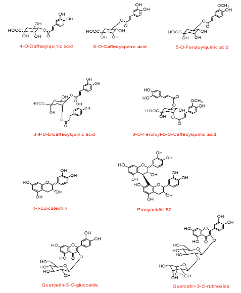
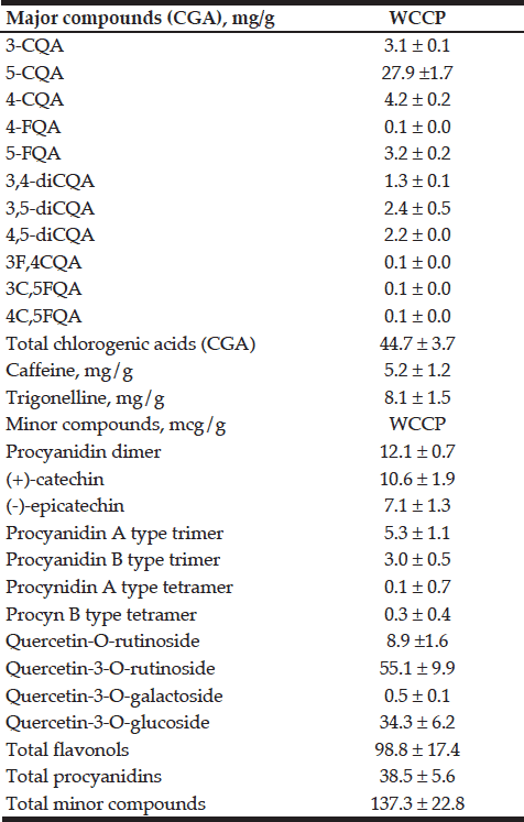
We identified and quantified the major phytochemicals present in WCCP. Structures of some of the major identified components are shown in Figure 1. As reported in table 1; total chlorogenic acids were the most abundant phytochemicals present in WCCP (44.7 ± 3.7 mg/g), of which 5-O-Caffeoylquinic acid (5-CQA) showed the highest concentration (27.9 ± 1.7 mg/g) which represents 62% of the total CGA content. Other chlorogenic acids were also detected, such as 4-O-caffeoylquinic acid (9%), 3-O-caffeoylquinic acid (6%), 3,4-O-dicaffeoylquinic acid (5%), 3,5-O-dicaffeoylquinic acid (5%). Caffeine and trigonelline were additionally detected (5.2 ± 1.2 mg/g and 8.1 ± 1.5 mg/g; respectively). Minor compounds such as procyanidin dimer, (+)-catechin and (-)-epicatechin were less abundant.


Figure 1 Structure of major hydroxycinnamates, flavan-3-ols and flavonols detected in WCCP samples

Figure 1
Structure of major hydroxycinnamates, flavan-3-ols and flavonols detected in WCCP samples


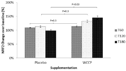
Nrf2 detected post supplementation with placebo and WCCP is shown in Figure 2. During the placebo supplementation non-significant Nrf2 increases were detected at T60 (109% ± 1%), T120 (113% ± 3%) and T180 (98% ± 3%). When treated with WCCP, Nrf2 was increased at T60 (114% ±2%), T120 (132% ± 4%) and T180 (145% ±6%). When compared to placebo, T60 and T120 showed no significance (P= 0.5 and P=0.3 respectively). However, T180 was statistically significant (P=0.03).


Table 1 Chemical composition of WCCP (dried whole coffee cherry powder). Results are displayed as mean ± SD (n = 3 true replicates), in mg/g. 3-CQA (3-O-Caffeoylquinic acid);5-CQA (5-OCaffeoylquinic acid); 4-CQA (4-O-caffeoylquinic acid); 4-FQA (4-O-Feruloyquinic acid); 5-FQA (5-O-Feruloyquinic acid); 3, 4-diCQA (3-4-O-Dicaffeoylquinica acid); 3, 5-diCQA (3-5-O-Dicaffeoylquinica acid); 4,5-diCQA (4-5-O-Dicaffeoylquinica acid); 3F, 4CQA (3-O-Feruloyl-4-O-caffeoylquinic acid); 3C, 5FQA (3-O-Caffeoyl-5-feruloyquinic acid); 4C, 5FQA (4-O-Caffeoyl-5-feruloyquinic acid); CGA (Chlorogenic acid)

Table 1
Chemical composition of WCCP (dried whole coffee cherry powder). Results are displayed as mean ± SD (n = 3 true replicates), in mg/g. 3-CQA (3-O-Caffeoylquinic acid);5-CQA (5-OCaffeoylquinic acid); 4-CQA (4-O-caffeoylquinic acid); 4-FQA (4-O-Feruloyquinic acid); 5-FQA (5-O-Feruloyquinic acid); 3, 4-diCQA (3-4-O-Dicaffeoylquinica acid); 3, 5-diCQA (3-5-O-Dicaffeoylquinica acid); 4,5-diCQA (4-5-O-Dicaffeoylquinica acid); 3F, 4CQA (3-O-Feruloyl-4-O-caffeoylquinic acid); 3C, 5FQA (3-O-Caffeoyl-5-feruloyquinic acid); 4C, 5FQA (4-O-Caffeoyl-5-feruloyquinic acid); CGA (Chlorogenic acid)


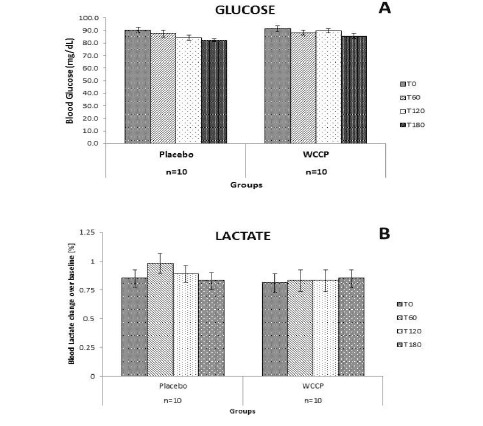
Glucose and lactate levels were also monitored in this 2 day study, as can be observed in Figure 3. Since subjects fasted for 12h prior to the supplementations, they were monitored for possible hypoglycemia. Also, we wanted to learn whether WCCP may affect blood glucose and lactate levels. There were no observed changes for glucose or lactate on placebo or WCCP. Blood glucose was monitored for placebo (Baseline: [90.3±2.21]; T60: [87.6±2.68]; T120: [84.3±2.12] and T180: [82.3±1.05]) and WCCP (Baseline: [91.8±2.26]; T60: [88.1±2.08]; T120: [89.8±1.81] and T180 [85.6±1.77]). Blood lactate was also monitored for placebo (baseline: [0.85±0.07]; T60: [0.98±0.08]; T120: [0.89±0.07] and T180: [0.83±0.07]) and for WCCP (baseline: [0.81±0.08]; T60: [0.83±0.09]; T120: [0.83±0.09] and T180: [0.85±0.07].It is important to reiterate that this study was conducted in healthy subjects and that any effect of WCCP has not been investigated in subjects with chronic conditions.


Figure 2 Nrf2 after supplementation with WCCP. Nrf2 was detected in nuclear extracts from isolated white blood cells. During the placebo supplementation, Nrf2 did not show any significant increase. When treated with WCCP, Nrf2 was increased at T60 (114% ±2%), T120 (132 ± 4%) and T180 (145% ±6%). When compared to placebo, T60 and T120 showed no significance (P= 0.5 and P=0.3 respectively). However, T180 was significant (P=0.03). Data are presented as Mean ± SE; n=10

Figure 2
Nrf2 after supplementation with WCCP. Nrf2 was detected in nuclear extracts from isolated white blood cells. During the placebo supplementation, Nrf2 did not show any significant increase. When treated with WCCP, Nrf2 was increased at T60 (114% ±2%), T120 (132 ± 4%) and T180 (145% ±6%). When compared to placebo, T60 and T120 showed no significance (P= 0.5 and P=0.3 respectively). However, T180 was significant (P=0.03). Data are presented as Mean ± SE; n=10

Figure 3 Glucose and lactate after placebo or WCCP supplementation. Subjects were fasted for 12h prior to the supplementation. Both glucose and lactate were monitored at the indicated time points (T0, T60, T120 and T180), for the duration of the study. Neither glucose nor lactate showed any significant changes on either supplementation day. Data are presented as Mean ± SE; n=10

Figure 3
Glucose and lactate after placebo or WCCP supplementation. Subjects were fasted for 12h prior to the supplementation. Both glucose and lactate were monitored at the indicated time points (T0, T60, T120 and T180), for the duration of the study. Neither glucose nor lactate showed any significant changes on either supplementation day. Data are presented as Mean ± SE; n=10


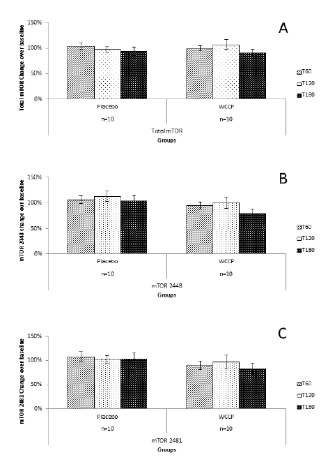
Total mTOR, 2448-mTOR and 2481-mTOR are shown in figure 4A. Total mTOR showed no changes at T60 (107% ±7%), T120 (98% ± 6%) and 180 min (93% ± 8%) in the placebo group. Supplementation with WCCP showed only as a slight non-significant change (99% ±5% at T60; 107% ±9% at T120, and 91% ± 6% at T180). When compared to placebo, neither T60 (P=0.23), T120 (P=0.31) nor T180 (P=0.09) were significant. For mTOR 2448; T60 and T180 showed no change, and for T120, there was a slight increase (13% above baseline). For the supplemented group, T60 and T120 showed no change or significance (P=0.39; P=0.38, respectively) when compared to placebo. At T180, mTOR showed a decrease of 20% below baseline, which is not significant when compared to placebo (P=0.07) (Figure 4B). For mTOR 2481, placebo showed no change. Supplementation showed a non-significant reduction when compared to placebo at T60 (90% ± 9%; P=0.22), T120 (97% ±14%, P=0.77) and T180 (83% ± 12%; P=0.19) (Figure 4C).
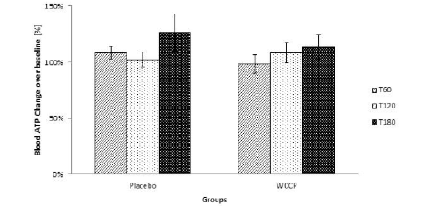
Blood ATP was detected by using a luciferase-based assay. As reported in Figure 6, ATP levels were not modified even though they  showed a tendency to increase at T180. However, when compared to placebo, supplemented group was not significant at any collection point (P=0.12; P=0.62; P=0.5 respectively). Data are presented as Mean ± SE; n=10 (Figure 6).

Figure 4 mTOR detection after supplementation with placebo or WCCP. Total mTOR (A) did not show any significant changes when compared to placebo at T60 (P=0.23), T120 (P=0.31) or T180 (P=0.09). mTOR 2448 (B) as well as mTOR 2481 (C) showed a reduction at T180 for the WCCP supplementation. However, when compared to placebo, neither mTOR 2448 (P=0.07) nor mTOR 2481 (P=0.19) were significant. Data are presented as Mean ± SE; n=10

Figure 4
mTOR detection after supplementation with placebo or WCCP. Total mTOR (A) did not show any significant changes when compared to placebo at T60 (P=0.23), T120 (P=0.31) or T180 (P=0.09). mTOR 2448 (B) as well as mTOR 2481 (C) showed a reduction at T180 for the WCCP supplementation. However, when compared to placebo, neither mTOR 2448 (P=0.07) nor mTOR 2481 (P=0.19) were significant. Data are presented as Mean ± SE; n=10


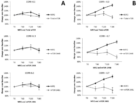
Pearson’s correlation was performed for placebo NRF2 and total mTOR (r= 0.5, CI -0.88 to 0.98; P=0.5, n=10), and mTOR 2481 (r=0.3, -0.93 to 0.97, P=0.7, n=10) where no correlation was observed, while mTOR 2448 showed a positive correlation (r=0.9, CI -0.47 to 0.99, P=0.1, n=10) (Figure 5A). NRF2 and total mTOR showed a negative correlation (r=-0.3, CI -0.98 to 0.94, P=0.7, n=10), as did mTOR 2448 (r=-0.7, CI -0.47 to 0.99, P=0.1, n=10) and mTOR 2481 (r=-0.7, CI -0.99 to 0.79, P=0.3, n=10).


Figure 5 Correlation between Nrf2 and mTOR levels within placebo and supplemented groups. Placebo (A) Nrf2 was compared to total mTOR (r=0.43, n=10), as well as mTOR 2448 (r=0.89, n=10) and mTOR 2481 (r=0.27, n=10). For the WCCP supplemented group (B), Nrf2 was also correlated with total mTOR (r=-0.32, n=10); mTOR 2448 (r=-0.68, n=10) and mTOR 2481 (r=-0.7, n=10)

Figure 5
Correlation between Nrf2 and mTOR levels within placebo and supplemented groups. Placebo (A) Nrf2 was compared to total mTOR (r=0.43, n=10), as well as mTOR 2448 (r=0.89, n=10) and mTOR 2481 (r=0.27, n=10). For the WCCP supplemented group (B), Nrf2 was also correlated with total mTOR (r=-0.32, n=10); mTOR 2448 (r=-0.68, n=10) and mTOR 2481 (r=-0.7, n=10)


Figure 6 Blood ATP was measured after supplementation. ATP levels were not significantly modified. Although the trend indicates that there was a tendency to increase when compared to placebo, WCCP supplemented group was not significant at any collection point (P=0.12; P=0.62; P=0.5 respectively). Data are presented as Mean ± SE; n=10

Figure 6
Blood ATP was measured after supplementation. ATP levels were not significantly modified. Although the trend indicates that there was a tendency to increase when compared to placebo, WCCP supplemented group was not significant at any collection point (P=0.12; P=0.62; P=0.5 respectively). Data are presented as Mean ± SE; n=10


Discussion

Dietary polyphenols such as flavones, isoflavones, flavonols, catechins and phenolic acids are mostly found in fruits and vegetables (20, 21). These compounds have shown antioxidant, anti-aging, anti-inflammatory, anti-atherosclerotic, and other biological abilities (11, 12). Chlorogenic acid (CGA) (5-caffeoylquinic acid) and caffeic acid, which have been reported to show antioxidant properties In vitro, (16, 20) are major components of coffee cherry (Table 1). The chemical composition of coffee beans (green and roasted) has been studied to a considerable extent (22, 23), as well as the proprietary green coffee fruit (24, 25). Recently, the proprietary composition for two coffee fruit extracts and two coffee powders has been reported (16). In all cases, chlorogenic acids have been reported to be the most abundant components (23). In WCCP, CGA is one of the main constituents (44.7 mg/g). When compared to roasted or green coffee, green coffee fruit, coffee fruit extracts and coffee powders, WCCP has a similar composition as of that described for “coffee fruit powder” 1 and 2 (CFP-1, CFP-2) (16). For WCCP; 5-O-Caffeoylquinic acid was the main chlorogenic acid, at 62% of the total content. Other chlorogenic acids were also detected, such as 4-O-caffeoylquinic acid (9%), 3-O-caffeoylquinic acid (6%), 3,4-O-dicaffeoylquinic acid (5%), 3,5-O-dicaffeoylquinic acid (5%). Trigonelline, detected in green coffee bean (23) was also found in WCCP, as well as caffeine. Average caffeine content of regular coffee goes from 9-13mg/g in Coffea arabiga to 15-25mg/g in Coffea canephora (23). Whilst coffee brew contains from 90-160mg caffeine per cup (26), WCCP contains only 5.2mg/g.

Several coffee constituents such as kahweol and cafestol have been reported to increase the nuclear Nrf2 protein level and modulating ARE-mediated gene expression. More recently, chlorogenic acid has been proposed as an activator of the Nrf2/ARE pathway, activating nuclear Nrf2-translocation as well as gene expression of different phase II enzymes in the colon carcinoma cell line HT29 (27, 28). In a human intervention study, an increase of transcripts of phase II genes in peripheral blood lymphocytes (PBL) after 4 weeks of daily consumption of either a coffee rich in CGA or one rich in N-methylpyridinium (NMP) was observed (29).
In this study, we examined the acute effect of WCCP on peripheral blood expression of markers related to healthy aging and longevity in healthy human subjects. Acute testing was performed following a single dose, oral administration of the supplement. No significant changes in peripheral WBC Nrf2 protein levels were seen over time following consumption of placebo. In contrast, a linear increase in peripheral WBC Nrf2 protein levels was seen during the 3 hours evaluated after oral supplementation with WCCP. A significant increase from baseline control was seen at 180 minutes. Neither the effect of the placebo nor the supplementation was evaluated beyond three hours. Fasting is associated with oxidative stress and can activate Nrf2 expression (30). It has been reported that activation of Nrf2 is associated with increased glucose uptake by the pentose phosphate pathway in fibroblasts (31). Similar observations have been made in animal models (32). Cells starved of glucose have decreased Nrf2 mediated detoxification of reactive oxygen species and decreased Nrf2 initiated expression of antioxidant defense proteins (31). Diabetic animal models also suggest Nrf2 expression can affect glucose metabolism (6). In this study, baseline fasting glucose levels were in the normal range in all the subjects. Also, no changes in blood glucose or lactate levels were seen in study subjects over time following supplementation with WCCP (Figure 3). Serum glucose and lactate levels were similar to those of placebo controls. These findings suggest acute oral WCCP intake did not have a measurable impact on the pentose phosphate pathway glucose uptake during the time period examined or that the glucose uptake was compensated for by glycogenolysis or other mechanisms, even though Nrf2 expression was increased.

In this study, a trend towards a decrease in total mTOR S2481, and mTOR S2448 expression was seen over time (Figure 4). These findings suggest that there was minimal effect of WCCP on mTOR expression and the two related pathways. No significant correlation was found between peripheral WBC expression of Nrf2 and total mTOR, mTOR 2481, or mTOR 2448 expression after treatment with placebo, although they did appear similar. This finding suggests that the two pathways are not interlinked closely during the time period examined. The expression of Nrf2 and all three mTOR proteins appeared to be opposed, increased for Nrf2and decreased for mTOR, after supplementation with WCCP (Figure 5A, 5B). These findings would be in line with current models of longevity
Activation of Nrf2 is associated with increased production of NADPH in fibroblasts (31). NADPH is responsible for providing reducing equivalents to allow glutathione or thioredoxin redox cycling, elements responsible for countering cellular redox stress. Increased NADPH synthesis is often linked to the generation of ATP, and Nrf2 expression has been correlated with ATP expression (33). These findings suggest increased ATP levels would be expected with increased Nrf2 expression. No significant changes in blood ATP levels were seen acutely after administration of placebo. In this study, a linear increase in blood ATP levels was seen after oral WCCP administration, although this increase was not statistically significant (Figure 6). It has been reported that cellular ATP levels were not altered in cells with genetically or pharmacologically activated Nrf-2 expression (31).
In summary, a single dose of coffee cherry powder WCCP was shown to increase Nrf2 expression. The highest Nrf2 expression was seen at the final time point of 180 minutes. Consequently, any higher subsequent expression could have been missed. Blood ATP levels remained unchanged, demonstrating that glucose is not diverted for energy production upon activation of Nrf2, whilst mTOR levels decreased. In this case, the small number of subjects evaluated may not have had enough power to detect differences in treatment outcomes. Further investigations are needed to verify whether WCCP affects Nrf2 directly or indirectly, as well as to investigate the acute and long term effect of WCCP on aging or related neurodegenerative conditions. Additional clinical testing is justified in order to further verify potency of WCCP to increase levels of Nrf2 and any potential correlation between reduced mTOR (S2448) and increased levels of Nrf2.


Funding: The present study was funded by Futureceuticals, Inc. The sponsors had no role in the design and conduct of the study; in the collection, analysis, and interpretation of data.

Conflict of interest disclosure: All authors declare that they have no conflict of interest.

Acknowledgments: We express our gratitude to John Hunter (FutureCeuticals, Inc.) for his comments and suggestions in the preparation of this paper. We would like to thank Michael Sapko for his help in editing and reviewing this manuscript.

Ethical standard: Ethical approval was granted by the Ethics Committee named Comité de Investigación Biomédica para el Desarrollo de Fármacos (Biomedical Research and Medicine Development Committee) México (Ref: FC-2015-N287).


References

1.     Balligand JL, Reducing damage through Nrf-2. Cardiovasc Res, 2013. 100:1-3.
2.    Gao B, Doan A and Hybertson BM, The clinical potential of influencing Nrf2 signaling in degenerative and immunological disorders. Clin Pharmacol 2014; 6:19-34.
3.    Dayalan Naidu S, Kostov RV and Dinkova-Kostova AT, Transcription factors Hsf1 and Nrf2 engage in crosstalk for cytoprotection. Trends Pharmacol Sci 2015; 36:6-14.
4.    Dinkova-Kostova AT and Abramov AY, The emerging role of Nrf2 in mitochondrial function. Free Radic Biol Med 2015; 88:179-88.
5.    Zhang M, An C, Gao Y, et al. Emerging roles of Nrf2 and phase II antioxidant enzymes in neuroprotection. Prog Neurobiol 2013; 100:30-47.
6.    Whitman SA, Long M, Wondrak GT, et al. Nrf2 modulates contractile and metabolic properties of skeletal muscle in streptozotocin-induced diabetic atrophy. Exp Cell Res 2013; 319:2673-2683.
7.    Sahin K, Orhan C, Tuzcu M, et al. Orally administered lycopene attenuates diethylnitrosamine-induced hepatocarcinogenesis in rats by modulating Nrf-2/HO-1 and Akt/mTOR pathways. Nutr Cancer 2014; 66:590-8.
8.    Harrison DE, Strong R, Sharp ZD, et al. Rapamycin fed late in life extends lifespan in genetically heterogeneous mice. Nature 2009; 460:392-5.
9.    Johnson SC, Rabinovitch PS and Kaeberlein M, mTOR is a key modulator of ageing and age-related disease. Nature 2013; 493: 338-45.
10.    Papaioannou AI, Rossios C, Kostikas K, et al. Can we delay the accelerated lung aging in COPD? Anti-aging molecules and interventions. Curr Drug Targets 2013; 14:149-57.
11.    Kishimoto Y, Tani M and Kondo K, Pleiotropic preventive effects of dietary polyphenols in cardiovascular diseases. Eur J Clin Nutr 2013; 67:532-535.
12.    Tomás-Barberán FA and Andrés-Lacueva C. Polyphenols and Health: Current State and Progress. J. Agric. Food Chem 2012; 60:8773–8775.
13.    Dinkova-Kostova AT, Talalay P. Direct and indirect antioxidant properties of inducers of cytoprotective proteins. Mol Nutr Food Res 2008; 52:S128-38.
14.    Jeong WS, Jun M and Kong AN, Nrf2: a potential molecular target for cancer chemoprevention by natural compounds. Antioxid Redox Signal 2006; 8:99-106.
15.    Nicoli, MC, Anese M, Manzocco L, et al. Antioxidant properties of coffee brews in relation to the roasting degree. Lebensm Wiss Technol 1997; 30:292–297.
16.    Mullen W, Nemzer B, Ou B, et al. The antioxidant and chlorogenic acid profiles of whole coffee fruits are influenced by the extraction procedures. J Agric Food Chem 2011; 59:3754-62.
17.    Mullen W, Nemzer B, Stalmach A, et al. Polyphenolic and hydroxycinnamate contents of whole coffee fruits from China, India, and Mexico. J Agric Food Chem 2013; 61:5298-309.
18.    Duarte GS, Pereira AA and Farah A. Chlorogenic acids and other relevant compounds in Brazilian coffees processed by semi-dry and wet post-harvesting methods. Food Chemistry 2010; 118:851-855.
19.    Reyes-Izquierdo T, Hammond, LE; Sikorski, RS; et al. Acute effect of HH2o on oxygen consumption rate, intracellular ATP and ROS in freshly isolated human peripheral blood mononuclear cells. Current Topics in Nutraceutical Research 2011; 9:139-146.
20.    Han X, Shen T, Lou H. Dietary polyphenols and their biological significance. Int J Mol Sci 2007; 8:950-988.
21.    Kim HS, Quon MJ and Kim JA, New insights into the mechanisms of polyphenols beyond antioxidant properties; lessons from the green tea polyphenol, epigallocatechin 3-gallate. Redox Biol 2014; 2:187-95.
22.    Crozier A, Jaganath IB and Clifford MN, Dietary phenolics: chemistry, bioavailability and effects on health. Natural Product Reports 2009; 26:1001-1043.
23.    Chu YF, Coffee: emerging health effects and disease prevention. 2012. John Wiley & Sons.
24.    Farah A, Monteiro M, Donangelo CM, et al. Chlorogenic acids from green coffee extract are highly bioavailable in humans. J Nutr 2008; 138:2309-15.
25.    Farah A, Donangelo CM. Phenolic compounds in coffee. Brazilian Journal of Plant Physiology, 2006. 18:23-36.
26.    Blauch JL and Tarka SM, HPLC determination of caffeine and theobromine in coffee, tea, and instant hot cocoa mixes. J Food Sci 1983; 48:745-747.
27.    Boettler U, Volz, N., Pahlke, G., et al. Coffees rich in chlorogenic acid or N-methylpyridinium induce chemopreventive phase II-enzymes via the Nrf2/ARE pathway in vitro and in vivo. . Mol. Nutr Food Res 2011; 55:798–802.
28.    Boettler U, Volz N, Pahlke G, et al. Coffee constituents as modulators of Nrf2 nuclear translocation and ARE (EpRE)-dependent gene expression. J Nutr Biochem 2011; 22:426-440.
29.    Volz N, Boettler U, Winkler S, et al. Effect of coffee combining green coffee bean constituents with typical roasting products on the Nrf2/ARE pathway in vitro and in vivo. J Agric Food Chem 2012; 60:9631-41.
30.    Zhang YK, Wu KC and Klaassen CD, Genetic activation of Nrf2 protects against fasting-induced oxidative stress in livers of mice. PLoS One 2013; 8:e59122.
31.    Heiss EH, Schachner D, Zimmermann K, et al. Glucose availability is a decisive factor for Nrf2-mediated gene expression. Redox Biol 2013; 1:359-365.
32.    Bardoux P, Zhang P, Flamez D, et al. Essential role of chicken ovalbumin upstream promoter-transcription factor II in insulin secretion and insulin sensitivity revealed by conditional gene knockout. Diabetes 2005; 54: 1357-63.
33.    Holmstrom KM, Baird L, Zhang Y, et al. Nrf2 impacts cellular bioenergetics by controlling substrate availability for mitochondrial respiration. Biol Open 2013; 2:761-770.