jarlife journal
Sample text

AND option

OR option

UTILITY OF POLYGENIC RISK SCORING TO PREDICT COGNITIVE IMPAIRMENT AS MEASURED BY PRECLINICAL ALZHEIMER COGNITIVE COMPOSITE SCORE


Q. Gao1, P. Daunt1, A.M. Gibson1, R.J. Pither1; for the Alzheimer’s Disease Neuroimaging Initiative*


1. Cytox Limited, Manchester, UK.

Corresponding Author: Qian Gao, Cytox Ltd., John Eccles House, Robert Robinson Avenue, Oxford Science Park, Oxford, OX4 4GP, United Kingdom. Email: qian.gao@cytoxgroup.com. Tel:+44 (0)1865 338018

J Aging Res & Lifestyle 2022;11:1-8
Published online February 23, 2022, http://dx.doi.org/10.14283/jarlife.2022.1


Abstract

Abstract: Background: The utility of Polygenic Risk Scores (PRS) is gaining increasing attention for generating an individual genetic risk profile to predict subsequent likelihood of future onset of Alzheimer’s disease (AD), especially those carry two copies of the APOE E3 allele, currently considered at neutral risk in all populations studied. Objectives: To access the performance of PRS in predicting individuals whilst pre-symptomatic or with mild cognitive impairment who are at greatest risk of progression of cognitive impairment due to Alzheimer’s Disease from the Alzheimer’s Disease Neuroimaging Initiative (ADNI) as measured by the Preclinical Alzheimer Cognitive Composite (PACC) score profile. Design: A longitudinal analysis of data from the ADNI study conducted across over 50 sites in the US and Canada. Setting: Multi-centre genetics study. Participants: 594 subjects either APOE E3 homozygotes or APOE E3/E4 heterozygotes who upon entry to the study were diagnosed as cognitively normal or with mild cognitive impairment. Measurements: Use of genotyping and/or whole genome sequencing data to calculate polygenic risk scores and assess its ability to predict subsequent cognitive decline as measured by PACC over 5 years. Results: Assessing both cognitively normal and mild cognitive impaired subjects using a PRS threshold of greater than 0.6, the high genetic risk participant group declined more than the low risk group over 5 years as measured by PACC score (PACC score reduced by time). Conclusions: Our findings have shown that polygenic risk score provides a promising tool to identify those with higher risk to decline over 5 years regardless of their APOE alleles according to modified PACC profile, especially its ability to identify APOE3/E3 cognitively normal individuals who are at most risk for early cognitive decline. This genotype accounts for approximately 60% of the general population and 35% of the AD population but currently would not be considered at higher risk without access to expensive or invasive biomarker testing.

Key words: Polygenic risk, cognitive decline, Alzheimer’s disease.


Introduction

Dementia describes an intra-individual pattern of decline in memory and thinking impairing at least two domains of cognition (1). Alzheimer disease (AD) is the most common cause of dementia. The majority of cases occur after age 65, constituting late-onset AD (LOAD), while cases occurring earlier than age 65 are considerably rarer, constituting less than 5% of all cases and are termed early-onset AD (EOAD) (2, 3). Approximately 1%–2% of AD is inherited in an autosomal dominant fashion (ADAD) and can present with very early age of onset and a more rapid rate of progression and is sometimes associated with other neurologic symptoms seen less frequently in sporadic AD (4). Sporadic or LOAD show a multifactorial heredity pattern caused by genetic and complex environmental interactions associated with several predisposing factors and age. The rate of cognitive deterioration during the development of AD varies among individuals (5, 6) and seems to be guided by a combination of genetic and environmental factors (7). Some genes, such as CLU, PICALM, and CR1, have been shown to be related to AD as indicated by genome-wide association studies (GWAS) (8, 9). However, only apolipoprotein E (APOE) polymorphisms have been established as consistent genetic susceptibility factors for LOAD in all populations studied in the world (10).
Development of polygenic risk scoring (PRS) algorithms that can capture all the genetic contribution towards the risk of developing AD (11) is an attractive strategy to allow for stratifying patients at risk prior to or as part of screening for clinical trial participation Furthermore understanding risk for future onset or progression of symptoms due to AD at a much earlier stage may lead to greater uptake of lifestyle interventions that have been shown to at least delay the progression of disease by several years. It is generally recognised that changes to lifestyle that will reduce risk for onset of AD are most effective when made earlier in life prior to any significant symptoms being displayed. A PRS test that can provide a cost-effective and widely accessible way of supporting the stratification of cognitively normal and MCI patients into those that are highest risk of developing AD will provide an additional tool for identifying individuals most likely to benefit from new disease modifying therapies or other patient management decisions.
Here we investigate the performance of our PRS in predicting cognitive decline with a particular focus on whether it can provide predictive information on identifying early changes of cognitive performance in cognitively normal individuals. As such, polygenic risk has been used here to predict cognitive changes using the modified PACC score (12, 13) over 5-year period. We have focussed on subjects who were either APOE E3 homozygotes and APOE E3/E4 heterozygotes (see Table 1-3). This accounts for approximately 80% of the general population but also that of the study population (sub-analyses of other APOE genotypes is compromised by low subject numbers).


Methods

Data used in the preparation of this article were obtained from the Alzheimer’s Disease Neuroimaging Initiative (ADNI) database (adni.loni.usc.edu). The ADNI was launched in 2003 as a public-private partnership, led by Principal Investigator Michael W. Weiner, MD. The primary goal of ADNI has been to test whether serial magnetic resonance imaging (MRI), positron emission tomography (PET), other biological markers, and clinical and neuropsychological assessment can be combined to measure the progression of mild cognitive impairment (MCI) and early Alzheimer’s disease (AD).
ADNI genotyping and/or whole genome sequencing data was used to calculate polygenic risk scores and assess their ability to predict subsequent cognitive decline as measured by the modified PACC score over 5 years.

Sample Description

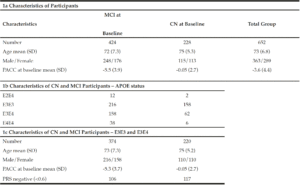
In order to understand the predictive performance of the PRS algorithm above and beyond that which is provided for by APOE status alone, we initially investigate data from 652 CN and MCI subjects selected from ADNI 1, ADNI GO, ADNI 2 and ADNI 3 studies examined between 2005 and 2017 (see Table 1a). Due to the low sample sizes (n≤50) of APOE E2E4 and E4E4 individuals in either CN or MCI groups (see Table 1b) at baseline (bl), further analyses were only carried out in APOE E3 homozygotes and APOE E3/E4 heterozygotes. Therefore, all results shown in this paper were based on 594 CN and MCI subjects who were either carried two copies APOE E3 allele or were APOE E3/E4 heterozygotes (see Table 1c) and had modified PACC score data at entry to the study in addition to having suitable genetic data and at least 5 years’ worth of follow up cognitive testing and imaging scans.

Table 1
Characteristics of participants

Genotyping Procedures and Quality Control

The ADNI samples were genotyped using with Whole Genome Sequencing and/or the Illumina Omni 2.5M BeadChip array. Quality control checks were performed using PLINK software (www.cog-genomics.org/plink/2.0/). Checks included the exclusion of SNPs with missingness greater than 0.02 and minor allele frequency of less than 0.01. SNPs with Hardy-Weinberg equilibrium p-value less than 1 x 10⁻⁶ were also excluded. After such checks 8,990,292 SNPs were left for analysis of which approximately 114,000 were used as part of the polygenic risk scoring algorithm (14).

The ADNI modified PACC score

PACC is a composite score which combines tests that assess episodic memory, timed executive function and global cognition which has been shown to be able to detect the first signs of cognitive decline before clinical signs of MCI manifest (15). In this study, we use a ADNI modified PACC with Digit Symbol Substitution (mPACCdigit) (12, 13) downloaded using R package “adnimerge” (https://adni.bitbucket.io/reference/pacc.html#references).-.
In ADNI, Free and Cued Selective Reminding Test (FCSRT) is not used and has been replaced Delayed Recall test that is included within the Alzheimer’s Disease Assessment Scale (ADAS) as a suitable proxy to be included in the modified PACC score. Furthermore, mPACCdigit score also includes the Digit Symbol Substitution Test (DSST) when available (ADNI1) and mPACCtrailsB uses (log transformed) Trails B as a proxy for DSST. Raw component scores standardized according to the mean and standard deviation of baseline scores of ADNI subjects with normal cognition to create Z scores for each component (Z=(raw – mean(raw.bl))/sd(raw.bl)). The Z scores are reoriented if necessary, so that greater scores reflect better performance. The composite is the sum of these Z scores. At least two components must be present to produce a score. If more than two components are missing, the PACC will be NA.

Calculation of Polygenic Risk Scores

A specifically built, proprietary software called SNPfitRTM was used for all subsequent PRS calculations. The PRS calculations are based on a pre-determined logistic regression model based on the modelling of the association between the incidences of variants within a large panel of SNPs with a known links to AD to the presence of the disease in a substantial cohort of subjects (Escott-Price et al.16). Subject age, sex and APOE status are included as covariates. The software calculates the normalised sum of the individual scores weighted by their effect sizes for each SNP, adds the values for the covariates and derives the predicted risk from the model equation.
Effect sizes were determined from the International Genomics of Alzheimer’s (IGAP) study. The score contribution from SNPs with missing values were imputed based on the population frequency of the effect allele for that SNP.

Statistical Analysis

The polygenic risk scores generated were exported for the analysis presented.
R version 4.0.4 (https://www.r-project.org/) was used to carry out all data processing and analysis. The receiver operating characteristic (ROC) analysis and AUC calculations were performed using R package “pROC”. Modified PACC data were obtained from R package “adnimerge” (https://adni.bitbucket.io/reference/pacc.html#references). T tests were performed in R using the t.test() function to determine whether there is significant different between high and low risk groups (see p-value in Results).
To determine whether applying a PRS approach would provide further accuracy for predicting cognitive decline as measured by a modified PACC, we analysed the cognitively normal APOE E3/E3 and APOE E3/E4 individuals, where both genetics and modified PACC score data were available (n=220, see Table 1c). PRS were calculated and individuals were assigned to either “high risk” (defined as a PRS ≥ 0.6, n=103) or “low risk” (PRS<0.6, n=117) groups (see Table 1c). A similar evaluation was performed on APOE E3/E3 and APOE E3/E4 individuals who entered the study with a diagnosis of MCI and for whom both genetic data and PACC score data were available (n=374 , See Table 1c). PRS were calculated and MCI individuals were assigned to “high risk” (defined as a PRS ≥ 0.6, n=268) or “low risk” (PRS<0.6, n=106) groups (see Table 1c). Note that not all subjects had follow-ups at each time point over the 5 years. Thus, the number of subjects varies at each follow-up check points. A PRS of 0.6 was chosen as a threshold based on an optimal balance between sensitivity and specificity in previous studies (17).


Results

The overall performance for predicting individuals who would decline by at least -1 PACC score within 5 years from a baseline diagnosis of either cognitively normal or mild cognitive impairment was 65.6% (CI:61.3-69.8) area under the curve (AUC), suggesting PRS could be an effective stratification tool to identify patients with a higher likelihood to decline cognitively over a period of 5 years.

PRS to predict early cognitive decline from a cognitively normal baseline

As expected, as measured by modified PACC score, those individuals who carry a copy of the APOE E4 allele are more likely to decline cognitively than those who are APOE E3 homozygotes over a 5-year period (see Figure 1a). The mean change in modified PACC score in APOE E3/E3 after 60 months was just -0.4 points ±4.1 whereas APOEE3/E4 individuals declined, on average, by 1.3 points ±4.7, on the modified PACC score scale after 60 months (see Figure 1a).
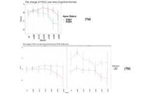
Figure 1
Time-course PACC scores for individuals carrying APOE E3E3 and E3E4 in CN Group: (1a) The change of PACC over time in individuals who entered as cognitively normal over 5-year period grouped by APOE status; (1b) The change of PACC over time in individuals who entered as cognitively normal over 5-year period grouped by risk score
(bl=baseline, m=month)


There was a significant difference in the average change of the modified PACC score approximate to 2 between the two groups observed from as early as 48 months (high risk n=77, low risk n=94; high risk average PACC=-1.2, low risk average PACC=0.7; p-value =0.003, see Table 2). When considering APOE E3 homozygotes alone, the difference in the change of PACC score between the high risk and low risk groups observed was 2 points over 60 months years (high risk n=20, low risk n=51; high risk average PACC=-1.7, low risk average PACC=0.2, p=0.12, see Table 2). Importantly, though sample size is smaller (see Table 2), low PRS risk E3/E4 individuals that entered the study as cognitively normal appeared more likely to remain cognitively stable compared with the high risk group (Figure 1b).

Table 2
Participants carrying APOE E3E3 and E3E4 in CN Group

bl: baseline; m: month. Thus month 6 is represented by m06


PRS to predict early cognitive decline from an MCI baseline

Again, as expected, those individuals carrying an E4 allele demonstrate greater cognitive decline, on average, compared to E3 homozygotes at all timepoints over the 5-year follow-up period (E3/E3 mean PACC change after 60 months -1.4 points ±5.6; E3/E4 mean PACC change after 60 months -8.9 points ±12.2; Figure 2a).
There were no individuals within the APOE E3/E4 MCI cohort (n=158) with a low PRS score (PRS <0.6). This is unsurprising, since these individuals who have already declined cognitively to an MCI diagnosis are likely to have a high PRS. Notwithstanding, this meant that a comparison between low and high PRS risk within the MCI group individuals could not be made. However, the APOE E3 homozygote MCI group contained both high PRS risk (≥0.6, n=110) and low PRS risk (<0.6, n=106) individuals (Figure 2b). Among this group, high PRS risk patients declined, on average, by approximately 1 point more than the low risk group after over 6 months (high risk n=105, low risk n=102; high risk average PACC=-3.9, low risk average PACC=-2.7, p=0.07, see Table 3) and a significant additional 5 points over 60 months (high risk n=59, low risk n=52; high risk average PACC=-8.2, low risk average PACC=-3.2, p-value<0.001, see Table 3) above those calculated as low risk, who did not decline further over the 5 year period studied (Figure 2b).
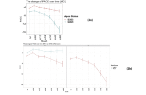
Figure 2
Time-course PACC scores for individuals carrying APOE E3E3 and E3E4 in MCI Group: (2a) The change of PACC over time in individuals who entered as MCI (EMCI or LMCI) over 5-year period grouped by APOE status; (2b) The change of PACC over time in individuals who entered as MCI (EMCI or LMCI) over 5-year period grouped by risk score (bl=baseline, m=month)

Table 3
Participants carrying APOE E3E3 and E3E4 in MCI group

bl: baseline; m: month. Thus month 6 is represented by m06


Discussion

PRS approaches have demonstrated accuracies of between 75 and 84% for predicting onset of AD when including APOE status, sex and age in addition to PRS (16). In particular, the PRS approach as developed by Escott-Price et al., (14) is built as a sum of the weighted contributed of 10,000s of Single Nucleotide Polymorphisms (SNPs) where the weights are the β-coefficients of each SNP association with the disease. In contrast to other PRS algorithms, where fewer SNPs have been used (for example just 31 SNPs (18)) this approach includes SNPs that are not considered as having genome wide significance in GWAS studies. However, inclusion of this vastly increased number of variants which alone carry sub-threshold significance provides an additive contribution to the overall performance that may be substantive and also reduce risk that performance is not
lost when being applied across different cohorts. Until now the analyses performed using this approach have been carried out to predict those individuals diagnosed with AD or MCI (19) versus those who are cognitively normal, though PRS algorithms have been used to look at a variety of AD pathology and risk by Altmann et al. (20).
Patients who present to clinicians with very mild or subjective cognitive complaints can provide a diagnostic and patient management challenge in terms of decisions on whether to progress to more expensive and/or invasive testing or to discharge. Easier access to risk evaluation data will help better patient management decisions in a cost-efficient manner and provide further basis for dialogue on risk mitigation through lifestyle changes. Furthermore, screening of large pre-symptomatic populations to identify potential clinical trial participants for prevention studies in AD is challenging. Genetic risk prediction can be generated from DNA simply extracted from saliva or blood samples, thus providing a viable route to wide-scale risk stratification to characterise potential clinical trial subjects.
We have previously reported (17) on the performance of a PRS algorithm for predicting those individuals, with a bassline diagnosis of MCI who would decline by at least 15 ADAS-Cog13 points in 4 years with an AUC of 72.8% (CI:67.9-77.7) increasing to 79.1% (CI: 75.6-82.6) when also including those at baseline who were considered cognitively normal. Furthermore, by designating MCI patients as either high or low risk as determined by a PRS threshold of 0.6 it was observed that the high risk group declined, on average, by 1.4 points more on the CDR-SB scale than the low risk group over a period of 4 years. This performance in predicting cognitive decline due to AD was similar to that when defining risk using a pTau/Ab1-42 ratio as measured in a cerebrospinal fluid (CSF) sample.
This study was designed to demonstrate the potential utility of a specific PRS algorithm for identifying individuals at highest risk of developing early or continued cognitive decline from either pre-symptomatic (CN) baseline or a relatively early stage of their disease (MCI). The results show the potential to use a PRS approach to identify those individuals most likely to decline cognitively. Importantly this includes identifying cognitively normal APOE E3 homozygous individuals who are at most risk for early cognitive decline due to AD. This genotype accounts for approximately 60% of the general population and 35% of the AD population but currently would not be considered at higher risk without access to expensive or invasive biomarker testing. PRS could therefore provide a useful tool for identifying individuals within this group who require additional monitoring, investigation or, with future developments, therapeutic intervention.
This study shows that PRS predictions can identify individuals with the highest risk of subtle cognitive decline, as measured by PACC scores, in patients who did not display any measurable symptoms upon entry to the ADNI study. The timeframe of 5 years used for the analysis is relevant in the context of both primary and secondary prevention trials and clinical practice. Furthermore, future work will be conducted to evaluate the predictive performance of our PRS algorithm in order to identify patients during mid-life (40-60 years old) at risk of future cognitive deficits due to AD which can provide a critical strategy for reducing the number. This genetic risk assessment represents an easily accessible intervention with the potential to reduce cost and patient burden through blood or mouth swab testing. Additionally, this genetic risk assessment provides an extremely valuable tool for expanding recruitment into secondary prevention trials which currently are typically limited to recruiting E4 carriers only. Furthermore, as disease modifying drugs enter clinical practice finding an easy to deploy risk prediction test to identify patients most likely to benefit from therapeutic intervention will be critical.
PRS does have its own challenges and limitations. For example, this work considers genetic risk together with age and sex in developing a model for predicting further development of cognitive symptoms but does not consider other risk factors that are known to influence onset and development of disease, for example, lifestyle and environment. Further studies will be required to combine both genetic and lifestyle risk factors to accurately identify those individuals at the most risk of Alzheimer’s disease.

Study Limitations

This study is not without limitations, with sample size being the primary shortcoming. This was particularly relevant in evaluating the APOE E4 carrier sub-group (E2/E4, E3/E4 and E4 homozygous, see Table 1b). Furthermore, studies with larger sample sizes across all diagnostic categories, including those declining from a cognitively normal baseline, will be important to understand broader utility. As with most studies of this nature, observing similar performance in alternative cohorts is important and is critical towards the understanding and confirmation of polygenic risk score assessment for use in clinical trial recruitment and in clinical practice.


*Data used in preparation of this article were obtained from the Alzheimer’s Disease Neuroimaging Initiative (ADNI) database (adni.loni.usc.edu). As such, the investigators within the ADNI contributed to the design and implementation of ADNI and/or provided data but did not participate in analysis or writing of this report. A complete listing of ADNI investigators can be found at: http://adni.loni.usc.edu/wp-content/uploads/how_to_apply/ADNI_Acknowledgement_List.pdf

Conflict of interest: Q. Gao, P. Daunt, A.Gibson and R. Pither are all employees of Cytox Ltd.

Ethical standard: The authors declare that the study was carried out according to all ethical standards.

Acknowledgments: Data collection and sharing for this project was funded by the Alzheimer’s Disease Neuroimaging Initiative (ADNI) (National Institutes of Health Grant U01 AG024904) and DOD ADNI (Department of Defense award number W81XWH-12-2-0012). ADNI is funded by the National Institute on Aging, the National Institute of Biomedical Imaging and Bioengineering, and through generous contributions from the following: AbbVie, Alzheimer’s Association; Alzheimer’s Drug Discovery Foundation; Araclon Biotech; BioClinica, Inc.; Biogen; Bristol-Myers Squibb Company; CereSpir, Inc.; Cogstate; Eisai Inc.; Elan Pharmaceuticals, Inc.; Eli Lilly and Company; EuroImmun; F. Hoffmann-La Roche Ltd and its affiliated company Genentech, Inc.; Fujirebio; GE Healthcare; IXICO Ltd.; Janssen Alzheimer Immunotherapy Research & Development, LLC.; Johnson & Johnson Pharmaceutical Research & Development LLC.; Lumosity; Lundbeck; Merck & Co., Inc.; Meso Scale Diagnostics, LLC.; NeuroRx Research; Neurotrack Technologies; Novartis Pharmaceuticals Corporation; Pfizer Inc.; Piramal Imaging; Servier; Takeda Pharmaceutical Company; and Transition Therapeutics. The Canadian Institutes of Health Research is providing funds to support ADNI clinical sites in Canada. Private sector contributions are facilitated by the Foundation for the National Institutes of Health (www.fnih.org). The grantee organization is the Northern California Institute for Research and Education, and the study is coordinated by the Alzheimer’s Therapeutic Research Institute at the University of Southern California. ADNI data are disseminated by the Laboratory for Neuro Imaging at the University of Southern California. We also acknowledge Prof. Julie Williams, Prof. Valentina Escott-Price, Dr Rebecca Sims and Dr Eftychia Bellou from the University of Cardiff for their advice on adaptation and implementation of the polygenic risk algorithm, and Mr Greg Davidson from Ledcourt Associates Ltd for his contribution on implementation of the algorithm. We thank Dr Simon Flint and Dr Vicky Jones from Cytox Ltd for comments on the manuscript.

Open Access: This article is distributed under the terms of the Creative Commons Attribution 4.0 International License (http://creativecommons.org/licenses/by/4.0/), which permits use, duplication, adaptation, distribution and reproduction in any medium or format, as long as you give appropriate credit to the original author(s) and the source, provide a link to the Creative Commons license and indicate if changes were made.


References

1. Mckhann GM, Knopman DS et al. The diagnosis of dementia due to Alzheimer’s disease: recommendations from the National Institute on Aging-Alzheimer’s Association workgroups on diagnostic guidelines for Alzheimer’s disease. Alzheimers Dement. 2011; 7: 263-269.
2. Wittenberg R, Hu B et al. Projections of older people with dementia and costs of dementia care in the United Kingdom, 2019–2040. Report commissioned by Alzheimer’s Society from the Care Policy and Evaluation Centre (CPEC) at the London School of Economics and Political Science. November 2019.
3. Alzheimer’s Association. Alzheimer disease facts and figures. Alzheimers Dement. 2019; 15: 321-387.
4. Bateman RJ, Xiong C et al. Clinical and biomarker changes in dominantly inherited Alzheimer’s disease. N. Engl. J. Med. 2012; 367: 795-804.
5. Storandt M, Grant EA et al. Rates of progression in mild cognitive impairment and early Alzheimer’s disease. Neurology. 2002 Oct 8; 59(7):1034-41.
6. Hui JS, Wilson RS, et al. Rate of cognitive decline and mortality in Alzheimer’s disease. Neurology. 2003 Nov 25; 61(10):1356-61.
7. Cosentino S, Scarmeas N et al. APOE epsilon 4 allele predicts faster cognitive decline in mild Alzheimer disease. Neurology. 2008 May 6; 70(19 Pt 2):1842-9.
8. Harold D, Abraham R et al. Genome-wide association study identifies variants at CLU and PICALM associated with Alzheimer’s disease. Nat Genet. 2009 Oct; 41(10):1088-93.
9. Lambert JC, Ibrahim-Verbaas CA et al. Meta-analysis of 74,046 individuals identifies 11 new susceptibility loci for Alzheimer’s disease. Nat Genet. 2013 Dec;45(12):1452-8. doi: 10.1038/ng.2802. Epub 2013 Oct 27. PMID: 24162737; PMCID: PMC3896259.
10. Avramopoulos D. Genetics of Alzheimer’s disease: recent advances. Genome Med. 2009 Mar 27; 1(3):34.
11. Stocker H, Möllers T, Perna L, et al, The genetic risk of Alzheimer’s disease beyond APOE ε4: systematic review of Alzheimer’s genetic risk scores. Transl Psych 2018;8;166-174.
12. Donohue MC, et al. The Preclinical Alzheimer Cognitive Composite: Measuring Amyloid-Related Decline. JAMA Neurol. 2014;71(8):961–970. doi: 10.1001/jamaneurol.2014.803. PMID: 24886908; PMCID: PMC4439182.
13. Donohue MC, Sperling RA, Petersen R, Sun C, Weiner MW, Aisen PS, for the Alzheimer’s Disease Neuroimaging Initiative. Association Between Elevated Brain Amyloid and Subsequent Cognitive Decline Among Cognitively Normal Persons. JAMA. 2017;317(22):2305–2316. doi: 10.1001/jama.2017.6669. PMID: 28609533; PMCID: PMC5736301.
14. Escott-Price V, Leonenko G, Sims R et al, Polygenic risk and hazard scores for Alzheimer’s disease prediction. Ann Clin Transl Neurol 2019;6;456-465.
15. Donohue MC, Sperling RA, et al. Australian Imaging, Biomarkers, and Lifestyle Flagship Study of Ageing, Alzheimer’s Disease Neuroimaging Initiative, and Alzheimer’s Disease Cooperative Study (2014) The preclinical Alzheimer cognitive composite: measuring amyloid-related decline. JAMA Neurol 71(8):961–970.
16. Escott-Price V, Sims R, Williams J et al, Common polygenic variation enhances risk prediction for Alzheimer’s disease. Brain 2015;138;3673-3684.
17. Daunt P, Ballard CG, et al. Polygenic Risk Scoring is an Effective Approach to Predict Those Individuals Most Likely to Decline Cognitively Due to Alzheimer’s Disease. J Prev Alzheimers Dis. 2021;8(1):78-83.
18. Desikan RS, Fan CC, Wang Y et al, Genetic assessment of age-associated Alzheimer disease risk: development and validation of polygenic hazard score. PLoS Med 2017 e1002258.
19. Escott-Price V, Myers A, Huentelman AJ, et al, Polygenic risk score analysis of pathologically confirmed Alzheimer’s disease. Ann Neurol 2017;82;311-314.
20. Altmann A, Schott JM, Scelsi et al, A comprehensive analysis of methods for assessing polygenic burden on Alzheimer’s disease pathology and risk beyond APOE. Brain Commun 2020;2; fcz047.

GENETIC VARIANTS RELATED TO LIPID METABOLISM AS A RISK FACTOR TO LATE-ONSET ALZHEIMER’S DISEASE


M.A.S. Pinhel1, A.M. Crestani2, G.F. Sousa-Amorim3, M.L. Gregório4, J.C. Cação3, W.A. Tognola3, D. Rossi Silva Souza3


1. Ribeirao Preto Medical School – University of Sao Paulo, USP – Brazil; 2. Universidade Estadual Paulista (UNESP), Brazil; 3. Sao Jose do Rio Preto Medical School, FAMERP, SP, Brazil; 4. – Franca University – UNIFRAN, Franca, SP, Brazil

Corresponding Author: Marcela Augusta de Souza Pinhel, Faculdade de Medicina de Ribeirão Preto – USP, Avenida dos Bandeirantes, 3900, Ribeirao Preto – SP / CEP: 14090-900 / Brazil, Phone: +55 16 3315 4810, marcelapinhel@yahoo.com.br

J Aging Res Clin Practice 2017;inpress
Published online February 8, 2017, http://dx.doi.org/10.14283/jarcp.2017.3


Abstract

Background: Genetic polymorphisms in genes regulating cholesterol metabolism have been suggested to risk factor of developing Alzheimer’s disease (AD). Objective: to analyze the frequency of polymorphisms apolipoprotein E (APOE-HhaI) and adenosine triphosphate binding cassette transporter 1 (ABCA1-StyI) in patients with late-onset AD. Design: case-control study. Participants: We studied 166 subjects (≥65 years old): Study Group (SG)- 88 patients and Control Group (CG)- 88 without dementia. Setting: The polymorphisms were determined using the polymorphism chain reaction and restriction fragment length polymorphism (PCR-RFLP) methods. It was applied Fisher’s exact/chi-square tests (P<0.05). Results: Genotypes with APOE*4 prevailed in SG. The genotypic combination between APOE-HhaI and ABCA1-StyI polymorphisms showed a prevalence of heterozygous genotypes of risk for AD. Conclusion: Although genetic variants for ABCA1-StyI alone does not differentiate patients and controls, the G allele in synergy with APOE*4 allele is highlighted in patients suggesting the influence of ABCA1 in the disease.

Key words: Genetic variants, Alzheimer’s disease, ABCA1, APOE.


Background

Alzheimer’s disease (AD) is the most common form of dementia (1), affecting millions of individuals of all ethnic groups (2 to 6 million of North-Americans and about 1 million Brazilians), with higher incidence in Caucasian European descendants than in Japanese and black Africans (2).  AD is characterized by deficits in short-term memory, language, visuospatial and executive functioning, eventually resulting in global cognitive impairment, with a mean disease duration of 5–15 years (3). Patients with AD present neuropathologic features like plaques formed by neurotoxic oligomeric aggregates of Ab42 peptide, produced by cleavage of the amyloid-beta precursor protein by β and γ secretases instead of by α secretase (3), and hyperphosphorylated forms of Tau protein aggregate into neurofibrillary tangles. The probable or possible diagnosis of AD is based on clinical criteria and may be supported by reduced amyloid-beta, increased total and hyperphosphorylated tau levels in the cerebrospinal fluid, hippocampal atrophy, reduced metabolism or retention of amyloid on image exams (4, 5).
Late-onset AD (starting from 65 years of age) corresponds to approximately 95% of the total cases, is well-defined by age factors as family history, Down syndrome and polymorphism of the apolipoprotein E (apo E) (2). In addition, there is crescent evidence that cholesterol plays an important role in the regulation of APP cleavage, held by the enzymes α and β secretase (6), besides the role of the APOE*4 allele in lipid metabolism (7).
These studies emphasize the critical involvement of cholesterol in the production of βA. However, there is few research about the mechanism by which cholesterol participates in this process. Studies suggest that the association between APP and platforms of lipids present in the plasma membrane determines the production of βA (6). These platforms are characterized as side groups of sphingolipids and cholesterol on the membrane (6). Together, these components build the platforms that are ordered and float in the lipid matrix of the cell membrane. Thus, APP is present in two cellular pathways, one associated with the platform of lipid in which the βA is generated, and other out of platforms, where the APP cleavage occurs by the β secretase enzyme (6). Therefore, it is possible that those platforms influence the production of βA.
Furthermore, genetic variants involved in the metabolism of lipoproteins and cholesterol, as well as the apoE, associated with receptors, proteins and enzymes have been highlighted as candidates for genetic risk factors for AD (8, 9). ApoE acts as a specific ligand for receptors such as the LDL receptor or apo B/E and LRP (LDL receptor-related protein), allowing the removal of particles, carrying them to the liver (10).  APOE gene polymorphism was observed by Zannis et al. (11) in the form of three main alleles APOE*2, APOE*3 and APOE*4, that encode the isoforms of apo E designated by apoE2, apoE3 and apoE4. The APOE*4 allele is recognized for important role on development of AD, however the mechanism by which influences the disease is complex and obscure.
Several members of the ABC transporter superfamily form a complex of proteins that regulate homeostasis of cholesterol in cells and tissues using ATP to transport several molecules across biological membranes (7). The ABCs function in AD is relevant, especially for two prospects. First, these transporters participate in the efflux of excess cellular cholesterol, distributing these lipids to molecules that accept extracellular lipids, including apoE in the brain (7). We highlight the recognized impact of apoE in the deposition or removal of βA, and the important role of ABC transporters that mediate the transport of apoE affecting the pathogenesis of AD in vivo. Furthermore, ABC transporters also participate in the regulation of intracellular cholesterol levels. Several studies support this hypothesis, using cell models, indicating that high intracellular concentration of cholesterol increases the amyloidogenic process of APP raising the production of βA (7, 12). Moreover, depletion of intracellular cholesterol is associated with reduced production of βA and tendency for the non-amyloidogenic process of APP (6).
ABCA1 (adenosine triphosphate binding cassette transporter 1) has the primary function of cellular cholesterol efflux and apolipoproteins, including apoA-I, which acts in the biosynthesis of HDL in the peripheral circulation, besides the secretion of apoE in astrocytes and microglia (15). In this context, ABCA1 is expressed in brain and apoE is the largest lipid acceptor that connects the efflux of cholesterol via ABCA1 in the CNS (7, 16, 17). The gene encoding ABCA1 is located on human chromosome 9q22 (18). A polymorphism for ABCA1, also known as R219K, promotes a nucleotide substitution between G>A in exon 7, resulting in the change from lysine to arginine at 219 position, a region in which occurs the first extracellular loop of ABCA1 protein.
Therefore, this study aimed to analyze the allelic and genotypic distribution of APOE-Hha I and ABCA1-Sty I polymorphisms in patients with late-onset AD; evaluating together the alleles, which represents risk, intending to identify a subgroup at risk of AD.


Methods

We studied 166 individuals of mixed race (19), over age 65, regardless of gender, divided into two groups, including Study Group (SG) – 88 patients with late-onset AD (76.6 ± 11.8 years and 68% female) and Control Group (CG) – 88 elderly patients without dementia (72.3 ± 6.76 years, 61% female). Patients were treated at the Geriatric Neurological Clinic of the Hospital Base of the Sao Jose do Rio Preto Medical School HB/FAMERP. The diagnosis of AD followed the criteria of service of Neurogeriatrics (NINCDS – ADRDA) (4, 5), including neuropsychological test  organized according to the protocol used in that service and at least one method of neuroimaging (computed tomography, magnetic resonance imaging cerebral or single photon emission) consistent with the diagnosis. Individuals with other possible etiologies for dementia were excluded. The control group, including elderly people with the same age in group of patients, without cognitive impairment, was from support groups, held in that institution. This study was established as part of a project evaluated and approved by Certificate of Appreciation Presentation Ethics (CEP-FAMERP – CAAE: 0046.0.140.000-08). All subjects signed an informed consent form. For genotyping, DNA was extracted from peripheral whole blood using a saline precipitation method or saling-out (20).
The APOE-HhaI (rs429358 and rs7412), and ABCA1-StyI (rs2230806) polymorphisms were determined using the polymorphism chain reaction and restriction fragment length polymorphism (PCR-RFLP) methods (Mastercycler – Eppendorf) with restriction enzymes HhaI and StyI, respectively (Fermentas). The PCR solution was comprised of 2.5 μL of desoxynucleotide (4mM), 2.5uL of dimethylsulfoxide 10%, 2.5 uL of each primer (25mM), 0,2 uL of Taq polymerase (5U/uL), 11 uL of Milli Q water an 2.5 uL of diluted genomic DNA (0.25 ug).
For APOE-HhaI polymorphism was amplified region of the 112 and 158 polymorphic codons. In this case, we used the primers P1:5′ ACAGAATTCGCCGGCCTGGTACAC3′ and P2:5′ TAAGCTTGGCACGGCTGTCCAGCA3′. The initial denaturation of DNA was obtained at 94°C for 5 minutes and the reaction mixture was then subjected to 40 cycles of 94°C for 30 seconds and 65°C for 2 minutes with the final cycle at 72°C for 7 minutes (21). The product of PCR amplification was subjected to HhaI restriction enzyme (5U per reaction tube) in water bath at 37 ° C, overnight, to divide the amplified sequences of APOE*2, APOE*3 and APOE*4 alleles in specific regions (GCGC), separating fragments with 91pb and 83bp (APOE*2), 91bp and 48pb (APOE*3) and 72bp and 48bp (APOE*4). Electrophoresis was performed in 6% polyacrylamide non-denatured gel under constant current of 200V for 1 hour and 30 minutes. As a control we used a sample of standard 100bp DNA (Fermentas) (21).
To the ABCA1-Sty I polymorphism, we used these primers in PCR: P1: 5’CCTGTCATTGTGCCTTGT-3 ‘and P2: 5′-GGATTGGCTTCAGGATGT-3’. The initial denaturation was performed at 98°C for 3 minutes, followed by 32 cycles of denaturation at 94°C for 45 seconds, annealing at 58 ° C for 1 minute and 30 seconds and extension at 72°C for 1 minute and 30 seconds, with the final cycle at 72°C for 10 minutes. The amplification product was subjected to enzymatic restriction for 4 hours at 37°C with StyI endonuclease. The digested DNA samples and a sample of standard DNA (100bp ladder – Fermentas Life Sciences) were applied to agarose gel 1% and subjected to electrophoresis to identify the polymorphisms. For ABCA1-Sty I polymorphism is possible to identify fragments with 183 base pairs (bp) (A allele) and G allele with fragments of 131 and 112 bp (22).
Biochemcial evaluation consisted of the determination of serum concentrations of total cholesterol (TC), low density lipoprotein cholesterol (LDLc), high density lipoprotein cholesterol (HDLc) and triglycerides (TG) using the reference values of the I Brazilian Directives for Cardiovascular Disease Prevention (23).

Statistical Methodology

Fisher’s exact test and chi-square (x²) were applied in the analysis of allelic and genotypic distributions of APOE-HhaI and ABCA1-StyI, as well as in calculations of Hardy-Weinberg equilibrium, considering the distribution of genotypes for these polymorphisms. The t test was applied in the analysis of lipidic profile. It was admitted an alpha error of 5%. Statistical Package for Social Science 20.0  software (SPSS [Inc. Chicago. IL]).


Results

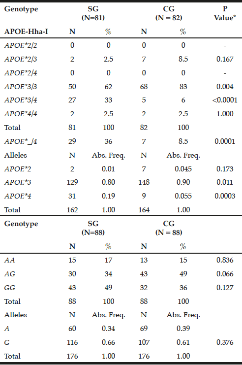
The APOE*3/3 genotype prevailed in GC (83%) compared to SG (62%, P = 0.004), while the mutant heterozygous model (APOE*_/4) stands out on patients (36% versus CG = 8.5%, P = 0.0001). Thus, the APOE*3 allele showed a higher frequency in controls (0.90 versus SG = 0.80, P = 0.011), while APOE*4 prevailed in SG (0.19 vs. CG = 0.055, P = 0.0003) (Table 1).


Table 1 Genotypic and allelic frequency distribution for APOE-Hha-I and ABCA1-Sty I polymorphisms in patients with late-onset Alzheimer's disease (SG) and individuals without clinical signs of the disease (CG)

Table 1
Genotypic and allelic frequency distribution for APOE-Hha-I and ABCA1-Sty I polymorphisms in patients with late-onset Alzheimer’s disease (SG) and individuals without clinical signs of the disease (CG)

*Chi-square or Fisher test; N= number of individuals; Abs. Freq.= absolute frequency; ABCA1= adenosine triphosphate binding cassette transporter 1; ApoE= apolipoprotein E; P= Level of significance <0.05; SG= study group; CG= control group.


For ABCA1-StyI polymorphism there was similarity in allelic and genotypic distribution between the groups (P> 0.05), highlighting the GG genotype in patients (49%) and AG in controls (49%), with prevalence of the G allele in both groups (0.66 and 0.61, respectively, p = 0.376, Table 1).
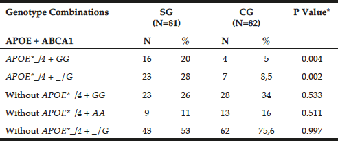
Table 2 shows the genotypic combination between APOE-HhaI polymorphism and ABCA1-StyI. The APOE*_/4 + GG genotypes prevailed in patients (20% versus GC = 5%, P = 0.0041), as observed for APOE*_/4 + _/G (SG = 28% versus GC = 8.5%, P = 0.0021).
It was found Hardy-Weinberg equilibrium for APOE-HhaI in patients (x²= 0.55; P= 0.50), which did not occur in controls (x2= 39.35; P <0.0001). For ABCA1-Sty I the Hardy-Weinberg equilibrium was maintained in controls (x2= 0.06; P = 0.90), but not in patients (x2= 5.13; P= 0.025).

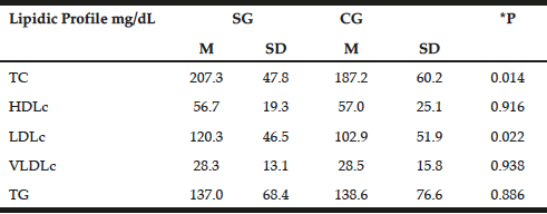
The lipid profile was similar between the groups with emphasis on increased levels of TC and LDLc in patients (207.3±47.8 mg/dL and 120.3±46.5 mg/dL, respectively) compared with the control group (187.2±60.2 mg/dL and 102.9±51.9mg/dL P=0.014, P=0.022, respectively). However, with the exception of TC in patients, lipid profile values remained within normal limits (Table 3) (23).


Table 2 Preferential combination between APOE-Hha I and ABCA1-Sty I polymorphisms in patients with late-onset Alzheimer's disease (SG) and individuals without clinical signs of the disease (CG)

Table 2
Preferential combination between APOE-Hha I and ABCA1-Sty I polymorphisms in patients with late-onset Alzheimer’s disease (SG) and individuals without clinical signs of the disease (CG)

*Chi-square or Fisher test; N= number of individuals; Abs. Freq.= absolute frequency; ABCA1= adenosine triphosphate binding cassette transporter 1; ApoE= apolipoprotein E; P= Level of significance <0.05; SG= study group; CG= control group.


Table 3 Distribution of mean values, standard deviations and mean differences for biochemist profile in patients with late-onset Alzheimer disease (SG) and individuals without clinical signs of disease (Control - CG)

Table 3
Distribution of mean values, standard deviations and mean differences for biochemist profile in patients with late-onset Alzheimer disease (SG) and individuals without clinical signs of disease (Control – CG)

t test; *P = significance level P <0.05; TC = total cholesterol, HDLc = concentration of high density lipoprotein cholesterol; LDLc = concentration of low-density lipoprotein cholesterol; VLDLc = concentration of very low density lipoprotein cholesterol; TG = triglycerides; M = Average; SD = standard deviation; SE = study group; CG = control group.


Discussion

The present study confirms the association of APOE-HhaI polymorphism with late onset AD, and suggests synergism of APOE*4 allele and G to ABCA1 in the metabolism of lipids in individuals with AD. In this case, the APOE-HhaI allelic and genotypic distribution discriminates patients with high frequency of APOE*4 allele (0.19) and controls (0.055, P= 0.0007). These results are consistent with other studies in Brazilian papers, whose frequency of the referred allele ranged from 0.22 to 0.34, whereas, in control group, the values remained between 0.07 and 0.11 (2, 24), and also consistent with results of studies with patient populations in Colombia, where the values for APOE*4 allele frequency was between 0.23 and 0.08 for study and control group, respectively (25).
The ABCA1-StyI polymorphism, however, was not associated with late onset AD. The G allele frequency was high in this casuistic study with AD (0.66), differing from the world’s population, including Chinese casuistry (0.48) (26), American and Canadian (0.27 for both) (27).
A study by Wavrant-De Vrieze et al. (28) in Spanish casuistic sample with AD, showed association between R219K polymorphism (ABCA1-StyI) and AD (P = 0.010), regardless of the presence of the APOE*4 allele. In addition, a sample of 1275 Swedish individuals with AD and control 2203 also discloses the influence of R219K polymorphism (AB 1.25, CI 1.12 to 1.40, P = 5.9 × 10 5), which is linked to G allele, associated with increased risk for AD (29). However, there is disagreement among authors.
Such polymorphism has been extensively studied in relation to risk for AD among other phenotypes, in particular those related to cardiovascular disease and plasma lipids (29). There is reference to the G allele for ABCA1 may amount to 2.77 times the risk for AD (12) and this variant can influence levels of 24S-hydroxycholesterol, which is essential for the transfer of cholesterol in blood-brain barrier increasing, thereby, the βA42 producing (30).
The present study shows an association between the synergism of APOE*_/4 and _/G genotypes and late onset AD, corroborating with the study conducted for the first time in Hispanic casuistic sample, showing that carriers of the APOE*4 allele and G allele for ABCA1 are at risk for increased AD by 3.7 times (15). In this case, the G allele represents the reduced activity of gene expression and thus could modify the risk for AD in synergism with allele APOE*4 (28, 29). On the other hand, Wang & Jia (26) revealed that the GG genotype for ABCA1 may reduce the risk for AD in Chinese sample. However, this association was only observed in women (P= 0.016) or individuals without the APOE*4 allele (P= 0.010).
In this context, there is reference of ABCA1 as a regulator of lipidation and apoE levels in brain. Deficiency of ABCA1 leads to loss of approximately 80% of apoE in the brain and the rest of residual apoE is poorly lipidated (17). In this case, studies in animal models of AD have shown that apoE poorly lipidated increases the amyloid load. This reinforces the hypothesis that apoE lipidation by ABCA1 affects deposition or removal of the amyloid protein, contributing to the formation of senile plaques in excess (17). Conversely, overexpression of the brain ABCA1 promotes apoE lipidation and eliminates the formation of mature amyloid plaques (17). These studies show that the capacity lipidic binding of apoE is the primary mechanism to the pathogenesis of AD.
This study highlights the demonstration of synergism between APOE and ABCA1, represented by the APOE*4 and G allele, respectively, with possible impact on the risk for late onset AD. The reduced expression of ABCA1 due to the presence of the G allele, may be compounded by low cholesterol efflux of glial cells to neurons due to the presence of the APOE*4 allele, which promotes the outflow less efficient compared to the APOE*2 and APOE*3 alleles (17).
Among patients with AD was found that the genotype distribution for APOE-HhaI was in agreement with Hardy-Weinberg equilibrium, the same was not true for the control group, as compared to ABCA1-Sty I polymorphism in patients, as was also observed in some studies of case-control analyzes of different genetic polymorphisms (29). The criterion for selecting the groups used in this study provided a group of older individuals, given that late-onset AD occurs in individuals of older age. Thus, the profile of patients, as well as the control group does not represent the general population by gender and age, which could influence the distribution of genotypes between the groups. Incidentally, the absence of Hardy-Weinberg equilibrium would be expected for a wide range of genetic diseases, considering the contribution of genes, though modest, for complex diseases. However, considering the large number of candidate gene studies in different cases, the number of genetic markers in imbalance is scarce. In this case, it is possible that researchers disregard the distribution of imbalanced genotypes, neglecting valuable information to identify casual polymorphisms (30).


Conclusion

The APOE-HhaI polymorphism is associated with late onset AD, however, genetic variants in ABCA1-StyI alone do not differentiate patients and controls. Furthermore, APOE*4 and G alleles in synergism are related to AD. Though, studies with more numerous series are needed to confirm this combination of genes with AD, as well as its influence on lipid metabolism.


Funding: This work was supported by The Sao Paulo Research Foundation (FAPESP) (#grant 2010/17476-0) and São Jose do Rio Preto Medical School (FAMERP – Laboratory of Biochemistry and Molecular Biology).

Acknowledgements: nothing to declare.

Ethical standard: This project was evaluated and approved by Certificate of Appreciation Presentation Ethics (CEP-FAMERP – CAAE: 0046.0.140.000-08). This is according to DECLARATION OF HELSINKI. Ethical Principles for Medical Research Involving Human Subjects


References

1.     Lupton MK, Proitsi P, Lin K, et al. The role of ABCA1 gene sequence variants on risk of Alzheimer’s disease. J Alzheimers Dis 2014;38:897-906.
2.    Pinhel MA, Nakazone MA, Cação JC, et al. Glutathione S-transferase variants increase susceptibility for late-onset Alzheimer’s disease: association study and relationship with apolipoprotein E epsilon4 allele. Clin Chem Lab Med 2008;46:439-445.
3.    Cohn-Hokke PE, Elting MW, Pijnenburg YA, et al. Genetics of dementia: update and guidelines for the clinician. Am J Med Genet B Neuropsychiatr Genet 2012; 159B:628-643.
4.    McKhann GM, Knopman DS, Chertkow H, et al. The diagnosis of dementia due to Alzheimer’s disease: recommendations from the National Institute on Aging-Alzheimer’s Association workgroups on diagnostic guidelines for Alzheimer’s disease. Alzheimer Dement 2011; 7:263-269.
5.    McKhann G, Drachman D, Folstein M, et al. Clinical diagnosis of Alzheimer’s disease: report of the NINCDS–ADRDA Work Group under the auspices of Department of Health and Human Services Task Force on Alzheimer’s Disease. Neurology 1984;34:939–944.
6.    Ehehalt R, Keller P, Haass C, et al. Amyloidogenic processing of the Alzheimer β-amyloid precursor protein depends on lipids rafts. J Cell Biol 2003;160:113-123.
7.    Hirsch-Reinshagen V, Burgess LB, Wellington CL. Why lipids are important for Alzheimer disease? Mol Cell Biochem 2009;326:121–129.
8.    Lupton MK, Proitsi P, Lin K, et al. The role of ABCA1 gene sequence variants on risk of Alzheimer’s disease. J Alzheimers Dis 2014;38:897-906.
9.    Xue-Shan Z, Juan P, Qi W, et al. Imbalanced cholesterol metabolism in Alzheimer’s disease. Clin Chim Acta 2016;456:107-114.
10.    Harris JR, Milton NG. Cholesterol in Alzheimer’s disease and other amyloidogenic disorders. Subcell Biochem 2010;51:47-75.
11.    Zannis VI, Just PW, Breslow JL. Human apolipoprotein E isoprotein subclasses are genetically determined. Am J Hum Genet 1981;33:11-24.
12.    Rodríguez-Rodríguez E, Mateo I, Infante J, et al. Interaction between HMGCR and ABCA1 cholesterol-related genes modulates Alzheimer’s disease risk. Brain Res 2009;1280:166-171.
13.    Refolo LM, Malester B, LaFrancois J, et al. Hypercholesterolemia accelerates the Alzheimer’s amyloid pathology in a transgenic mouse model. Neurobiol Dis 2000;7:321-331.
14.    Hirsch-Reinshagen V, Wellington CL. Cholesterol metabolism, apolipoprotein E, adenosine triphosphate-binding cassette transporters, and Alzheimer’s disease. Curr Opin Lipidol 2007;18:325-332.
15.    Wahrle SE, Jiang H, Parsadanian M, et al. ABCA1 is required for normal central nervous system ApoE levels and for lipidation of astrocyte-secreted apoE. J Biol Chem 2004;279:40987-40993.
16.    Burns MP, Vardanian L, Pajoohesh-Gangi A. The effects of ABCA1 on cholesterol efflux and Ab levels in vitro and in vivo. J Neurochem 2006;98:792–800.
17.    Blacker D, Bertram L, Saunders AJ, et al. Results of a high-resolution genome screen of 437 Alzheimer disease families. Hum Mol Genet 2007;12:23-32.
18.    Pena SD, Bastos-Rodrigues L, Pimenta JR, et al. DNA tests probe the genomic anscestry of Brazilians. Braz J Med Biol Res 2009;42:870-876.
19.    Salazar LA, Hirata MH, Cavalli SA, et al. Optimized procedure for DNA isolation from fresh and cryopreserved clotted human blood useful in clinical molecular testing. Clin Chem 1998;44:1748-1750.
20.    Hixson JP, Vernier DT. Restriction isotyping of human apoliprotein E by gene amplification and cleavage with Hhal. J Lipid  Res 1990;31:545-548.
21.    Fidani L, Goulas A, Crook R, et al. An association study cholesteryl ester transfer protein TaqI B polymorphism with late  onset Alzheimer’s disease. Neurosci Lett 2004;357:152-154.
22.    Simão AF, Precoma DB, Andrade JP, et al. I Diretriz Brasileira de Prevenção Cardiovascular. Arq Bras Cardiol 2013;101(6 Suppl 2):1-63.
23.    Souza DRS, Godoy MRP, Hotta J, et al. Association of apoliprotein E polymorfism in late-onset Alzheimer’s disease and vascular dementia in Brazilians. Bras J Med Biol Res 2003;36:1-5.
24.    Jacquier M, Arango D, Villareal E, et al. Apo e4 and Alzheimer’s disease. Positive association in a Colombian clinical series and review of the Latin-American studies. Arq Neuropsiquiatr 2001;59:11-17.
25.    Wang F, Jia J. Polymorphisms of cholesterol metabolism genes CYP46 and ABCA1 and the risk of sporadic Alzheimer’s disease in Chinese. Brain Res 2007;1147:34-38.
26.    Li Y, Tacey K, Doil L, et al. Association of ABCA1 with late-onset Alzheimer’s disease is not observed in a case–control study. Neurosci Lett 2004;366:268–271.
27.    Wavrant-De Vrièze F, Compton D, Womick M, et al. ABCA1 polymorphisms and Alzheimer’s disease. Neurosci Lett 2007;416:180-183.
28.    Reynolds CA, Hong MG, Eriksson UK, et al. A survey of ABCA1 sequence variation confirms association with dementia. Hum Mutat 2009;30:1348-1354.
29.    Kölsch H, Lüjohann D, Jessen F, et al. Polymorphism in ABCA1 influences CSF 24Shydroxycholesterol levels but is not a major risk factor of Alzheimer’s disease. Int J Mol Med 2006;17:791–794.
30.    Xu J, Turner A, Little J, et al. Positive results in association studies are associated with departure from Hardy-Weinberg equilibrium: hint for genotyping error? Hum Genet 2002;111:573-574.

KNOWLEDGE OF ALZHEIMER’S DISEASE AND TRAINING NEEDS IN FINAL YEAR MEDICAL AND PHARMACY STUDENTS


C. Scerri


Department of Pathology, Faculty of Medicine and Surgery, University of Malta, Msida, Malta MSD 2080.

Corresponding Author: Charles Scerri Ph.D., Department of Pathology, Faculty of Medicine and Surgery, University of Malta, Msida MSD 2080, Malta; Tel.: +356 23402905; Fax: +356 21320281; E-mail: charles.scerri@um.edu.mt

J Aging Res Clin Practice 2017;6:9-13

Published online November 3, 2016, http://dx.doi.org/10.14283/jarcp.2016.122


Abstract

Abstract: Although a significant number of medical and pharmacy professionals come into contact with an increasing number of individuals with Alzheimer’s disease and other dementias, there is concern on the lack of knowledge and skills received during their undergraduate training programmes with the consequence of not providing the required hospital and community care for these individuals following programme completion. The aim of this report is to describe the results of a small scale study investigating the level of knowledge of Alzheimer’s disease and training needs in medical and pharmacy students at the end of their final year of undergraduate training. The findings indicated a lack of in-depth knowledge for both categories of students, in particular on risk factors and pharmacotherapeutic management highlighting an urgent need of refining existent training programmes that equip future medical and pharmacy professionals with the necessary skills in providing adequate care and management for individuals with the disease.

Key words: Alzheimer’s disease, dementia, medical students, pharmacy students, knowledge.


Introduction

Alzheimer’s disease (AD) is the most common form of dementia accounting to 50-70% of all the cases (1). In 2015, it was estimated that 1.5% of the population in Malta had dementia, a figure that is projected to more than double in the next 30 years (2). This, in conjunction with the changing demographics favouring a progressive increase in the elderly population, will invariably put greater demands and challenges on medical and pharmacy professionals who, in the majority of cases, are at the forefront in providing formal care to these individuals both in hospital and the community. Concerns however exist that such healthcare professionals do not possess the required knowledge and skills to offer the required care and advice to these patients and their caregivers (3) with shortcomings in undergraduate curricula being one of the main reasons (4, 5). Such knowledge gaps were recently highlighted by general practitioners working in Malta as a motive towards their lack of understanding of dementia diagnosis and pharmacotherapeutic management of the cognitive and behavioural symptoms in individuals with dementia (6).
The aim of this study was to investigate the level of knowledge of AD, including its pharmacotherapeutic management, and training needs in final year medical and pharmacy undergraduate students.  The scope was to use the information gathered to support evidence on the need of enhancing dementia training in the current medical and pharmacy undergraduate curriculum in Malta with the latter being one of the priorites highlighted in the recent launch of the national dementia strategy (7).


Method

The targeted population consisted of all full-time undergraduate medical (MD) and pharmacy (BPharm) students in the final year of their studies at the University of Malta, the latter being the sole higher education institution licensed to offer tertiary education in both medicine and pharmacy. The Maltese medical degree programme is a five-year university course organised by the Faculty of Medicine and Surgery. Dementia-specific topics are covered in teaching modules under the specialties of neurology, geriatric medicine, pharmacology and principles of good practice and psychological and social aspects of healthcare for a total of five hours throughout the whole training course. The Maltese undergraduate pharmacy 4-year programme is also organised by the Faculty of Medicine and Surgery with dementia training being delivered in a three-hour session focusing mainly on neuropathology, prevalence and pharmacotherapy.
The study consisted of a survey, in English language, composed of an anonymous closed-ended questionnaire and distributed to the students following the completion of all taught and clinical placement modules but prior to sitting for final assessment. The measurement tools included the Alzheimer’s Disease Knowledge Scale (ADKS) (8) and the Alzheimer’s Disease Pharmacotherapy Measure (ADPM). ADKS contains 30 true/false items aimed to assess knowledge on AD. It is designed to be used by healthcare professionals, students, patients, caregivers and laypeople to pinpoint educational needs (8). The questions focus on 7 subscales that characterise knowledge about AD namely on: risk factors, assessment and diagnosis, symptoms, course of the disease, life impact, caregiving, and treatment. The percentage correct score is calculated using the equation: sum of correct items/30 x 100. The ADPM was developed by the author. It consists of 18 true/false items focusing on knowledge about pharmacological management in AD including available treatment options, role of supplementation, drug efficacy, dose titration, adverse effects, augmentation therapy and the use of antipsychotics for the behavioural and psychological symptoms of dementia (BPSD). Face validity was ascertained by asking three local clinical medical specialists in the field of dementia to independently evaluate the relevance of the topics selected. Similar to ADKS, the percentage correct score for ADPM was found by calculating the percentage of dividing the number of correct items by the maximum possible score.
The survey also contained a section that requested participants to rate their educational and training needs using a 3-point Likert scale (ranging from 1: least, 2: moderately and, 3: most needed) from a list of 20 topics related to the care of individuals with AD and related dementias (9).
Background characteristics of participating students were collected via closed-ended questions that looked for information about age, gender, presence of and/or caring for a family member with dementia, and history of caring for persons with dementia during clinical placement. The study was approved by the Faculty of Medicine and Surgery Ethics Committee as well as the Research Ethics Committee of the University of Malta.
Descriptive statistics including mean data, standard deviations and percentages were used to describe socio-demographic data and the ADKS and ADPM scores. Following data analysis for normality of distribution by the Shapiro-Wilk test, the parametric independent sample t-test and the non-parametric Mann-Whitney U test were used for group comparisons. Mean data was expressed as mean ± SD (standard deviation). The significance level was set at 0.05. All statistical analyses were performed using PASW Statistics (Version 20).


Results

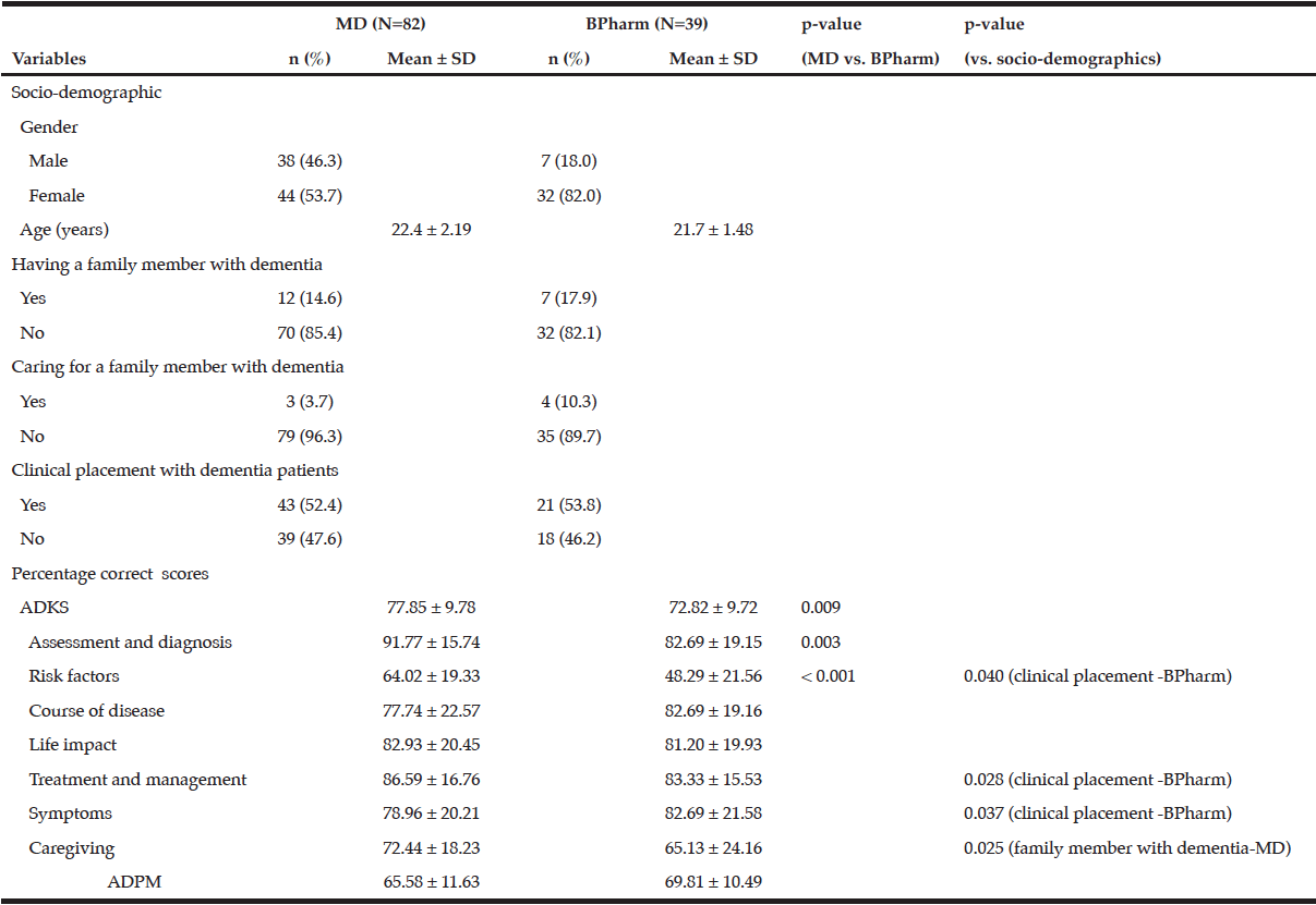
A total of 82 medical (74.6% response rate) and 39 pharmacy (92.9% response rate) final year students replied to the questionnaire with the female gender being over represented in the pharmacy but not the medical programme. The majority of respondents indicated that they don’t have or care for a family member with dementia. Approximately half of the students in both programmes attended optional clinical placements with AD and related dementias patients (Table 1).

Table 1 Socio-demographic data, ADKS and ADPM scores of final year undergraduate medical and pharmacy students

Table 1
Socio-demographic data, ADKS and ADPM scores of final year undergraduate medical and pharmacy students

ADKS: Alzheimer’s Disease Knowledge Scale, ADPM: Alzheimer’s Disease Pharmacotherapy Measure, BPharm: pharmacy students, MD: medical students

Students who attended the medical programme had significantly higher percentage of correct ADKS scores compared to pharmacy students (t = 2.647, P = 0.009). Moreover, analysis of the ADKS subscales revealed that medical students were more knowledgeable in assessment and diagnosis (z-score = -2.987, p = 0.003) and risk factors (z-score = -3.712, p < 0.001) for AD. BPharm students who reported to have had exposure to dementia patients during their clinical placements had significantly higher scores in knowledge of risk factors (z-score = -2.041, p = 0.040), treatment and management (z-score = -2.194, p = 0.028) and symptoms (z-score = -2.091, p = 0.037) than their counterparts who did not have such exposure. A significant effect on the caregiving construct of the ADKS was reported in MD students having a family member with dementia (z-score = -2.241, p = 0.025) and in BPharm students caring for a family member with dementia (z-score = -2.036, p = 0.041). No significant differences were reported in the percentage correct ADPM scores between the two undergraduate training programmes (Table 1).
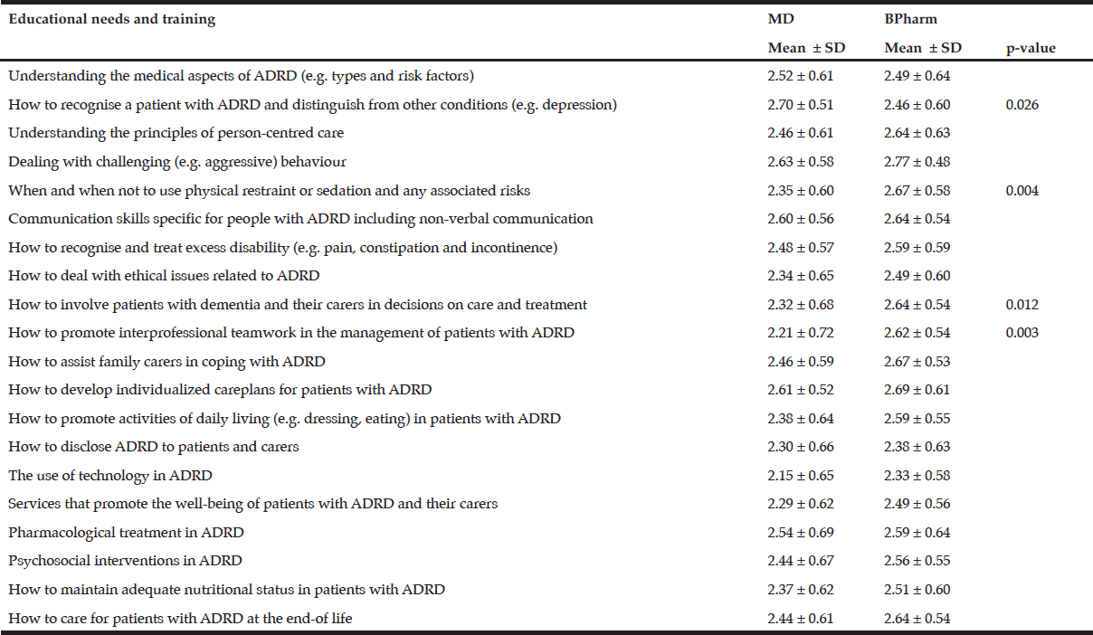
The mean scores in all educational and training needs topics were greater than 2 indicating that the majority of students in both undergraduate programme categories necessitate more knowledge in these subject areas. Compared to BPharm students, MD students expressed a greater need of having more training in recognising a patient with AD and related dementias and how to distinguish such medical conditions from others (z-score = -2.232, p = 0.026). Conversely, BPharm students scored significantly higher in educational and training needs involving the use of physical restraint and sedation (z-scores = -2.906, p = 0.004), the involvement of patients with dementia and their caregivers in decision taking on care and treatment (z-score = -2.514, p = 0.012), and how to promote interprofessional teamwork in managing patients with AD and related dementias (z-score = -3.004, p = 0.003) compared to their medical counterparts. The use of technology scored the lowest in both undergraduate training programmes whereas dealing with challenging behaviour scored the highest in BPharm students and second-highest in MD students (Table 2).

Table 2 Perceived educational and training needs of final year undergraduate medical and pharmacy students

Table 2
Perceived educational and training needs of final year undergraduate medical and pharmacy students

ADRD: Alzheimer’s disease and related dementias


Discussion

The present study has shown that albeit the limited number of hours directed towards undergraduate dementia training, both final year medical and pharmacy students had adequate knowledge on AD with ADKS scores higher than US college students (8). However, analysis of ADKS subscales revealed that students following both undergraduate training programmes were unfamiliar with risk factors associated with the disease with a possible consequence of such students not being able to provide the required advice in terms of disease prevention upon graduation. Conversely, topics related to presenting symptoms and assessment and diagnosis were well recognised suggesting that they have the necessary skills to refer such patients to the appropriate support services for clinical assessment. However, whether such knowledge will eventually be translated into practice post-registration remains unclear. A recent national survey investigating practices in diagnosis, disclosure and pharmacotherapeutic management of dementia by general practitioners in Malta has found that referral to a dementia specialist was routinely adopted only by a limited number of practitioners (6). Interestingly, clinical placement played an important role in enhancing knowledge of risk factors, symptoms and treatment and management in pharmacy undergraduate students. It may be that learning through informal interaction with the care-recipient overcame any existing curriculum shortcomings and that hidden curricular activities can play a significant role in enhancing knowledge, shaping values and professional identity (10). A possible reason of why this has been observed in pharmacy and not medical students may lie in the different nature of their clinical placements. In MD students, these are mostly composed of ward rounds where students work in groups under the close guidance of medical specialists and with little hands-on experience. In addition, pharmacy students also attend pharmacy practice sessions designed to interact with clients on an individual basis thus enhancing the possibilty of building a closer relationship leading to a better understanding of their needs and concerns.  As previously reported with undergraduate nursing students (9), medical and pharmacy undergraduates who had or cared for a family member with dementia were found to have higher scores in the caregiving subscale of the ADKS.
Closer analysis of the ADPM scores revealed that, similar to what has been reported with general practitioners (6), the majority of final year medical and pharmacy students believed that early pharmacotherapeutic management of AD could postpone institutionalisation. This is expected due to the strong national emphasis that early management and diagnosis of AD could prevent dependency onto long-term services (7). Only a minority of students (36.6% MD; 47.8% BPharm) correctly indicated that AD pharmacotherapy does not stop the decline in activities of daily living. The observation that the lowest scores obtained for both undergraduate progammes (23.2% MD; 25.6% BPharm) was to a question related to the efficacy to acetylcholinesterase inhibitors was strongly indicative of the lack of in-depth knowledge of how these pharmacotherapeutic agents work. Questions related to drug management in challenging behaviour also yielded low scores possibly reflecting on the limited training that medical and pharmacy undergraduate students receive in this field during their study programmes. This is of particular concern due to an increase risk of mortality and morbidity following the use of antipsychotic drugs in managing BPSD (11).
Compared to their medical counterparts, pharmacy students significantly valued the need of enhancing training that promotes interprofessional teamwork in the management of patients with AD and related dementias. There is a strong case of incorporating interprofessional education interventions in undergraduate medical and pharmacy dementia curricula as this has the potential of improving knowledge related to dementia recognition and care (12, 13). Notwithstanding the recent  efforts by a number of national dementia plans together with individuals with dementia and their caregivers towards an increase in awareness and use of assistive technology (14), both medical and pharmacy students rated educational and training needs in this particular area as the least important. The lack of knowledge of how assitive technologies could support individuals with dementia may limit such healthcare professionals in providing advice on support systems that aim at enhancing autonomy, independence and quality of life.
In conclusion, this small scale study revealed that medical and pharmacy undergardaute training in the field of AD is not providing in-depth knoweldge and skills necessary to provide adequate care for these individuals. As a result, there is an urgent need to develop and implement pre-registration training modules designed to enhance the skills and better prepare the next generation medical and pharmacy professionals in the field of AD management and care.


Acknowledgements: The authors would like to thank the students who participated in the study.

Conflict of interest: none

Funding: none

Ethical Standards: Ethical approval for the study was granted by the Faculty of Medicine and Surgery Ethics Committee and the Research Ethics Committee of the University of Malta.


References

1.    Hofman A, Ott A, Breteler MMB et al. Atherosclerosis, apolipoprotein E, and prevalence of dementia and Alzheimer’s disease in the Rotterdam Study. The Lancet 1997;349:151-154.
2.    Scerri A, Scerri C. Dementia in Malta: new prevalence estimates and projected trends. Malta Med J 2012;24:21-24.
3.    Tsolaki M, Papaliagkas V, Anogianakis G et al. Consensus statement on dementia education and training in Europe. J Nutr Health Aging 2010;14:131-135.
4.    StClair Tullo E, Gordon AL. Teaching and learning about dementia in UK medical schools: a national survey. BMC Geriatr 2013;13:29.
5.    Oakley R, Pattinson J, Goldberg S et al. Equipping tomorrow’s doctors for the patients of today. Age Ageing 2014;43:442-447.
6.    Caruana-Pulpan O, Scerri C. Practices in diagnosis, disclosure and pharmacotherapeutic management of dementia by general practitioners – a national survey. Aging Ment Health 2014;18:179-186.
7.    Scerri C. Empowering change: A national strategy for dementia in the Maltese islands. 2015. Parliamentary Secretariat for the Rights of Persons with Disability and Active Ageing, Valletta.
8.    Carpenter BD, Balsis S, Otilingam PG et al. The Alzheimer’s Disease Knowledge Scale: development and psychometric properties. Gerontologist 2009;49:236-247.
9.    Scerri A, Scerri C. Nursing students’ knowledge and attitudes towards dementia – a questionnaire survey. Nurse Educ Today 2013;33:962-968.
10.    Annear MJ, Lea E, Lo A, Tierney L, Robinson A. Encountering aged care: a mixed methods investigation of medical students’ clinical placement experiences. BMC Geriatr 2016;16:38.
11.    Banerjee S. The use of antipsychotic medication for people with dementia: time for action. 2009. http://www.rcpsych.ac.uk/pdf/Antipsychotic%20Bannerjee%20Report.pdf. Accessed 5 July 2016.
12.    Brody AA, Galvin JE. A review of interprofessional dissemination and education interventions for recognizing and managing dementia. Gerontol Geriat Educ 2013; 34:225-256.
13.    McCaffrey R, Tappen RM, Lichtstein DM, Friedland M. Interprofessioanl education in community-based Alzheimer’s disease diagnosis and treatment. J Interprof Care 2013;27:534-536.
14.    Newton L, Dickinson C, Gibson G, Brittain K, Robinson L. Exploring the views of GPs, people with dementia and their carers on assistive technology: a qualitative study. BMJ Open 2016;13:6(5).

ALZHEIMER’S DISEASE, CEREBROVASCULAR DISEASE AND DEMENTIA: THEIR ASSOCIATION AND PREVENTION


D.A. Davey


Corresponding Author: Faculty of Health Sciences, University of Cape Town, Anzio Road, Observatory, Western Cape, South Africa 7925, e-mail profdad@eject.co.za, Tel +27 21 712 1314


Abstract

Abstract: Alzheimer’s disease (AD) and cerebrovascular disease (CVD) frequently co-exist and CVD acts additionally and synergistically with AD in ageing–related impairment of cognitive function and dementia. A significant number of men and women with normal cognition at the time of death have the neurodegenerative and cerebrovascular changes of AD and CVD and are regarded as having high cognitive reserve or cognitive resilience. Many measures used to prevent and treat cardiovascular disease, decrease the incidence, or delay the onset of ageing-related cognitive impairment and dementia. Ageing-related cognitive impairment and dementia are increased by adverse psycho-social factors and can be prevented or mitigated by appropriate psycho-social measures. There is now more than sufficient evidence to implement, as a matter of urgency, personal health and life-style measures and public health initiatives in the endeavor to prevent, postpone or ameliorate ageing-related cognitive impairment and dementia and to decrease its burden world-wide.

Key words: Alzheimer’s disease, cognitive impairment, cerebrovascular disease, dementia, prevention.


Nomenclature

Alois Alzheimer in 1906 described a “peculiar severe disease process of the cerebral cortex” with “miliary foci” (β-amyloid plaques) and “fibrils” (neurofibrillary tangles) in a patient with dementia praecox and the condition was named “Alzheimer’s Disease” (1). The term “Alzheimer’s Disease” is currently used in several different senses:
(a) specifically, by neurologists, psychiatrists and others to mean the form of neurodegeneration characterized by β-amyloid plaques and neurofibrillary tangles in the brain as described by Alzheimer. The term “vascular dementia” (VaD) is used for dementia attributed to cerebrovascular disease
(b) loosely, to include all forms of ageing-related cognitive impairment and dementia with varying cerebral pathologies
(c) generally, in non-medical circles instead of the word “dementia”.
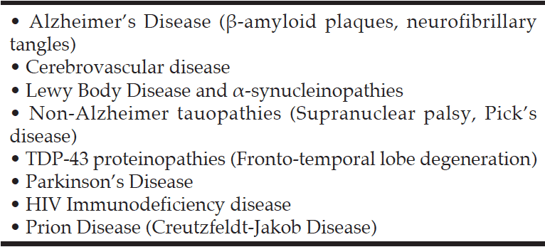
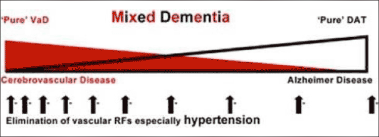
The different uses of the term “Alzheimer’s Disease” have led to misunderstanding and the meaning may only be clear from the context. Alzheimer’s disease (AD) as first described by Alzheimer is but one of several causes of Ageing-Related Cognitive Impairment and Dementia (ARCID) (Table 1). The commonest are AD, cerebrovascular disease (CVD) and Lewy Body Disease (LBD) which frequently co-exist .It has been proposed that ARCID and dementia should be regarded as a syndrome i.e. a complex of symptoms with multiple causes, similar to other chronic diseases (2).
The purpose of this review is to substantiate the evidence that:
(A) AD and CVD are commonly associated and act additively and synergistically in ARCID
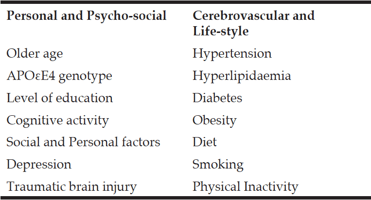
(B) Many risk factors for ARCID and measures that may prevent or postpone its development are very similar to the risk factors and measures to prevent and treat cardiovascular and cerebrovascular disease.
(C) Adverse psycho-social factors are significant risk factors for ARCID and psycho-social measures that increase cognitive reserve and resilience may prevent, delay the onset or ameliorate ARCID
(D) In the current absence of effective disease-modifying treatments, primary prevention combining all possible protective measures is the best hope to prevent, delay the onset and ameliorate ARCID


Association of Alzheimer’s disease and cerebrovascular disease

The cerebral pathology in men and women with dementia and of those with normal cognition at the time of death has been investigated in at least four major post-mortem studies: the Religious Orders Study and Rush Memory and Aging Project, the Medical Research Council Cognitive and Ageing Study, the Vienna Trans-Danube Aging Study, and The National Alzheimer’s Coordinating Centre USA Study (3-6). The main conclusions were very similar in all four studies, namely that the changes of AD and CVD (a) frequently co-exist in late-onset dementia (b) overlap to varying degrees and have additive and synergistic effects on cognitive decline (c) are sometimes found in persons with normal cognition at the time of death (7). The neurodegenerative and cerebrovascular changes associated with dementia form a spectrum from “pure” AD to “pure” CVD and most commonly are combined and result in “mixed dementia” (8) Fig1. The separation of AD as described by Alzheimer, and “vascular dementia” has been claimed to be a false dichotomy (8).

Table 1 Brain pathologies associated with cognitive impairment and dementia

Table 1
Brain pathologies associated with cognitive impairment and dementia

Figure 1 Conceptual Diagram of Mixed Dementia

Figure 1
Conceptual Diagram of Mixed Dementia


Cognitive reserve and cognitive resilience

A significant proportion of men and women with normal cognition at the time of death have the neurodegenerative and cerebrovascular changes of the brain associated with AD, CVD and dementia. The discordance between neuropathology and lack of cognitive impairment constitutes prima facie evidence for the role of some type of brain, neural or cognitive reserve (9). The absence of impaired cognition and dementia in such cases has been ascribed to high cognitive reserve and cognitive resilience. Cognitive reserve” implies high cognitive ability from early in life and its maintenance in mid and later life with the consequent prevention or postponement of ARCID (10). “Cognitive resilience” refers to the prevention or delay of ARCID in spite of the development of the pathological changes of AD, CVD and LBD.


Risk factors for ageing-related cognitive impairment and dementia

Ageing-related cognitive impairment and dementia has been associated with a large number of risk factors. A recent extensive meta-analysis of 323 papers including 93 factors considered suitable for epidemiological analysis, identified nine potentially modifiable risk factor; type-2 diabetes, obesity, hypertension, homocystinaemia, frailty, depression, current smoking, carotid artery narrowing, low educational achievement (11). The calculated population attributable risk combining all nine factors was 0.66 and it was claimed that two third of AD cases could be explained by these factors. In another study, potentially modifiable risk factors have been estimated to be present in approximately 50% of individuals with AD in the USA and worldwide (12). The seven modifiable risk factors included in these estimates were midlife hypertension, midlife obesity, diabetes mellitus, physical inactivity, smoking, depression and low education. The estimates do not take into account the non-independence of risk factors and the combined population-attributable risk factors have been estimated to be about 30% in the USA and Europe (13). Risk Factors can be divided into (a) Personal and Psycho-Social and (b) Cerebrovascular and Lifestyle (Table 2).

Table 2 Risk Factors for Ageing-related cognitive impairment and dementia

Table 2
Risk Factors for Ageing-related cognitive impairment and dementia


Personal factors

Personal factors including age, family history and the presence of the lipoprotein APOEε4 allele, are not modifiable but their effects can be mitigated or postponed by favourable environmental factors. Age is the most important factor determining the incidence and prevalence of cognitive impairment and dementia; the incidence of all-cause dementia increases exponentially from about 5/1,000 person-years in the 65-69 years age group to about 85/1,000 person-years in the age 90+ years (14). The most common genetic risk factor is the ε4 allele of the lipoprotein APOE4 and the APOEε4 allele has been estimated to increase the risk of AD about 3 times in heterozygotes and 15 times in homozygotes (15).

Psycho-social factors

Psycho-social factors often play an important part in ARCID and measures that increase cognitive reserve and cognitive resilience may be of considerable benefit in preventing, delaying or ameliorating ARCID. In an analysis of more than 20 studies involving 29,000 individuals followed for a median of 7.1 years, higher brain reserve was associated with a lower risk for incident dementia OR 0.54 (0.49–0.59) (10). The psycho-social factors that have been studied include, level of education, continuing cognitive activity and cognitive interventions, social and personality factors, depression and traumatic injury.

Level of education

The relative risks for low versus high education in a meta-analysis of 13 cohort and 6 case-control studies were, for AD 1.80 (1.43–2.27), for non-AD 1.32(0.92–1.88) and for all dementias 1.59(1.26–2.01) (16). In a meta-analysis of 31 studies with incident AD the pooled relative risk for lower education was RR 1.99(1.30–3.04)(17). In an analysis of 22 longitudinal studies including 21,456 individuals and 1,733 cases of dementia, the risk of dementia was lower for those with higher education OR 0.53 (0.45–0.62) (17). Low level of education is one of the biggest contributors to the high prevalence of AD world-wide (12).

Continued Cognitive Activity and Cognitive Interventions

A systematic review of 22 cohort studies including 29,000 individuals concluded that complex patterns of mental activity in early and mid-life was associated with a significant reduction in the incidence of dementia in later life RR 0.54(0.49–0.59) (10). In the Rush Memory Project frequent participation in cognitive stimulating activities was associated with less rapid decline in cognitive function and a lower incidence of AD, HR 0.58 (0.44–0.77) after controlling for a low baseline cognitive function, past cognitive activity, socioeconomic status and current social and physical activity (18). A Cochrane review in 2011 concluded that cognitive training interventions significantly improved immediate and delayed recall in healthy older adults and that more studies in other cognitive domains were necessary (19).

Social and Personality Factors

Social isolation and loneliness increase cognitive decline and the risk of late-life dementia (20, 21). Conscientiousness and purpose in life have been associated with a reduced risk of ARCID (22, 23). In the MRC-CFAS Study a combined Cognitive Lifestyle Score (CLS) based on educational attainment, occupational complexity and social engagement found that those who maintained a high CLS throughout life had a 40% reduced risk of developing dementia (24).

Depression

Depression may be a cause or consequence of cognitive impairment and dementia. A systematic review and meta-analysis of 20 studies including1,020,172 individuals found that history of depression increased risk of developing AD with a pooled OR of 2.03(1.73–2.38) for case control studies and of 1.90 (1.55–2.33) for cohort studies (25).

Traumatic brain Injury

Moderate and severe traumatic brain injury increases the risk of cognitive decline and is estimated to increase the risk of dementia in later life two to three fold (26). There is an increased risk of cognitive impairment and later onset of dementia in military veterans who have suffered brain injuries and in those involved in sports such as boxing and football of all forms, particularly in players who have experienced multiple concussions (27).

Cerebrovascular and life-style factors

Many cerebrovascular and lifestyle factors that predispose to ageing-related MCI and dementia are potentially preventable or modifiable (7). Measures that may prevent CVD are similar to those that prevent cardiovascular disease and include active treatment of hypertension, hyperlipidaemia and diabetes. The extensive study of 5,715 cases with a single neurodegenerative disease in the National Alzheimer’s Coordinating Centre USA database concluded that “in the absence of any specific disease-modifying treatments for Alzheimer’s disease in the near future, we urge, based on the high prevalence on cerebrovascular disease described in our data here, that aggressive management of vascular risk factors and encouragement of healthy life styles in mid-life may have benefit for Alzheimer’s disease or α-synucleinopathies individuals at increased risk to become clinically symptomatic, and probably to those with other causes of cognitive impairment. Indeed, even those who already manifest the clinical features of Alzheimer’s disease or α-synucleinopathy may benefit from effective therapies that mitigate vascular risk factors and cerebrovascular disease” (6).

Hypertension

Mid-life, but not late-life, hypertension is associated with an increased risk of AD and dementia with a calculated OR of 1.61(1.16–2.24) (28). A cohort of a random, population-based sample of 1449 individuals in Sweden was followed for an average of 21 years. Those with a raised systolic pressure in midlife (BP>160mm Hg) had a significantly higher risk of AD in later life OD 2.3 (1.0–5.5), after adjusting for age, body mass index, education, vascular effects, smoking and alcohol consumption (29). A quantitative meta-analysis of 14 studies of subjects without cognitive impairment or dementia, 32,658 with and 36,905 without hypertensive medication found no significant difference in the incidence of AD between the two groups but that those who had received anti-hypertensive medication has a significantly lower incidence of vascular dementia RR 0.67 (0.52–0.87) and of all-cause dementia RR 0.87 (0.7–-0.96) (30)

Hyperlipidaemia

A systematic review of 18 prospective studies found a significant association between high mid-life total cholesterol (TC) and an increased risk of AD and all-cause dementia but there was only weak evidence of an association between TC and cognitive decline (31).

Diabetes

A number of systematic reviews and meta-analyses have reported an increased risk of impaired cognition or dementia in association with Type-II diabetes (11). A meta-analysis of prospective 28 observational studies found that the pooled relative risk of developing AD was 1.56 (1.41–1.73) of VaD was 2.27 (1.96–2.66) and all-cause dementia was 1.73 (1.65-1.82) (32). Diabetes increased the risk of conversion of mild cognitive impairment to dementia and Mediterranean diet decreased the risk (33).

Obesity

In prospective studies and meta-analyses mid-life obesity has been found to be associated with a significant increase of all-cause dementia with a pooled estimate of RR of 1.60 (1.34–1.92) (17).
In addition to the specific effect of obesity on ARCID, obesity is associated with an increased incidence of hypertension, diabetes and cardiovascular disease.

Diet

A Mediterranean diet – high intake of vegetables, fruits, nuts and olive oil, relatively low intake of dairy products and red meat, and a moderate intake of wine – has been claimed in several observational studies to slow cognitive decline and to lower the risk of AD (34). In a prospective study of a similar “MIND” diet, high adherence was reported to be associated with a reduced risk of AD (35).

Smoking

A review of 37 studies found that compared with never smokers, current smokers had an increased risk of AD (RR1.40 (1.13–1.73), VaD (RR 1.38 (1.15-1.66) and all cause dementia (RR1.30 (1.13-1.73) (36). The risk of all-cause dementia increased by 34% for every 20 cigarettes smoked per day but was not increased in former smokers. In a study of a cohort of 21,123 people, heavy smoking in mid-life was associated during two decades of follow-up with a more than 100% increase in AD, VaD and all–cause dementia (37).

Physical Inactivity

A review and meta-analysis of 16 prospective studies on the association between physical activity and dementia found that comparing highest v lowest activity groups the combined RR for AD was 0.55 (0.36–0.84) and for all-cause dementia was 0.72 (0.60–0.80) (38). These values have been reversed to reflect the risks with inactivity as 1.82 (1.19–2.78) for AD and 1.39 (1.6–1.67) for all cause dementia (12). A review and meta-analysis of physical activity in 21 prospective cohorts comparing higher with lower levels of activity the RR on cognitive decline was 0.65 (0.55–0.76) and on dementia was 0.86 (0.76–0.97) (39). A Cochrane analysis in 2015 found that healthy, sedentary elders who begin exercise have a significant improvement in cognitive function, particularly mental processing speed (40).


Prevention of ageing-related cognitive impaiment and dementia: Combined measures

In the absence of disease-modifying treatments, measures to prevent or postpone the onset of ARCID should include measures to prevent cerebrovascular disease and improve physical health and to ensure an optimum psycho-social environment. In view of the long preclinical phase of both CVD and AD these measures need to be actively instituted as early in life as possible and not later than mid-life. Many studies of individual risk factors have been published but there have to date been virtually no long-term, randomized, controlled studies of combined measures to prevent or postpone ARCID. A recent Finnish Geriatric Intervention Study to Prevent Cognitive Impairment and Disability (FINGER) reported the results of a double-blind, randomized, controlled trial of 2,654 individuals aged 60-77 years assigned in a 1:1 ratio to multidomain intervention (cognitive training, diet, exercise and vascular risk monitoring) or a control group (general health advice) (41). The primary outcome was a change in cognition in a neuro-psychological test battery score (NTB). The difference in NTB between the two groups after 2 years was statistically significant (p=0.03). There was also a significant difference in the secondary outcomes of executive functioning (p=0.04) and processing speed (0.04) but not in memory (p=0.36). It was concluded that a multidomain intervention can improve cognitive function in at-risk elderly people.


Conclusion

Nine population-based studies of dementia incidence and prevalence have reported a declining prevalence and age-specific incidence of dementia in England, Sweden, The Netherlands and the USA (42). The decreases have been attributed to rising levels of education, better prevention and treatment of cardiovascular disease and healthier life-style including exercise. It is uncertain whether these favourable trends will continue in the face of rising levels of obesity and diabetes in these populations, and whether they will manifest in low income countries. Although the age-specific incidence of dementia may be decreasing in some countries, the population of the world, the number living to advanced old age and the number with dementia world-wide is increasing. The incidence of dementia rises rapidly over the age of 75 and it has been estimated that the total number of people with dementia will triple from 2015 to 2050. The best hope for reducing the incidence and prevalence of ageing-related dementia currently lies in primary prevention, and in particular better education, continued mental and physical exercise and strict control of vascular risk factors. The evidence is now more than sufficient evidence to urge the immediate implementation of both personal health and life-style measures and public health initiatives to prevent or delay the onset of ARCID and to decrease the burden world-wide.


Acknowledgements: I wish to acknowledge the authors M. Valenzuela, M Esler, K Ritchie and H Brodaty (8) and the publishers Translational Psychiatry ©Macmillan Publishers Limited for permission to publish Fig 1.

Conflict of interest: There are no conflicting interests. I have received no funds or writing assistance in preparation of the paper.


References

1.     A. Alzheimer A. Uber eine eigenartige Erkankung der Hinrninde. Allegmine Zeitschrift fϋr Psychiatrie und Psychisch-GEritliche Medizin. 1907;64:146-8.
2.    E.B. Larson, K. Kaffe,, K.M. Langa, New insights into the Dementia epidemic. N Engl J Med 2013;369:2275-7
3.    S. Negash, D.A. Bennett, R.S. Wilson, J.A. Schneider, S.E. Arnold, Cognition and neuropathology in aging: multidimensional perspective from the Rush Religious Orders Study and Rush Memory Aging Project. Curr Alzheimer Res 2011;8:336-40.
4.    S.B. Wharton, C. Brayne, G.M. Savva et al. Epidemiological neuropathology: the MRC Cognitive Function and Aging experience. J Alzheimers Dis 2011;25:359-72.
5.    G.G. Kovacs, I. Milenkovic, A. Wohrer et al. Non-Alzheimer neurodegenerative pathologies and their combinations are more frequent than commonly believed in the elderly brain: a community-based autopsy series. Acta Neuropathol. 2013;126:365-84.
6.    J.B. Toledo, S.E. Arnold, K. Raibie, Contribution of cerebrovascular disease in an autopsy confirmed neurodegenerative disease case in the National Alzheimer’s Coordinating Centre. Brain 2013;136:2697-706
7.    D.A. Davey. Alzheimer’s Disease, Cerebrovascular Disease and Dementia: a Potentially Preventable and Modifiable Syndrome, J Alzheimer’s Dis Parkinsonism 2015;5:1-5.
8.    M. Valenzuela, M. Esler, K. Ritchie, H. Brodaty, Antihypertensives for combating dementia? A perspective on candidate molecular mechanisms and population-based prevention. Transl Psychiatry 2012 2,e107 doi: 10.1038/tp.2012.28.
9.    D.A. Bennett, S.E. Arnold, M. J. Valenzuela, C. Brayne, J. A. Schneider, Cognitive and social life-style: links with neuropathology in late life. Acta Neuropathol 2014 127:137-50.
10.    M.J. Valenzuela, P. Sachdev, Brain Reserve and dementia: a systematic review. Psychological Medicine, null441-4545 doi10.1017/S0033291705006264.
11.    W. Xu, L. Tan, H-F. Wang H-F et al. Meta-analysis of modifiable risk factors for Alzheimer’s disease. J Neurol Neurosurg Psychiatry 2015 doi:10.1136/jnnp-2015-310548.
12.    D. Barnes D, K. Yaffe. The projected impact of risk factor reduction on Alzheimer’s disease prevalence. Lancet Neurol 2011;10:819-28.
13.    S. Norton, F.E. Matthews, D.E. Barnes, K. Yaffe, C. Brayne. Potential for primary prevention of Alzheimer’s disease: an analysis of population-based data. Lancet Neurol 2014;13:788-94.
14.    W.A. Kukull, R.Higdon, J.D. Bowen et al. Dementia and Alzheimer disease incidence: a prospective cohort study. Arch Neurol 2002;59:1737-46.
15.    K. Blennow, M.J. de Leon, H. Zetterberg, Alzheimer’s disease. Lancet 2006;368:387-403.
16.    F. Caamano-Isorna, M. Corral. A. Monyes-Matrinez, B. Takkouche, Education and dementia: a meta-analytic study. Neuroepidemiology 2006;26:226-32.
17.    M.A. Beydoun, H.A. Beydoun, A.A. Gamaldo, A. Teel, A.B. Zondeman, Y. Wang. Epidemiological studies of modifiable factors associated with cognition and dementia: systematic review and meta- analysis. BMC Public Health 2014;14:643-76.
18.    R.S. Wilson, P.A. Scherr, J.A. Schneider, Y. Tang, D.A. Bennett. Relation of cognitive activity to risk of developing Alzheimer’s disease. Neurology 2007;69:1911-20.
19.    M. Martin, L. Clare, A.M. Altgassen, M.H. Cameron, F. Zehnder. Cognition-based interventions for healthy older people and people with mild cognitive impairment. Cochrane database Syst Rev 2011(1):CD006220.
20.    R.S. Wilson, K.R. Krueger, S.E. Arnold et al. Loneliness and risk of Alzheimer’s disease. Arch Gen Psychiatry 2007;64:234-40.
21.    L. Fratiglioni. S. Paillard-Borg, B. Winblad, An active and socially integrated lifestyle in late life might protect against dementia. Lancet Neurol 2004;3:343-53.
22.    P.A. Boyle, A.S. Buchman, L.L. Barnes, D.A. Bennett, Effect of a purpose in life on risk of incident Alzheimer disease and mild cognitive impairment in community-dwelling older persons. Arch Gen Psychiatry. 2010;67:304–310.
23.    R.S. Wilson, J.A. Schneider, S.E. Arnold, J.L. Bienas, D.A. Bennett, Conscientiousness and the incidence of Alzheimer disease and mild cognitive impairment. Arch Gen Psychiatry 2007;64:1204-12.
24.    M. Valenzuela, C. Brayne, P. Sachdev, G. Wilcock, F. Matthews, Cognitive lifestyle and long-term risk of dementia and survival after diagnosis  in a multicenter population-based cohort. Am J Epidemiol 2010;173:1004-12.
25.    R.L. Ownby, E. Crocco, A. Acevedo, V. John, D. Loewenstein. Depression and risk for Alzheimer disease: systematic review, meta- analysis, and metaregression analysis. Arch Gen Psychiatry 2006;63:530-8.
26.    S. Shively, A.L. Scher, D.P. Perl, R. Dias-Arrastia, Dementia resulting from traumatic brain injury: what is the pathology? Arch Neurol 2012;69:1245-51.
27.    D.E. Barnes, A. Kaup, K.A. Kirby, A.L. Byers, R. Diaz-Arrastia, K. Yaffe, Traumatic brain injury and risk of dementia in older veterans. Neurology 2014;83:312-9.
28.    M. Baumgart, H.M. Snyder, M.C. Carrillo, S. Fazio, H. Kim, H. Johns, Summary of the evidence on modifiable factors for cognitive decline and dementia: A population-based perspective. Alzheimer’s & Dementia 2015;11:718-726.
29.    M. Kivipleto, E.L. Helkala, M.P. Laakso et al. Midlife vascular risk factors and Alzheimer’s disease in later life: Longititudinal , population based study. BMJ 2011;322:1447-51.
30.    H. Chang-Quan, W. Hui, W. Chao-Min et al. The association of antihypertensive medication use with risk of cognitive decline and dementia: a meta-analysis of longitudinal studies. Int J Clin Pract 2011 65:1295 -305.
31.    K.J. Anstey, D.M. Lipnicki, F. Low F, Cholesterol as a risk factor for dementia and cognitive decline: a systematic review of prospective studies with meta- analysis. Am J Geriatr Psychiatry 2008;16:343-354.
32.    K. Guadala, D. Bansal, F. Schifano, A. Bhansal. Diabetes Mellitus and risk of dementia: A meta-analysis of prospective observational studies. J Diabetes Investig 2013;4:3640-50.
33.    C. Cooper, A. Sommerland, C.G. Lyketsos, G. Livingston. Modifiable predictors of dementia in mild cognitive impairment: a systematic review and meta-analysis. Am J Psychiatry 2015;172:323-34.
34.    I. Lourida, M. Soni, J. Thompson-Coon et al. Mediterranean diet, cognitive function and dementia: a systematic review. Epidemiology 2013. 24:479-89.
35.    M.C. Morris, C.C. Tagney, Y. Wang, T.M. Sacks, D.A. Bennett, N.T. Aggarwal, MIND diet associated with reduced incidence of Alzheimer’s disease. Alzheimers Dement 2015 doi;10.1016/J.ALZ.2014.11.009.
36.    G. Zhong, Y. Wang, Y. Zhang, J.J. Guo, Y. Zhao. Smoking is associated with an increased risk of dementia: a meta-analysis of prospective cohort studies with investigation of potential effect of modifiers, PLoS One 2015.10;e118333. Doi10.1371/journal.pone0118333.
37.    M. Rusanen, M. Kivipelto, C.P. Quesenberry Jr, J. Zhou, R.A. Whitmen, Heavy smoking in midlife and long-term risk of Alzheimer disease and vascular dementia. Arch Int Med 2011. 171;333-9.
38.    M. Hamer, Y. Chida, Physical activity and risk of neurodegenerative disease: a systematic review of prospective evidence. Psychol Med. 2009;39:3-11.
39.    S.J. Blondell, R. Hammersley-Mather, J.L. Veerman. Does physical activity prevent cognitive decline and dementia? A systematic review and meta- analysis of longitudinal studies. BMC Public Health 2014;14:510.
40.    M. Angevaren, G. Aufdemkampe, H.J. Verhaar, A. Aleman, L. Vanhees. Physical activity and enhanced fitness to improve cognitive function in older people without known cognitive impairment. Cochrane database of systematic reviews 2008 Jul 16;(3):CD005381. doi:1002/14651858.CD005391.pub2. Review. Update in Cochrane database of systematic reviews 2015 .4.CD00538
41.    T. Ngandu, J. Lehtisalo, A. Solomons et al. A 2 year multidomain intervention of diet, exercise, cognitive training and vascular risk monitoring versus control to prevent cognitive decline in at-risk elderly people (FINGER): randomized controlled trial Lancet 2015;385:2255-63.
42.    K.M. Langa. Is the risk of Alzheimer’s disease and dementia declining Alzheimers Res Ther 2015:7;34.

ALZHEIMER’S DISEASE IS, AT LEAST IN PART, A COPPER-2 TOXICITY DISEASE


G.J. Brewer

University of Michigan, USA

Corresponding Author: George J Brewer, University of Michigan, USA, brewergj@umich.edu


Abstract

Developed countries have a raging epidemic of Alzheimers disease, with prevalence around 20% by age 70. Good evidence indicates this epidemic is new, with an Alzheimer’s-like dementia being rare prior to 1900. Prevalence of Alzheimer’s in undeveloped countries is still quite low, around 1%. These sets of facts strongly indicate that environmental change is causing the epidemic in developed countries. It is hypothesized here, with very good evidence, that the new environment change in developed countries is ingestion of inorganic copper, copper-2, from drinking water and supplement pills. There is good evidence that copper toxicity plays a major role in the pathogenesis of Alzheimer’s, with the size of the free copper pool intimately tied to cognition and cognition loss. Studies in AD animal models show that tiny amounts of inorganic copper in drinking water greatly enhance Alzheimers-type pathology and memory loss. Studies in humans show that those ingesting supplement pills containing copper, if they also eat a high fat diet, suffer rapid loss of cognition. Drinking water copper, and pill copper are both divalent copper, or copper-2. A recent study shows that food copper is primarily copper-1. There is an intestinal transport system specific for copper-1, and this copper goes to the liver, and is put into safe channels. Because mammals, including humans, ingested only copper-1, their systems evolved to safely handle copper-1. With development came copper plumbing and supplement pill ingestion, and copper-2 is now ingested. Some of it bypasses the liver, ends up in the blood free copper pool, and is toxic to cognition.

Key words: Alzheimer’s disease, copper-2, copper toxicity, drinking water copper, supplement pill copper. 


Introduction 

Considerable evidence has been developed that copper toxicity plays an important role in Alzheimer’s disease (AD). But the puzzle has been that the brain toxicity of copper in AD causes cognition loss, while the brain toxicity of copper in Wilson’s disease (WD), a disease known to be caused by copper toxicity, causes a movement disorder. This is puzzle 1. Secondly, considerable evidence has developed that inorganic copper, the divalent copper found in drinking water and supplement pills, is especially damaging to cognition and causative of AD in AD animal models, compared to the organic copper in foods. Why? This is puzzle 2.

Recently a paper was published which brings clarification and solutions to both puzzles 1 and 2 (1). Most exciting, it indicates much of the epidemic of AD can be aborted by simply stopping the ingestion of inorganic copper (divalent copper, or copper-2). This paper, which studied the speciation of copper in food, found that food copper was almost entirely monovalent copper, or copper-1. This finding was very surprising, because it had always been assumed food copper was a mixture of copper-1 and copper-2, since the two valence forms make a redox doublet that allow the catalysis of many redox reactions critical to life. Apparently, after death or harvest, in the absence of oxygen transport, copper-2 is reduced to copper-1 in foods. This finding changes everything. It means that mammals, including the human, evolved ingesting only copper-1. In line with this, the body has a transport system for copper-1, first passing it through the liver to keep it in safe channels. No such system exists for copper-2, and some bypasses the liver, appearing immediately in the blood, where it is toxic to cognition. So this solves puzzle 2. Inorganic copper, which is copper-2, is especially toxic because it bypasses the liver. It also solves puzzle1. The diseases, AD and WD, show different forms of copper toxicity- because AD is a copper-2 toxicity disease and WD is a general copper overload disease with copper-2 playing no special role. In this review we will elaborate on these new and exciting features.


Copper Toxicity in Alzheimer’s Disease

In this section the evidence for copper toxicity in AD will be reviewed without reference to the valence of copper. The group in Italy led by Professor Rosanna Squitti has amassed a large amount of data showing copper toxicity is intimately involved with AD pathogenesis. They have focused on what is called the “blood free copper pool”. Blood copper can be thought of as existing in two pools. The larger, comprising about 85%, is copper covalently bound to the ceruloplasmin (Cp) molecule. The smaller pool, comprises copper more loosely bound to albumin and various small molecules, and is called the free copper pool. Because of its more loose binding it is available to meet tissue needs in the body. If this pool becomes expanded, as it does greatly in WD, this copper becomes toxic.

The Squitti group has shown in AD patients: 1) The free copper pool is larger than in age-matched controls (2); 2) The size of the free copper pool correlates negatively with  measures of cognition (3); 3) The size of the free copper pool correlates positively with the rate of cognition loss over time (4), and; 4) The size of the free copper pool correlates positively with the risk of conversion of patients from mild cognitive impaired status, the precursor state to AD, to full AD (5). These studies tie copper toxicity closely to the pathogenesis of loss of cognition in AD. 

Additionally a paper by James et al (6) shows copper toxicity directly in the AD brain. They find what they call ”labile” copper is increased in the AD brain, and causing toxicity.

A study from China is also relevant. Shen et al (7) have studied the correlation of AD prevalence to soil copper concentration across the provinces of China. They find the two positively correlated.


Cognitive and Alzheimer’s Disease-Related Toxic Effects of Inorganic Copper (Copper-2)

In 2003 Sparks and Schreurs (8) published a ground-breaking paper which showed that trace amounts (0.12 ppm) of copper added to the drinking water of rabbits in a rabbit model of AD greatly enhanced amyloid plaque formation (one of the pathologic hallmarks in the AD brain) and memory loss. A 25 fold greater increase in food copper of the animals would have no toxic effect. This study has been amply confirmed in other AD animal models (9) and in another laboratory (10). For reference, the US Environmental Protection Agency (EPA) allows up to 1.3 ppm copper in human drinking water, ten times the amount found toxic in AD animal models (11). In the discussion of the relevance of AD causation in humans by consumption of copper-2 from drinking water, the important work of Sparks and Schreurs (8) will be further discussed.

In 2006 another groundbreaking paper was published, this one by Morris et al (12). They studied nutrient intake and cognition over several years in a large Chicago population. They found that those in the highest quintile of copper intake, who were there because they ingested daily supplement pills containing copper, if they also at a high fat diet, lost cognition at six times the rate of other groups. This is extraordinary! Ingesting a pill containing copper rapidly damages cognition, and many Americans, and others in developed countries, take these multimineral pills every day. This, too, will be further discussed in more detail.


Gastrointestinal Absorption of Copper-2 Versus Copper-1

What the 2003 studies of Sparks and Schreurs (8) and the 2006 studies of Morris et al (12) had in common was that they both showed cognitive damage from ingestion of inorganic copper, which is, of course, copper-2. A good clue as to why inorganic copper might behave differently than organic copper (food copper, or copper-1) was provided in earlier copper-64 studies in WD (13). Radiolabeled copper-64, as an inorganic salt, was given orally to WD patients to see if zinc therapy was effectively blocking copper absorption. At baseline, before zinc administration, 15-25% of the radiolabel would appear in the blood 1-2 hours after being given orally, much too soon for passage through the liver. In contrast, if food copper was labeled with a longer lived radiolabel, the label wouldn’t appear in the blood for 1-2 days, and then it would appear covalently bound in proteins secreted into the blood by the liver. What these studies show is that organic (food) copper is first transported to the liver, where it is put into safe channels, while a portion of inorganic copper bypasses the liver and appears immediately in the blood, adding to the free copper pool, where it is damaging to cognition.

Of course, inorganic copper is copper-2. And, as mentioned earlier, it has always been assumed that organic (food) copper is a mixture of copper-1 and copper-2, since in living tissue they form a redox doublet, catalyzing reactions critical to life. As mentioned in the Introduction, the surprising findings of Ceko et al (1) changes this assumption dramatically, and explains why radiolabeled inorganic copper and radiolabeled organic copper are absorbed so differently, as discussed above. Organic (food) copper is all copper-1, while inorganic copper is copper-2. This means that as mammals evolved they were ingesting copper-1, and weren’t exposed to much copper-2. So a system evolved to safely handle copper-1, involving a specific carrier (Ctr1) (14) in the intestine, which results in routing the copper to the liver for safe handling. Exposure to copper-2 was undoubtedly quite limited until humans in developed countries started using copper plumbing and taking multimineral pills containing copper. As a consequence, there is no special system for safe handling of copper-2. Ctr1 can’t absorb copper-2. Some copper-2 can be absorbed by the nonspecific divalent cation transporter, and some can be absorbed by diffusion. Some of this absorbed copper-2 bypasses the liver and goes immediately into the blood, as shown in the copper-64 studies mentioned earlier (13), and is toxic to cognition.


The Epidemiology of Alzheimer’s Disease Versus the Epidemiology of Copper-2 Ingestion in Drinking Water and Pills

Although not everyone agrees, there is good evidence that the epidemic of AD in developed countries has only emerged over the last 100 years, and it doesn’t involve undeveloped countries. That there is currently an epidemic in developed countries is clear. For example, 10% of those age 60 and over, 20% of those 70 and over, and 30% of those 80 and over develop AD in the U.S (15). These high prevalences of AD don’t exist in undeveloped countries. For example, in rural India, in those age 65 and over, AD prevalence is 1.07% (16). As another example, in Nigeria, Africa, in those aged 65 to 74, AD prevalence is 0.52% (17). Interestingly, in the latter study, the prevalence of AD in African-Americans of the same age group in Indianapolis, Indiana, USA, was 8.02%, 15 fold higher. This great increase in AD prevalence in people of the same ethnic group in the US compared to Nigeria shows the strong effect of Westernization on increasing the prevalence.

The evidence that this epidemic, restricted to developed countries, has come on in the last 100 years, is based on the writings of various type of expert physicians in the late 1800 and early 1900s, who didn’t see an AD-like dementia. For example, Osler, an internist, gathered all medical knowledge into a series of volumes, including one volume devoted to the brain, published in the late 1800s, and an AD-like dementia was not mentioned (18). Gowers, a neurologist, wrote a textbook of neurology in this period, and didn’t mention an AD-like dementia (19). Freud, a psychiatrist, published extensively during this period, and didn’t describe an AD-like dementia (20). Finally, Boyd, a pathologist wrote a textbook of pathology during this period, updated until 1938, and didn’t describe the amyloid plaques and neurofibrillary tangles, hallmarks of AD brain pathology, in the brains at autopsy (21). 

Some have argued that there weren’t enough elderly people back then to show AD, since AD is a disease of aging. But Waldmann and Lamb (22) showed that half the French population lived to age 60 in 1911, and the US population census in 1900 shows 3.6 million people age 60 or over, which would generate 360,000 cases at today’s rate; more than enough to be seen in the clinics, and to show up at autopsy.

Others argue that people just accepted AD as a type of aging, and didn’t recognize it as a special disease. This conceivably could explain the clinicians, Osler (18), Gowers (19), and Freud (20), not noting it, but it wouldn’t explain Boyd (21) and other pathologists not seeing plaques and tangles at autopsy. 

Some have said that perhaps the staining and other pathology techniques 100 years ago were inadequate to detect plaques and tangles. If brain autopsy material were available from, say, 1875 to 1925, it would be valuable to restudy them with modern techniques. It would be good to settle this definitively, because if the disease was truly rare back then, it strengthens the argument that a new environmental agent, or agents, in developed countries, is responsible for the epidemic of AD in developed countries. 

Here it will be assumed that the data supporting the idea that the epidemic of AD in developed countries is new (over the last 100 years) is strong, and that new environmental causative agent or agents should be rigorously searched for. Given the foregoing section on the toxicity of inorganic copper ingestion on human cognition and in AD animal models, it is reasonable to examine the hypothesis that ingestion of inorganic copper is the new key environmental factor causing the epidemic. 

With this in mind, the “epidemiology” of the spread of copper plumbing in developed countries needs to be examined along side the epidemiology of the AD epidemic. Copper plumbing began to be used in the early 1900s, was curtailed by the two world wars, and then exploded after 1950, such that now, 80-90 % of US homes have copper plumbing (23). The timing of the AD epidemic is quite similar, with a few cases appearing in the early 1900s, and then exploding after 1950 (22).

At this point it is fair to ask – does enough copper leach from copper plumbing to be potentially toxic? In a study of drinking water from 280 homes all across N. America, it was found that about one third were over 0.1 ppm, the level toxic in animal models, about one third were below 0.01 ppm, a level deemed safe, and about one third were between 0.1 and 0.01 ppm, a level of unknown safety (24). Thus, in summary, one third to two thirds of N American drinking water samples were at the toxic level, or of unknown safety, if the animal models are a good guide. In conclusion, there is enough copper in drinking water in N America to be a major factor in causing the AD epidemic. 

In regard to the hypothesis that copper from copper plumbing is a major factor in the AD epidemic, Japan provides interesting data. Japan is a developed country with a low incidence of AD (25). And Japan has shunned copper plumbing for fear of toxicity. But when Japanese migrate to Hawaii, where copper plumbing is used, their AD prevalence increases as in developed countries (26).

The other major source of copper-2 ingestion is from copper supplement pill ingestion. Most multivitamin/multimineral and multimineral pills contain about 1.0 mg of copper, which equals the average dietary intake of copper. Probably half the population in the U.S. and other developed countries over age 50 takes one of these pills. This great use of these supplement pills began in the last half of the 20th century, and is hypothesized here, in view of the data of Morris et al [12], to be another contributor to the AD epidemic. 


Putting it All Together

Summarizing, there is strong evidence that the current epidemic of AD in developed countries is new in the sense that it has happened over the last 100 years. Probably, 100 years ago, there was an AD prevalence of 1% or so in those 60 and over in developed countries, just as there is currently in undeveloped countries. These circumstances indicate, in fact virtually “shout out”, that environmental changes in developed countries are fueling the current epidemic, where prevalence has gone up to 10-20%. This very likely scenario seems to have escaped the attention of the AD scientific community. There is no strong effort to find what has changed about the environment that could be causing this dramatic upsurge in AD prevalence. It seems likely that the views raised earlier, that the increase is due to an increasingly aged population, or AD was simply viewed as a type of senility, hold sway. The refutation of those arguments, as done here earlier, may be something the scientific community is completely unaware of.

This should change, and the search for responsible environmental agents should be pursued urgently, so that changes can be made to greatly reduce the prevalence, if possible. Here, the hypothesis that ingestion of inorganic copper is a major environmental change that is a strong factor in the epidemic of AD is put forward. First it was shown that AD pathogenesis is tightly linked to copper toxicity as reflected by cognition and cognition deterioration being intimately linked to the size of the blood free copper pool (2-5). The size of this pool is itself closely linked to copper toxicity in other diseases, as shown, for example, in Wilson’s disease (27). Second, it was shown, that inorganic copper, copper-2, is exquisitely toxic to the brain when given in drinking water to AD animal models (8-10). Tiny amounts of copper in the drinking water, an amount so small that 25 times as much would be inconsequential if given as food copper, caused greatly enhanced AD brain pathology and memory loss. Inorganic copper, or copper-2, given as supplement pills, was extremely toxic to cognition when given to humans (12). Third, it was shown that inorganic copper given orally as copper-64, was partially absorbed directly into the blood, bypassing the liver (13), whereas food copper all passes through the liver and is put into safe channels. Fourth, understanding of the differences in absorption of inorganic copper-2 and food copper came about with the publication of Ceko et al (1), where it became known for the first time that food copper is almost all copper-1. This explains why evolution developed the Ctr1 transport system (14)that handles only copper-1, and not copper-2, and the copper-1 ends up all going through the liver. Copper-2 is absorbed by other systems, and some of it bypasses the liver. Fifth, and finally, it was shown that the epidemic of AD (22) closely parallels the “epidemic” of use of copper plumbing (23), except Japan, which shunned copper plumbing and has a low prevalence of AD (25). It was also shown that there is plenty of copper leached from copper plumbing to be triggering AD, if the animal models are a good guide (24). 

Another factor that may be important in causing the AD epidemic is a high fat diet which is, of course, associated with development, due to increased meat eating. The original AD animal model that showed the effects of small amounts of copper in the drinking water was a cholesterol-fed rabbit model (8). (Although other AD animal models that showed the effect of copper on the drinking water were not fed high cholesterol or high fat diets). In the work of Morris et al (12), in which the highest quintile of copper intake lost cognition at six times the usual rate, these people also ate a high fat diet. Grant (28) has shown that AD prevalence across countries is correlated with average dietary fat intake. High intake of copper and fat together may act synergistically to damage the brain. Copper is known to oxidize cholesterol and other lipids into molecules that are damaging to neurons. 

There are various genetic risk factors for AD, such as hemochromatosis (29) and transferrin alleles (30), but these wouldn’t be expected to have changed in frequency with development over the last 100 years. An interesting example of genetic risk factors is the increased frequency of certain ATP7B alleles in AD patients (31-34), suggesting that these alleles increase risk. ATP7B is the Wilson’s disease gene, and when disease causing mutations are homozygous, serious copper accumulation and toxicity occur in the first two or three decades of life (27). Heterozygous carriers of one disease causing mutation have minor accumulations of copper which don’t require treatment. The question is, why does homozygous Wilson’s disease, with major copper toxicity, not cause cognition loss, while carriers of one Wilson’s allele, with a minor extra burden of copper, appear to have an increased risk of AD? The answer might be that Wilson’s disease patients are either adequately treated to eliminate the toxic free copper burden, or die, while untreated carriers expose the patient to a small amount of extra copper for a lifetime, eventually affecting cognition. So, possibly, AD can be triggered by many years of exposure to copper-2 ingestion, or to a lifetime of exposure to an extra overall copper burden.

The question can be asked, how does the copper-2 hypothesis fit into current hypotheses of AD causation? The current consensus hypothesis for AD causation is called the amyloid cascade hypothesis (35). In this hypothesis, a small piece of the amyloid precursor protein, called beta amyloid, is clipped off by the beta secretase enzyme. This goes on in the normal brain, but the function is unknown. In the normal brain the beta amyloid is cleared, and doesn’t accumulate. In the AD brain, for unknown reasons, the beta amyloid accumulates and aggregates into amyloid plaques. These plaques are toxic to neurons, particularly if they bind copper on iron, in which case they give off damaging oxidant radicals. The hypothesized role of copper-2 in this scenario, is first, copper is known to stimulate the aggregation of beta amyloid into plaques (36), and second, copper binds to the plaques and makes them more toxic by causing release of oxidant radicals (37). So copper-2 is a triggering agent, enhancing beta amyloid aggregation and toxicity.

A second hypothesis for AD causation is the primary oxidant damage hypothesis (38-40). In this hypothesis, the primary event is oxidant damage (39). Beta amyloid is an antioxidant, and is made in increased amounts to be protective. The accumulating beta amyloid aggregates into plaques, but in this hypothesis, the plaques are viewed as the proverbial cart (the result) rather than the proverbial horse (the cause) (38). In this hypothesis, copper-2 could be part of the cause of the disease, causing, at least in part, the initial oxidant damage. Beta amyloid is known to reduce copper-2 to copper-1, so part of its protective function could be to reduce toxic copper-2. 

At this point the copper-2 hypothesis, namely that ingestion of copper-2 is playing a very important role in triggering or causing AD, is just that, a hypothesis. It is very well supported by the evidence, and it seems prudent to accept it simply from the standpoint that removing copper-2 ingestion is not very difficult, and if the hypothesis is correct, great benefit ensues, and there is very little cost if it turns out not to be true. So in the following section, how to stop the AD epidemic by stopping the ingestion of copper-2, is discussed.


How to Stop the Epidemic of AD

The first step is to examine the label of all supplement pills being ingested. If they contain copper, throw them out. And don’t purchase supplement pills containing copper. Multivitamin pills without minerals are readily available. if a mineral is needed, such as calcium or iron, it can be taken as an individual supplement. The general population doesn’t require copper supplementation. Idiopathic copper deficiency is extremely rare. There are a few types of patients (extensive gastrointestinal surgery, malabsorption syndromes, large doses of zinc) that may require copper, but they are easily identified and treated.

The second step is to test drinking water for copper levels. Avoid “first draw” water. Let the tap run five minutes before collecting the sample for testing. There are many companies that offer copper tests. If values are 0.01ppm or lower, it is safe. If it is over 0.01 ppm, it is not necessary to remove copper plumbing. A device, such as a reverse osmosis device, can be placed on the tap used for drinking and cooking water, and it will lower copper to safe levels. Even if copper plumbing isn’t in use, it is a good idea to test the water. Sometimes source water has a high copper level.

Finally, it is probably a good idea to lower fat intake. The easiest way to do that is to lower meat intake, which is generally a healthy thing to do, because in one large study, all cause mortality was positively correlated with meat intake (41).

In conclusion, the situation with respect to the copper-2 hypothesis is much like the hypothesis that cigarette smoking causes lung cancer and cardiovascular diseases was 60-75 years ago. Those who believed the early observational data benefitted greatly from stopping, or never starting, smoking. Now, with the massive collection of ever more observational data, even the most ardent smoker is aware of the risk. The copper-2 hypothesis, as with the smoking hypothesis, can’t be finally proven by administering the putative agent to see if it causes the disease, because of ethical concerns. But if it proves to be correct, those who take the steps now to avoid copper-2 ingestion greatly decrease their risk of this very serious disease that robs the elderly of their “golden years”. 


Conflicts of interest:  The author declares no conflict of interest


References

1. Ceko MJ, Aitken JB, Harris HH. Speciation of copper in a range of food types by x-ray absorption spectroscopy. Food Chemistry 2014;164:50-54.

2. Squitti R, Pasqualetti P, Dal Forno G, Moffa F, Cassetta E, Lupoi D, et al. Excess of serum copper not related to ceruloplasmin in Alzheimer disease. Neurology 2005;64:1040–6.

3. Squitti R, Barbati G, Rossi L, Ventriglia M, Dal Forno G, Cesaretti S, et al. Excess of nonceruloplasmin serum copper in AD correlates with MMSE, CSF [beta]-amyloid, and h-tau. Neurology 2006;67:76–82.

4. Squitti R, Bressi F, Pasqualetti P, Bonomini C, Ghidoni R, Binetti G, et al. Longitudinal prognostic value of serum “free” copper in patients with Alzheimer disease. Neurology 2009;72:50–5.

5. Squitti R, Ghidoni R, Siotto M, Ventriglia M, Benussi L, Paterlini A, Magri M, Binetti G, Cassetta E, Caprara D, Vernieri F, Rossini PM, Pasqualetti P.  Value of serum nonceruloplasmin copper for prediction of mild cognitive impairment conversion to Alzheimer disease. Ann Neurol 2014;75:574-580.

6. James SA, Voritakis I, Adlard PA, Duce JA, Masters CL, Cherny RA, Bush AI. Elevated labile Cu is associated with oxidative pathology in Alzheimer disease. Free Radic Biol Med 2012;52:298-302.

7. Shen XL, Yu JH, Zhang DF, Xie JX, Jiang H. Positive relationship between mortality from Alzheimer’s disease and soil metal concentrations in mainland China. J Alzheimers Disease 2014.

8. Sparks DL, Schreurs BG. Trace amounts of copper in water induce beta-amyloid plaques and learning deficits in a rabbit model of Alzheimer’s disease. Proc Natl Acad Sci USA 2003;100:11065–9.

9. Sparks DL, Friedland R, Petanceska S, Schreurs BG, Shi J, Perry G, et al. Trace copper levels in the drinking water, but not zinc or aluminum, influence CNS Alzheimer-like pathology. J Nutr Health Aging 2006;10:247–54.

10. Singh I, Sagare AP, Coma M, Perlmutter D, Gelein R, Bell RD, Deane RJ, Zhong E, Parisi M, Ciszewski J, Kasper RT, Deane R. Low levels of copper disrupt brain amyloid-beta homeostasis by altering its production and clearance. Proc Natl Acad Sci USA 2013;110:14471-14476.

11. National Research Council. Copper in Drinking Water 2000.

12. Morris MC, Evans DA, Tangney CC, Bienias JL, Schneider JA, Wilson RS, et al. Dietary copper and high saturated and trans fat intakes associated with cognitive decline. Arch Neurol 2006;63:1085–8.

13. Hill GM, Brewer GJ, Juni JE, Prasad AS, Dick RD. Treatment of Wilson’s disease with zinc. II. Validation of oral 64 copper with copper balance. Am J Med Sci 1986;292:344–9.

14. Ohnik H, Thiele DJ. How copper transverses cellular membranes through the copper transporter 1, Ctrl. Annals of the New York Academy of Sciences 2014;1314:32-41.

15. Alzheimer’s Association. Alzheimer’s Disease Facts and Figures 2010;1–74. 

16. Chandra V, Ganguli M, et al. Prevalence of Alzheimer’s Disease & Other Dementias in Rural India. Neurology 1998;51:1000-1008.

17. Hendrie H, Osuntokun O, et al. Prevalence of Alzheimer’s Disease and Dementia in Two Communities: Nigerian Africans and African Americans. Am J Psychiatry 1995;152:1485-1492.

18. Osler W. Modern medicine in theory and practice. Philadelphia and New York: Lea and Febiger 1910.

19. Gowers WR. A manual of diseases of the nervous system. Philadelphia: P Blakiston, Son, and Co 1888.

20. Strachey J, Freud A, Strachey A, Tyson A. 24 volumes entitled, the standard edition of the complete psychological works of Sigmund Freud, written between 1895 and 1939. London: The Hogarth Press and the Institute of Psycho-Analysis 1966.

21. Boyd WA. Textbook of pathology: an introduction to medicine. Philadelphia: Lea and Febiger 1938.

22. Waldman M, Lamb M. Dying for a hamburger: modern meat processing and the epidemic of Alzheimer’s disease. 2005.1st US ed New York: Thomas Dune Books/St Martin’s Press.  

23. Foley PT. International Copper Demand Patterns – The Case of Plumbing Tube, CRU Consultants Inc., New York, NY, Economics of Internationally  Traded Minerals, Economics of Copper, 1995; Section 5.2, 183-186.

24. Brewer GJ. Copper excess, zinc deficiency, and cognition loss in Alzheimer’s disease. Biofactors 2012;38:107-113. 

25. Ueda K, Kawano H, Hasuo Y, Fujishima M. Prevalence and etiology of dementia in a Japanese community. Stroke 1992;23:798–803.

26. White L, Petrovitch H, Ross GW, Masaki KH, Abbott RD, Teng EL, et al. Prevalence of dementia in older Japanese-American men in Hawaii: the Honolulu-Asia Aging Study. J Am Med Assoc 1996;276:955–60.

27. Brewer GJ. Wilson’s disease In Harrison’s Principles of Internal Medicine, 17th Edition. Fauci AS, Braunwald E, Kasper DL, Hauser SL, Lango DL, Jameson JL, Loscalzo J (Eds). McGraw-Hill Companies, Inc, New York 2008.

28. Grant WB. Dietary links to Alzheimer’s disease. Alzheimers Dis Rev 1997;2:42–55.

29. Moalem S, Percy ME, Andrews DF, Kruck TP, Wong S, Dalton AJ, et al. Are hereditary hemochromatosis mutations involved in Alzheimer disease? Am J Med Genet 2000;93:58–66.

30. Zambenedetti P, De Bellis G, Biunno I, Musicco M, Zatta P. Transferrin C2 variant does confer a risk for Alzheimer’s disease in caucasians. J Alzheimers Dis 2003;5:423–7.

31. Bucossi S, Polimanti R, Mariani S, Ventriglia M, Bonvicini C, Migliore S, Manfellotto D, Salustri C, Vernieri F, Rossini PM, Squitti R. Association of K832R and R952K SNPs of Wilson’s disease gene with Alzheimer’s disease. J Alzheimers Dis 212;29, 913-919.

32. Bucossi S, Polimanti R, Ventriglia M, Mariani S, Siotto M, Ursini F, Trotta L, Scrascia F, Callea A, Vernieri F, Squitti R. Intronic rs2147363 variant in ATP7B transcription factor-binding site associated with Alzheimer’s disease. J Alzheimers Dis 2013;37, 453-459.

33. Squitti R, Polimanti R, Bucossi S, Ventriglia M, Mariani S, Manfellotto D, Vernieri F, Cassetta E, Ursini F, Rossini PM (2013) Linkage disequilibrium and haplotype analysis of the ATP7B gene in Alzheimer’s disease. Rejuvenation Res 2013;16, 3-10.

34. Squitti R, Polimanti R, Siotto M, Bucossi S, Ventriglia M, Mariani S, Vernieri F, Scrascia F, Trotta L, Rossini PM. ATP7B variants as modulators of copper dyshomeostasis in Alzheimer’s disease. Neuromolecular Med 15, 515-522. 

35. Hardy J, Selkoe DJ. The amyloid hypothesis of Alzheimer’s disease: Progress and problems on the road to therapeutics. Science 2002;297:353-356.

36. Sarell CJ, Wilkinson SR, Viles JH. Sub-stochiometric levels of copper ions accelerate the kinetics of fibre formation and promote cell toxicity of amyloid beta from Alzheimer’s disease. J Biol Chem. 2010;285, 41533-41540.

37. Sayre LM, Perry G, Harris PL, Liu Y, Schubert KA, and Smith MA. In situ oxidative catalysis by neurofibrillary tangles and senile plaques in Alzheimer’s disease: a central role for bound transition metals. J Neurochem 2000;74, 270-279.

38. Lee H-G, Castellani RJ, Zhu X, Perry G, Smith, MA. Amyloid-B in Alzheimer’s disease: the horse or the cart? Pathogenic or protective? Intl J of Exp Pathol 2005;86:133-138.

39. Nunomura A, Tamaoki T, Motohashi N, Nakumura M, McKeel Jr DW, Tabaton M, Lee H-g, Smith MA, Perry G, Zhu X. The earliest stage of cognitive impairment in transition from normal aging to Alzheimer’s disease is marked by prominent RNA oxidation in vulnerable neurons. J Neuropathol Exp Neurol 2012;71:233-241.

40. Schrag M, Muellar C, Zabel M, Crofton A, Kirsch WW, Chribi O, Squitti R, Perry G.  Oxidative stress in blood in Alzheimer’s disease and mild cognitive impairment: a meta-analysis. Neurobiol Dis 2013;59:100-110.

41. Sinha R, Cross AJ, Graubard BI, Leitzmann MF, Schotzkin A. Meat intake and mortality: a prospective study of over half a million people. Arch Int Med 2009;167:562-571.

MIDLIFE CIGARETTE SMOKING AND NEUROPSYCHIATRIC SYMPTOMS AMONG DEMENTED OUTPATIENTS


A. McMurtray1,2,3, V. Krishna4, B. Nakamoto5,6, N. Diaz1,2,3, B. Mehta1,2,3, S. Aboutalib2, E. Saito1


1. Neurology Division, Los Angeles Biomedical Research Institute, Torrance, CA, USA; 2. Neurology Department, Harbor-UCLA, Torrance, CA, USA; 3. Neurology Department, David Geffen School of Medicine, University of California at Los Angeles, Los Angeles, CA, USA; 4. Boston College, Chestnut Hill, MA; 5. Neurology Department, John A. Burns School of Medicine, University of Hawaii, Honolulu, HI, USA; 6. Neurology Department, Straub Hospital and Clinics, Honolulu, HI, USA.

Corresponding Author: Aaron McMurtray, M.D., Ph.D., Neurology Department Building N-25, Harbor-UCLA Medical Center, 1000 West Carson Street, Torrance, CA 90509. USA. Phone (310) 222-3897. Fax: (310) 533-8905. E-mail: amcmurtray@dhs.lacounty.gov.


Abstract

Background: Midlife cigarette smoking is associated with increased risk for both midlife neuropsychiatric symptoms and development of dementia later in life. Objective: This study was designed to extend knowledge of these relationships by assessing for increased risk of neuropsychiatric symptoms among demented outpatients related to midlife smoking habits. Design: A retrospective cross-sectional analysis. Setting: Patients seen in a community based outpatient clinic for treatment of dementia during a one year period. Participants: A total of 38 participants were included in this study, 22 with a history of midlife smoking and 16 lifetime non-smokers. Results: Midlife cigarette smoking was associated with midlife alcohol use (p = 0.023) and presence of delusions (p = 0.031) among demented outpatients. Conclusions: A history of midlife smoking is associated with increased frequency of delusions later in life among demented outpatients and may help identify those at higher risk for developing neuropsychiatric symptoms.


Key words: Dementia, Alzheimer’s disease, smoking, delusions.


Introduction

High rates of cigarette smoking have previously been reported among individuals with psychiatric disorders such as depression, anxiety, and psychosis (1). Previous estimates suggest that individuals with psychiatric disorders account for up to 46% of all cigarette use in the United States (2), and that smoking prevalence among individuals with psychiatric disorders may be as high as 41%, compared to only 22% among individuals without psychiatric disorders (3).

The high co-morbidity between cigarette smoking and psychiatric disorders has been suggested to occur due to significant alterations in functioning of brain cholinergic and other neurotransmitter systems (1, 4, 5). Nicotine is reported to affect several neurotransmitter systems involved in development of psychiatric disorders. The dopamine system is the most well studied neurotransmitter system affected by nicotine intake from cigarette smoking, with brain dopamine levels altered through activation of nicotinic acetylcholine receptors on mesolimbic interneurons (6-8). Other neurotransmitter systems altered by activation of nicotinic cholinergic neurons due to smoking are less well studied and include: endogenous opioid peptides, gama- aminobutyric acid, glutamate, norepinephrine, and serotonin systems (8). Smoking induced modulation of these neurotransmitter systems links cigarette smoking to neurotransmitter imbalances associated with psychiatric disorders. For this reason, cigarette smoking has even been described as a possible form of attempted self- medication as these patients try to correct dysfunction of cholinergic and other neurotransmitter systems that may be associated with their psychiatric symptoms (1, 4).

This study was designed to extend knowledge of these relationships by assessing for increased risk of neuropsychiatric symptoms among demented outpatients related to midlife smoking habits. We hypothesized that there is an increased prevalence of neuropsychiatric symptoms such as depression, anxiety, delusions, and hallucinations among dementia patients who have a history of cigarette smoking compared to those who did not smoke. We were particularly interested in patients with Alzheimer’s Disease as the etiology of their dementing illness since this condition is associated with a gradually developing alteration of brain cholinergic function that may precede development of cognitive symptoms by many years or even start during midlife.


Methods

Subjects: Participants were adults, over the age of 18 years, who presented sequentially to a community-based dementia subspecialty clinic during a one-year period from January 1st 2012 to January 1st 2013 for evaluation and treatment of dementia. De-identified data was obtained and analyzed in a retrospective fashion for all participants. Local Institutional Review Board approval was obtained.

Standardized Evaluation: All patients underwent a standardized workup consisting of a detailed history, general physical and neurological examinations, a brain imaging study with either computed tomography or magnetic resonance imaging, laboratory blood tests for treatable causes of cognitive impairment including serum vitamin B12, folate, and TSH levels, as well as RPR and HIV testing, and a Mini-Mental State Examination (9). Midlife smoking status was determined by either self- report or report by family members or caregivers if self- report was considered unobtainable or unreliable by the examining neurologist.

Dementia Diagnosis: Diagnoses of dementia were made by a board certified neurologist according to established clinical criteria (10). Dementia severity was determined by MMSE score with mild dementia indicated by scores ranging from 21 to 25, moderate dementia by scores ranging from 10 to 20, and severe dementia by scores below 10. Presence of neuropsychiatric symptoms including depression, anxiety, delusions and hallucinations were determined by a board certified neurologist during the history and physical examination.

Statistics: Normally distributed continuous demographic factors and other continuous variables were compared between groups using two-tailed t-tests. Continuous non-parametric data was compared between groups using the Mann-Whitney U test. Frequency of occurrence of categorical variables was compared between groups using chi-square analysis or Fisher’s exact test as appropriate. All statistical calculations were performed using IBM SPSS Statistics for Windows, version 21.0.


Results

A total of 79 patients presented to the community based dementia subspecialty clinic during the one-year retrospective study period. Of these, 38 patients met clinical diagnostic criteria for either possible or probable Alzheimer’s disease and were included in this retrospective analysis. Of the study participants, 22 had a history of midlife smoking and 16 were lifetime non- smokers. A total of 28 patients were excluded because they did not meet clinical criteria for either dementia and a total of 13 patients were excluded because an adequate and reliable midlife smoking history could not be obtained.

Overall midlife smoker and lifetime non-smoker groups were very similar and did not significantly differ in demographic factors such as mean age, dementia onset age, gender distribution, ethnicity distributions, or presence of other significant co-morbid medical problems such as hypertension, diabetes, coronary artery disease or previous stroke (See Table 1).

There was a trend towards lower mean MMSE scores among the lifelong non-smokers (mean = 13.86, S.D. = 9.670, p = 0.074) compared to midlife smokers (mean = 17.76, S.D. = 6.796). While midlife cigarette smoking was positively associated with midlife alcohol use (p = 0.023), no significant relationship was detected with midlife illicit substance use (p = 0.372). Neuropsychiatric symptoms were present in 22 of the 38 patients (57.89%), including 13 of the 22 midlife smokers (59.09%) and 9 of the 16 lifelong non-smokers (56.25%). Overall for the entire group, anxiety was present in 26.32% of the participants, delusions in 31.58%, depression in 57.89%, hallucinations in 31.58%, paranoia in 21.05%, disruptive behaviors in 44.74%, and wandering in 26.32% of the patients (See Table 2). There was no significant relationship between midlife cigarette smoking and presence of anxiety (p = 0.399), depression (p = 0.646), hallucinations (p = 0.743), or disruptive behaviors (p = 0.224). Midlife cigarette smoking, however, was positively associated with presence of delusions (p = 0.031). Additionally, trends were noted towards greater frequency of paranoia (p = 0.056) and wandering behaviors (p = 0.099) among midlife smokers compared to non-smokers (See Table 2).


Table 1: Demographic Factors of Midlife Smokers and Non-Smokers.

†S.D. = Standard Deviation; ‡AA = African American, A = Asian, C = Caucasian, H = Hispanic.


Table 2: Neuropsychiatric Symptoms among Midlife Smokers and Non-Smokers.


Discussion

In this study we identified an association between midlife cigarette smoking and increased frequency of delusions later in life among a community-based sample of demented outpatients. Because cortical cholinergic deficits are known to occur in both Alzheimer’s disease and vascular dementia, it is possible that midlife cigarette smoking may represent a form of self-medication used by these patients to treat subtle cholinergic disturbance present years or even decades before frank dementia is evident. The findings of this study suggest that a history of midlife cigarette smoking may help identify dementia patients at increased risk for development of neuropsychiatric symptoms.

Presence of neuropsychiatric symptoms among dementia patients is associated with greater cost of providing care at all stages of dementia severity (11). The greater care costs occur primarily due to increased time needed for providing direct help and supervision to these patients. One recent study quantified the average increase in direct help and supervision related to the number of neuropsychiatric symptoms present, reporting an additional 10.0 hours of active help and 12.4 hours of supervision required per week for those with 1-2 neuropsychiatric symptoms. Those with 3 or more neuropsychiatric symptoms required 18.2 hours of active help and 28.7 hours of supervision per week (11). Consequently, determining factors that may predict development and identify risk of neuropsychiatric symptoms among dementia patients could allow for earlier intervention and possibly reduce care costs.

In this study, 22 of the 38 demented individuals included (58%) reported a positive midlife smoking history. This is somewhat greater than the percentage described in previous reports which typically range between 46-48% (12, 13). Similarly, the prevalence of neuropsychiatric symptoms identified in this study was lower than previous reports. Recent prevalence reports for rates of at least one neuropsychiatric symptom among demented individuals range from 69-82% (14,15), which is greater than the prevalence rate of 58% described in this study. The greater prevalence of neuropsychiatric symptoms identified in the prior studies is likely due to differences in the populations being studied, with our study including relatively fewer nursing home residents and more community dwelling individuals.

This study has several limitations. First, the cohort studied consisted of a small convenience sample that was heavily over-represented by Caucasian participants. Consequently the generalizability of the results to ethnicities not better represented in the study would require further investigation. The small sample size may have also contributed to the lack of associations detected between midlife cigarette smoking and other neuropsychiatric symptoms, and it is possible that a similar assessment

tadalafil drugcialis genericlevitra onlinehttp://sildenafilgeneric-citrate.com/doxycycline dosagecialis dosagefurosemide side effectsdoxycycline dosagehttp://tadalafilgeneric-pharmacy.com/gabapentin 300 mgaugmentinlevitrasildenafil citrateside effects to lisinoprilfurosemide dosageaugmentinviagra onlinelisinopril

of a larger database may yield different results. This study was also hampered by the lack of a standardized tool for assessment of neuropsychiatric symptoms as well as for assessment and quantification of midlife smoking habits. Additionally, the diagnosis of dementia was made by a single investigator and was based on clinical examination only, consequently the inter-rater reliability of the diagnosis could not be determined and there is no pathological confirmation of the clinical diagnosis.

As rates of dementia are expected to continue to rise in coming years, understanding and predicting occurrence of neuropsychiatric symptoms in this population is likely to be a topic of increasing importance. The association identified in this study between midlife smoking habits and presence of delusions suggests that midlife smoking history may help to identify dementia patients at increased risk for future development of neuropsychiatric symptoms. Further study is needed to confirm and better understand the association described in this report. Particularly useful would be a study of a larger, well characterized demented population that includes quantitative assessment of midlife smoking habits such as pack-year smoking histories and data obtained from use of standardized assessment tools for detection of neuropsychiatric symptoms.


Funding: This work was supported by CTSI Grant UL1TR000124.

Conflicts of interest: None of the authors report any conflicts of interests.


References

  1. D’Souza MS, Markou A. Schizophrenia and tobacco smoking comorbidity: nAChR agonists in the treatment of schizophrenia-associated cognitive deficits. Neuropharmacology 2012;62:1564-1573.

  2. Grant BF, Hasin DS, Chou SP, Stinson FS, Dawson DA. Nicotine dependence and psychiatric disorders in the United States: results from the National Epidemiologic Survey on Alcohol and Related Conditions. Arch Gen Psychiatry 2004;61:1107-1115.

  3. Lasser K, Boyd JW, Woolhandler S, Himmelstein DU, McCormick D, Bor DH. Smoking and Mental Illness: a population-based study. JAMA 2000;284:2606- 2610.

  4. Kalman D, Morissette SB, George TP. Co-Morbidity of Smoking in Patients with Psychiatric and Substance Use Disorders. AM J Addict 2005;14:106-123.

  5. Mineur YS, Picciotto MR. Nicotine receptors and depression: revisiting and revising the cholinergic hypothesis. Trends Pharmacol Sci 2010;31:580-86.

  6. Moran LV, Sampath H, Kochunov P, Hong LE. Brain circuits that link schizophrenia to high risk of cigarette smoking. Schizophr Bull 2013;39:1373- 1381.

  7. George TP, O’Malley SS. Current pharmacological treatments for nicotine dependence. Trends Pharmacol Sci 2004;25:42–48.

  8. Picciotto MR. Nicotine as a modulator of behavior: beyond the inverted U. Trends Pharmacol Sci 2003;23:494–499.

  9. Folstein MF, Folstein SE, McHugh PR. Mini-mental state. A practical method for grading the cognitive state of patients for the clinician. J Psychiatr Res 1975;12:189–198.

  10. Sorbi S, Hort J, Erkinjuntti T, et al. EFNS-ENS Guidelines on the diagnosis and management of disorders associated with dementia. Eur J Neurol 2012;19:1159-1179.

  11. Okura T, Langa KM. Caregiver Burden and Neuropsychiatric Symptoms in Older Adults with Cognitive Impairment: The Aging, Demographics, and Memory Study (ADAMS). Alzheimer Dis Assoc Disord 2011;25:116-121.

  12. Kivipelto M, Helkala EL, Laakso MP, et al. Midlife vascular risk factors and Alzheimer’s disease in later life: longitudinal, population based study. BMJ 2001;322:1447-1451.

  13. Van Duijn CM, Hofman A. Relation between nicotine intake and Alzheimer’s disease. BMJ 1991;302:1491-1494.

  14. Selbaek G, Engedal K, Bergh S. The prevalence and course of neuropsychiatric symptoms in nursing home patients with dementia: a systematic review. J Am Med Dir Assoc 2013;14:161-169.

  15. D’Onofrio G, Sancarlo D, Panza F, et al. Neuropsychiatric symptoms and functional status in Alzheimer’s disease and vascular dementia patients. Curr Alzheimer Res 2012;9:759-771.


AN OVERVIEW ON ASSESSMENT TESTS FOR ALZHEIMER’S DISEASE IN MEXICO. THE NATIONAL DEMENTIA SURVEY: A STUDY FROM THE MEXICAN GROUP OF SPECIALISTS IN DEMENTIAS


S.P. Ramírez Díaz1, G.Albert Meza2, R.E. Albrecht Junghanns3, I.C. Zúñiga Gil4, M.A. Bedia Reyes5, L.A. Barba Valadez6, E. Almanza Huante6 and Mexican Group of Specialists in Dementias7


1. Instituto Biomédico de Investigación, Aguascalientes, México; 2. Hospital Español, Distrito Federal, México; 3. Hospital Ángeles, Puebla, México; 4. Hospital General de Tijuana, Baja California, México; 5. Hospital General de Guanajuato, Guanajuato, México; 6. Universidad Autónoma de Aguascalientes, Aguascalientes, México; 7. Other investigators of the Mexican Group of Specialists in Dementia that are listed in the acknowledgements.

Corresponding Author: Santiago Paulino Ramírez Díaz. MD, PhD, Instituto Biomédico de Investigación, Sierra Fría 218, Bosques del Prado Norte, Aguascalientes, Ags. México 20127, Tel. +52 (449) 912 3881 & 914 6994, Fax +52 (449) 153 3488, E-mail: ramirezdiazsp@gmail.com


Abstract

Abstract: Objective: To know the current status of the clinical assessment tests used to evaluate Alzheimer’s disease (AD) and memory-related dementias in specific regions throughout Mexico. Design, patients and settings: Patients with objective memory impairment were subjected to a clinical survey in medical centers specializing in memory loss. Each patient’s consultation was conducted like a routine clinical practice. Patient’s data were recorded using an anonymous patient survey. The most prominent behavioral problems were recorded. Results: 1350 patients were tested, 65.19% female (n=880). Out of 1350 patients, 76.59% (n=1034) had been previously diagnosed with any kind of dementia. The most common diagnosis concerning cognitive impairment was AD (54.2%, n=560) and Vascular Dementia (VaD, 19.7%, n=204). Minimental State Examination (MMSE) was performed in all patients and the average score was of 18±7. Katz scale for Activities of Daily Living (ADL) was performed in 49.41% (n=667) of patients, Lawton and Brody scale for Instrumental activities of daily living (IADL) in 35.78% (n=483), and Geriatric Depression Scale (GDS-Yesavage) in 32.89% (n=444). The most prominent behavioral symptom was apathy (12.15%, n=164).The most frequent concomitant diseases were: high blood pressure in 52.3%, diabetes in 27.0% and Dyslipidemia in 23.4%. Conclusions: Through the assessment of clinical surveys throughout Mexico, it was found that the most common form of dementia is AD and it is followed by VaD. Commonly, the Katz, Lawton and Brody, and the GDS-Yesavage scales are clinical assessment tests that are the most commonly used. There are many differences in the use of tests to evaluate patients with dementia across Mexico. For the first time, we were able to identify tendencies in the assessment of dementias by Mexican physicians.

Key words: Dementia, Alzheimer’s disease, mild cognitive impairment.

Abbreviations: AD: Alzheimer’s disease; MMSE: Mini-Mental State Examination; CDT: Clock Drawing Test; ADL: Activities of Daily Living; IADL: Instrumental Activities of Daily Living; CDR: Clinical Dementia Rating.


Introduction

Dementia is an acquired and progressive syndrome that affects several cognitive capacities such as memory, recognition, behavior and independence; therefore limiting the daily living activities of patients with the disease. Furthermore, dementia has also become a serious public health problem because it strikes directly into the economy with the needs that these patients generate for an appropriate life quality (1).

Epidemiological studies of dementia in various developing countries have been published (2), however not many studies have been conducted in México. In population above 20 years old, the prevalence of dementia in Mexico is 6.1-7.9% (4, 34) and it was reported the fifth country with the highest prevalence of the disease in Latin America (2, 3).

The Mexican Group of Specialist in Dementia is a group of physicians and health professionals (geriatricians, psychiatrists, neurologists) that are dedicated to the diagnosis, treatment, research and spreading awareness of AD and other dementias common in Mexico. These specialists join the group by invitation of other members, regarding their experience in the management of dementia.

One problem that has arisen in several comparative studies that include in their methodology a follow up of the dementia development, is the diversity and number of instruments used to assess it (5, 6). This shows the lack of consensus to assess the evolution of the cognitive impairment, and it gives the physician the freedom to use whichever scale they want, making the comparison in the response to the treatment between patients impossible.

The neuropsychological assessment is one of the most important tests in the process of differential diagnosis of AD (7, 8). This assessment can be done structured and standardized, using questionnaires or tests, or in a personalized way by selecting tests based on the specific deficit found in each patient. Currently there are many scales for assessing the performance of different types of dementia in every aspect that affects the patient, evaluating the cognition, function, behavior, quality of life, depression, caregiver burden and overall dementia severity (9; 10). These scales are used to reduce false positive diagnosis when screening specifically for cognitive impairment, making the diagnosis for dementia, its follow up, and the response to treatment quantifiable. Therefore, these instruments should be reliable, practical, and objective to let the physician perform a complete and fast examination in the subsequent visits (11).

The most known test worldwide used to measure cognition is the Mini-Mental State Examination (MMSE) with sensitivity of 79% and specificity of 95% (12). The Clock Drawing Test (CDT) with sensitivity of 86% and specificity of 96%, is another good instrument to measure cognitive dysfunction if it is well employed (13-15). The Katz scale for Activities of Daily Living (ADL) (16), the Lawton and Brody scale for Instrumental Activities of Daily Living (IADL) (17) and the Clinical Dementia Rating (CDR) (18, 19) are tools that are often used to evaluate patients with cognitive impairment in Mexico.

There is no doubt that the applications of these tests are vital to properly manage any type of dementia. Unfortunately, despite their reliability, only a few physicians and specialists use them. Based on a standardized questionnaire administered at specialized centers for dementias, this study aims to analyze the clinical background, diagnosis, behavioral problem and the use of different neuropsychological tests for clinical assessment of the patients with dementia in Mexico.

Methods

This is a descriptive, prospective, cross-sectional multicenter study. Forty-two researchers worked in 34 different memory-specialized centers around Mexico (Figure 1). From February 1st to December 1st, 2012, each center recruited randomly a minimum of 5 new subsequent patients with subjective cognitive impairment.

All patients that visited a memory center or memory specialist (hospital or ambulatory) and had cognitive impairment or moderate to severe memory loss were included in this study. Patients with subjective memory complaints associated with non-central nervous system disease or intracranial occupant causes, or whose diagnosis after consultation was different from dementia were excluded. In addition, those who were not able to complete a MMSE or patients with a life expectancy of less than 6 months were also excluded.

Figure 1 Distribution of centers across Mexico that participated in the study

Each patient’s visit was carried out according to a standardized clinical practice of each investigator, in which the investigator made an interview, physical exam, reviewed the laboratory and imaging tools and administered a questionnaire. This standardized questionnaire was used to acquire the patient’s clinical background and the patient’s demographics: gender, age, educational level, socioeconomic status, occupation, comorbidity, type of dementia, duration of symptoms, MMSE score, rating scales, diagnostic imaging and laboratory tools used (i.e. complete blood count or serum Vitamin B12 levels), and psychiatric symptoms. Out of these variables, the qualitative ones were analyzed by their frequency of appearance in each patient. The SPSS software version 20.0 was used to obtain and quantify our data. 

Results

Thirty-four centers throughout Mexico were used and 42 researchers (24 geriatricians, 12 neurologists and 6 psychiatrists) actively participated in the study (Table 1). On average, 39.7 patients were studied per center. The minimum number of patients seen at some centers was five, while some centers included a maximum of 113 patients. A total of 1350 patients participated in this study and 65.19% were female (n=880) and 34.81% were male (n=470). The mean age was 78.3 ± 10.7 years (Table 2).

Table 1 List of members from the mexican group of specialists in Dementias and number of patients studied in each centre

Table 2 Demographic characteristics of the population

The patients that participated in the study had variety of diseases and the most prevalent were: high blood pressure (52.37%, n=707), diabetes (26.96%, n=364) and dyslipidemia (23.41%, n=316) (Figure 2).

The MMSE was performed on all patients, the score was corrected for age and years of education and the average score was 18 ± 7. 23.41% (n=316) of the patients studied had mild cognitive impairment and the remaining 1034 patients (76.59%) were diagnosed with some type of dementia. The most commonly diagnosed dementia among the patients was Alzheimer’s disease (54.2%, n=560), followed by various forms of dementia: vascular dementia (19.7%, n=204), mixed dementia (6.2%, n=64), frontotemporal dementia (5.9%, n=61), Lewy body dementia (4.2%, n=43) and other types of dementia (9.7%, n=102).

Figure 2 Distribution of patient’s background diseases: HBP, High blood pressure; DM, Diabetes; DL, Dyslipidemia; COPD, Chronic obstructive Pulmonary Disease; MI, Myocardial Infarction; TBI, Traumatic Brain Injury; RF, Renal Failure; OAD, Osteoarthritic diseases; HT, Hypothyroidism

The duration of symptoms varied: 7.52% (n=142) had cognitive symptoms lasting from 0 to 6 months before the visit, 13.85% (n=187) for 6 to 12 months, 24.59% (n=332) for 1 to 2 years, 29.70% (n=401) for 2-5 years, and 21.33% (n=288) over 5 years with the symptoms.

Eighty-eight percent of the patients (n=1188) had a diagnostic imaging test, either Brain Computed Tomography (CT) or Brain Magnetic Resonance Imaging (MRI).

Each investigator had the option to use whichever assessment scale they preferred. A variety of assessment tools were used, the most common was the Katz Scale for ADL (16) (49.41%, n=667), followed by Lawton and Brody Scale for IADL (17) (35.78%, n=483), the Geriatric Depression Scale (GDS-Yesavage) (20) (32.89%, n=444), the Clinical Dementia Rating (CDR) (18) (15.93%, n=215), the Clock Drawing Test (CDT) (14) (14.22%, n=192), the Zarit Burden Interview (ZBI) (scale performed in the caregiver) (21) (14.15%, n=191), the Global Deterioration Scale (GDS-Reisberg) (22) (12.74%, n=172) and lastly, the Neuropsychiatric Inventory(NPI) (23) (8.22%, n=111). Other scales less commonly used were: the Hachinski Ischemic Scale (HIS) (24) (6.44%, n=87), the Informant Questionnaire on Cognitive Decline in the Elderly (IQCODE) (25) (6.14%, n=83), the Alzheimer’s Disease Assessment Scale Cognitive Subscale (ADAS-Cog) (26) (4.44%, n=60) and the Cornell scale for depression in dementia (27) (4.37%, n=59). The assessment scales used for the evaluation of the patients are summarized in Table 3.

With an interview and without using any clinical scale; almost three quarters of the patients (72.07%, n=973) did not show any psychiatric symptoms at the time of consultation. However, as noted by the interviewers using the NPI scale, the most common symptom noted was apathy in 16.9% (n=164) of patients, followed by anxiety in 15.3% (n=149) and lastly, hallucinations in 14.2% (n=138) (Figure 3).

Figure 3 Distribution of prominent associated psychiatric problems

Table 3 Distribution of the main neuropsychological assessment tools used in specialized centers across Mexico

Discussion

The National Dementia Survey outlines a general approach of the diagnosis and management of dementias in Mexico, the sample size is significant and capable to define most of the habits and practices among specialists of dementia in this country for screening, follow-up and treatment. Distrito Federal and central area of the country gather the centers with most patients analyzed because most of the Mexican population lives in that area (34).

It is well established that dementia rates are growing in high speed. Worldwide there are some descriptions of epidemiological surveys of this disease; most of them describe its prevalence, risk factors and neurological conditions (32, 33).

The most common comorbidities and background diseases in our study were cardiovascular, similar to that reported in the national literature (28).This study shows a well-supported diagnosis of cognitive disorders linked to dementia thanks to the laboratory and imaging tests performed by most centers to support the diagnosis. This tests are necessary to exclude a space-occupying lesion or any other condition outside neurodegenerative patterns, they improve accuracy on differential diagnosis and can be considered for monitoring the disease process or disease progression in its follow up (7). In addition, Alzheimer’s disease and vascular dementia were the most common diseases, as reported in previous studies (2, 4).

Many articles show that despite the broad and extensive development of evaluation tools, particularly in Alzheimer’s disease and other dementias, their appropriate use in monitoring and screening for clinical practice is not used properly (29). In common clinical practice we cannot perform all of the tests described previously because their use and application is not interchangeable. Some tests were designed for particular conditions associated with the cognitive impairment or to evaluate just a part of it. The assessment of the activities of daily living were the most used scales in patients with dementia. It is noteworthy that the third most commonly used scale was depression screening in patients with dementia, which supports the relationship between these conditions (30).

The characteristics of the scales used for dementia assessment may limit their application; conditions as language, time to perform the test, training of the rater; and other different circumstances concerning the patient, like psychiatric condition, can make the physician or specialist unable to implement correctly a neuropsychological assessment (31). In the sample, most of the patients did not complain of behavioral and psychological symptoms at the time of consultation contrary to what is stated in the literature, where over a half of the patients do present with these symptoms (35).

Even though we had a very good response from all over the country with over 1300 patients in the database (Figure 1) we did not cover all the country. We are now working in getting more centers involved for a better sample of the country.

Conclusions

Only a few clinical assessments scales are used for the evaluation of dementia. The Katz for ADL, Lawton-Brody for IADL and the GDS-Yesavage scales are the most common tests used for the assessment of dementia in México. There are many differences in the tests used in the centers to evaluate patients with dementia across Mexico. Unifying the way we use diagnostic criteria and the use of neuropsychological assessment tools to evaluate cognitive impairment is required. Nevertheless, it is important to mention that this work is the first attempt to get the whole country represented. The National Group of specialists in dementia are a group of physicians (geriatricians, neurologists and psychiatrists) working together to unify criteria for diagnose, treatment and follow-up patients with dementia.

Acknowledgments: Investigators that participated in the survey: Acero J (Aguascalientes),Albert G (Distrito Federal), Albrecht R (Puebla), Almanza E (Aguascalientes), Barba V (Aguascalientes), Bazaldua H (Chihuahua), Becerra I (Distrito Federal), Becerra M (Distrito Federal), Caldera J (Aguascalientes), Carrillo P (Distrito Federal), Carvajal R (Querétaro), Corona D (Distrito Federal), Cruz L (Jalisco), Flores F (Nuevo León), Gómez C (Aguascalientes), González A (Tamaulipas), González D (Chihuahua), González I (Nuevo León), Gutiérrez M (Distrito Federal), Gutiérrez R (San Luis Potosí), Iracema L (San Luis Potosí), Longoria E (Distrito Federal), Macías A (Nuevo León), Mena F (Distrito Federal), Montiel A (Distrito Federal), Navarrete H (Baja California), Ordoñez L (Distrito Federal), Orozco E (Distrito Federal), Pedraza S (Sinaloa), Pedrero L (Distrito Federal), Peña E (Distrito Federal), Pérez A (Distrito Federal), Ramírez S (Aguascalientes), Rangel L (Distrito Federal), Rendón J (Aguascalientes),Reyes S (Aguascalientes), Rivera D (Estado de México), Robles E (Tamaulipas), Rodríguez I (San Luis Potosí), Rosas O (Distrito Federal), Solís B (Baja California), Sosa A (Distrito Federal), Trujillo Z (Distrito Federal), Valdez M (Coahuila), Ville-Corona J (Jalisco), Viveros M (Yucatán), Zúñiga C (Baja California).

Conflict of interest: All authors declare that they have no conflicts of interest. No competing financial interests exist.

References

1. Holsinger T, Deveau J, Boustani M, Williams JW. Does this patient have dementia? JAMA 2007;297: 2391–2404.

2. Llibre Rodríguez JJ, Ferri CP, Acosta D, Guerra M, Huang Y, Jacob KS, Krishnamoorthy ES, Salas A, Sosa AL, Acosta I, Dewey ME, Gaona C, Jotheeswaran AT., LiS, Rodríguez D, Rodríguez G, Kumar PS, Valhuerdi A y Prince M. Prevalence of dementia in Latin America, India, and China: a population-based cross-sectional survey. Lancet, 2008;372: 464-474.

3. Prince M, Acosta D, Albanese E, Arizaga R, Ferri CP, Guerra M, Huang Y, Jacob KS, Jiménez-Velázquez IZ, Rodríguez JL, Salas A, Sosa AL, Sousa R, Uwakwe R, van der PR, Williams J, Wortmann M. Ageing and dementia inlow and middle income countries-using research to engage with public and policy makers. Int Rev Psychiatry. 2008;20 (4):332-43.

4. Mejía-Arango S & Gutiérrez LM. Prevalence and Incidence Rates of Dementia and Cognitive Impairment No Dementia in the Mexican Population: Data from the Mexican Health and Aging Study. J Aging Health. 2011;23: 1050–1074.

5. Robert P, Ferris S, Gauthier S, Ihl R, Windblad B, Tennigkeit F. Review of Alzheimer’s disease scales: is there a need for a new multi-domain scale for therapy evaluation in medical practice? Alzheimers Res Ther 2010;26;2(4):24.

6. Ramirez Diaz S, Gregorio P, Ribera Casado J, Reynish E, Ousset PJ, Vellas, B. The need for a consensus in the use of assessment tools for Alzheimer’s disease: the Feasibility study(assessment tools for dementia in Alzheimer Centers across Europe), a European Alzheimer’s Disease Consortium’s (EADC) survey. Int J Geriatr Psychiatry 2005;20: 744–748.

7. Gutierrez-Robledo L, Zúñiga-Gil C, Resnikoff-Fischer. Diagnostico de las demencias. Arch Neurocien 2006;1: 14-26.

8. Renaud D, Mulin E, Mallea P, Robert P. Measurement of Neuropsychiatric Symptoms in Clinical Trials Targeting Alzheimer’s Disease and Related Disorders. Pharmaceuticals 2010;3: 2387-2397.

9. Brodaty H, Low LF, Gibson L, Burns K. What Is the Best Dementia Screening Instrument for General Practitioners to Use? Am J Geriatr Psychiatry 2006;14:391-401.

10. Zahinoor I, Tarek R, Shulman K. Brief cognitive screening instruments: an update. Int J Geriatr Psychiatry 2010;25: 111–120.

11. Sheehan B. Assessment scales in dementia. Ther Adv Neurol Disord 2012;5(6): 349–358.

12. Folstein M, Folstein S, McHugh P. Mini Mental State: a practical method for grading the cognitive state of patients for the clinician. J Psychiatr Res 1975;12:189-98.

13. Brodaty H & Moore C. The Clock Drawing Test for dementia of the Alzheimer’s type: a comparison of three scoring methods in a memory disorders clinic. Int J Geriatr Psychiatry 1997;12: 619–627.

14. Shulman K, Shedletsky R, Silver I. The challenge of time. Clock drawing and cognition function in the elderly. Int J Geriatr Psychiatry 1996;1: 135-40.

15. Shulman K. Clock-drawing: is it the ideal cognitive screening test? Int J Geriatr Psychiatry 2000;15: 548–561.

16. Katz S, Ford A, Moskowitz R, Jackson B, Jaffe M. Studies of illness in the aged. The index of ADL: a standardized measure of biological and psychological function. JAMA 1963;185: 914– 919.

17. Lawton M, Brody E. Assessment of older people: self-maintaining and instrumental activities of daily living. Gerontologist 1969;179–186.

18. Hughes CP, Berg L, Danziger WL, Coben LA, Martin RL. A new clinical scale for the staging of dementia. Br J Psychiatry 1982;140: 566–572.

19. Morris JC. The Clinical Dementia Rating (CDR): current version and scoring rules. Neurology 1993;43: 2412–2414.

20. Yesavage JA, Brink TL, Rose TL, Lum O., Huang V., Adey M., Leirer V. Development and validation of a geriatric depression screening scale: A preliminary report. J Psychiatr Res 1983;17, 37-49.

21. Zarit S, Reever K, Bach-Peterson J.1980. Relatives of the impaired elderly: correlates of feeling of burden. Gerontologist 1980;20: 649–655.

22. Reisberg B, Ferris S, De Leon M, Crook T. The global deterioration scale for assessment of primary degenerative dementia. Am J Psychiatry 1982;139:1136-9.

23. Cummings J, Mega M, Gray K, Rosenberg-Thompson S, Carusi D, Gornbein J. The Neuropsychiatric Inventory: comprehensive assessment of psychopathology in dementia. Neurology 1994;44:2308-2314.

24. Hachinski V, Oveisgharan S, Romney A, Shankle W. Optimizing the Hachinski Ischemic Scale. Arch Neurol. 2012;69:169-75.

25. Jorm A, Jacomb A. The Informant Questionnaire on Cognitive Decline in the Elderly (IQCODE): Socio-demographic correlates, reliability, validity and some norms. Psycol Med 1989;19: 1015-1022.

26. Rosen WG, Mohs RC, Davis KL. A new rating scale for Alzheimer’s disease. Am J Psychiatry 1984;141: 1356–1364.

27. Alexopoulous GS, Abrams RC, Young RC, Shamoian CA. Cornell scale for depression in dementia. Biol Psychiatry 1988;2 3:271–284.

28. Mejía-Arango S, Miguel-Jaimes A, Villa A, Ruiz-Arregui L, Gutiérrez-Robledo L. Deterioro cognoscitivo y factores asociados en adultos mayores en México. Sal Púb Méx 2007;49: 475–481.

29. Shulman KI, Herrmann N, Brodaty H, et al. IPA survey of brief cognitive screening instruments. Int Psychogeriatr 2006;18: 281-294.

30. Milne A, Culverwell A, Guss R, Tuppen J, Whelton R. Screening for dementia in primary care: a review of the use, efficacy and quality of measures. Int Psychogeriatr 2008;20: 911–926.

31. Iracleous P, Nie JX, Tracy CS. Primary care physicians’ attitudes toward cognitive screening: findings from a national postal survey. Int J Geriatr Psychiatry 2009;25: 23–29.

32. Prince M, Bryce R, Albanese E, Wimo A, Ribeiro W, Ferri CP. The global prevalence of dementia: a systematic review and metaanalysis. Alzheimers Dement. 2013;9(1):63-75.

33. Rizzi L. Rosset I, Roriz-Cruz M. Global Epidemiology of Dementia: Alzheimer’s and Vascular Types. BioMed Res Int vol. 2014, ID 908915, doi:10.1155/2014/908915.

34. Gutiérrez JP, Rivera-Dommarco J, Shamah-Levy T, Villalpando-Hernández S, Franco A, Cuevas-Nasu L, Romero-Martínez M, Hernández-Ávila M. 2012. Encuesta Nacional de Salud y Nutrición 2012. Resultados Nacionales. Cuernavaca, México: Instituto Nacional de Salud Pública (MX).

35. Mirakhur, A, Craig D, Hart, DJ, McIlroy, SP and Passmore, AP. Behavioural and psychological syndromes in Alzheimer’s disease. Int J Geriatr Psychiatry 2004;19, 1035- 1039.

MARRIAGE AND ATTACHMENT IN ALZHEIMER’S DISEASE: A LITERATURE REVIEW


P. Velotti1, R. Castellano1, M. Canevelli2, G. Bruno2


1. Department of Educational Sciences, University of Genoa, Italy; 2. Department of Neurology and Psychiatry, Memory Clinic, University of Rome “Sapienza”, Italy

Corresponding Author: Patrizia Velotti, C.so Andrea Podestà 2, 16126 Genoa, Italy; Phone: +3901020953722; Fax: +3901020953728; Email: patrizia.velotti@unige.it.


Abstract

This article presents an overview of the role of attachment in the couple’ relationship in presence of a partner with Alzheimer’s Disease. The diagnosis of Alzheimer Disease has profound repercussions on the individual and family system. The first objective of this report is to discuss literature on the association between Alzheimer Disease and couple’s relationship, through the lens of attachment perspective. The usefulness of attachment framework is proposed for a deeper understanding of couple functioning in presence of AD. The methodology used was a systematic search on electronic databases for published literature. A detailed search of the databases was conducted for articles published between January 1st 1993 and October 10th 2013: MEDLINE (via Pub Med), PsycINFO and PsycARTICLES (via EBSCO). It is shown that promising studies from the attachment perspective can be useful for the understanding of marital relationship in presence of AD. Finally, the interlacement among attachment, caregiving and sexuality systems in the couple managing this diagnosis is proposed.


Key words: Alzheimer’s disease, attachment, couples, elderly.


Introduction

Alzheimer’s disease (AD) is recognized as the most common and devastating of the neuro-degenerative diseases in the world. Recent data estimated that 15–18 million individuals suffer from dementia and in 2025 this number will increase to 34 million people. Commonly it is sustained AD regards elder patients and, consequently, it has scarce repercussion of marital functioning. However, in line with the recent literature, the necessity of investigating the impact of AD on marriage is examined in this paper.

Studies investigating the impact of the diagnosis for the couple’s system when one spouse had recently been diagnosed with Alzheimer’s disease have stressed that both partners need to be supported in front of this diagnosis (1). Both partners, in fact, have to be helped to create a joint construction which would enable them to make sense of their situation, have to find ways of adjusting to the changes experienced in their roles and identity, have to manage the losses they face in the early stages of dementia. Findings have suggested how the presence of AD in one partner can significantly affect ‘marital quality’ (2, 3). Intimacy levels tend to decrease (4, 5), communication becomes more difficult, enjoyment of each other’s companionship and reciprocity tends to diminish (6). Other studies have focused on the presence of distorted perceptions of interactions with their caregiver spouses, with a tendency to deny problems, perceptions of tension and disagreement over sexual issues (7). Overall, literature suggests a negative correlation between the progress of AD symptomatology and marital relationship quality, although the exact nature of this association is still unclear.

Attachment theory and AD

Attachment theory (8, 9) is widely recognized of an enormous importance for the establishing and maintaining of intimate relationships, playing a vital role throughout the life cycle.

Growing literature is focusing on the features of attachment system in later life (10-13) and in presence of chronic disease. The main rationale for the investigations on attachment in elderly and in presence of chronic disease can be found in the Bowlby’s consideration that attachment behaviour is especially evident in times of ill health or loss, circumstances that become more likely and/or frequent with ageing. In fact, under these conditions of elderly and illness, the need to seek closeness and proximity to attachment figures seems to be more natural and pronounced (12, 14).

In this paper the attachment framework is proposed to deeply analyze the complex issues linked to the marital functioning in presence of a partner affected by Alzheimer’s Disease. Attachment contributions have, in fact, the potential to further examine the changes in the couple’s system in presence of this complex disease, with a focus on the interlacement among the attachment motives that are activated by the illness condition, the sexual changes that are directly and indirectly linked to the peculiarities of this disease, and finally the caregiving role that is assumed by the healthy partner.

Methods

The methodology used was a systematic search on electronic databases for published literature on Alzheimer’s Disease and attachment. A detailed search of the databases was conducted for articles published between January 1st 1993 and October 10th, 2013: MEDLINE (via Pub Med), PsycINFO and PsycARTICLES (via EBSCO). The review used the following key words: “Alzheimer” AND “attachment” OR “sexuality” OR “couples” OR “partners”. We mainly included the studies that were published in

when to take cialis, online pharmacy reviews, plavix with grapefruit

peer reviewed journals. Once the searches were completed, the title, key words, and abstracts were reviewed for final selection. Unpublished data were excluded from these analyses. In total, 63 articles were initially identified as potentially fitting the selection criteria. From the initial searches, articles were excluded where the title and abstract made it clear that the paper did not fulfill the inclusion criteria. Collectively, these search strategies resulted in a total of 7 articles fitting the study inclusion criteria. Examing articles, only 3 were completely fitting the criteria.

Literature syntesis

Few studies have focused on the specific links between attachment and Alzheimer’s Disease. Miesen (15) has sustained that the experience of dementia erodes feelings of safety and security and activates attachment behaviors. In dementia, those attachment behaviors eliciting the proximity of caregiver, such as calling out or crying, are used in order to seek reassurance from familiar others. However, as dementia progresses, orientation to the outside world would tend to diminish and known others may begin to appear strange or unfamiliar. In an increasingly unfamiliar environment, the activation of attachment behaviors becomes a less useful way of finding safety and well-being. Wright and colleagues (16) sustained the increasing importance of attachment behavior among ill elders and their family members in presence of Alzheimer’s Disease. The authors sustain that the phenomenon of attachment links ailing older people to their environment, and that attachment is vital if human development is to continue. Browne and Shlosberg (17) have provided further evidence for the value of studying dementia in terms of reorganizations in the attachment bonds. The authors have sustained that ‘the growing interest in attachment theory in relation to this population reflects the recent wider shift away from medical models in dementia care and the increased emphasis on person-centered models, which address the subjective experience of the person with dementia (p. 137).

In the same line, a growing body of research is interested in examining the interlacement between attachment, sexuality and caregiving systems in couple relationships in elderly (18, 19). According to Magai (18), in elderly, individuals tend to activate the care-seeking dimension in more pronounced ways for the following reasons: ‘1) the various forms of chronic illness with which persons are afflicted in late life that require constant monitoring and/or care (e.g., diabetes, poor vision, and kidney disease); 2) growing limitation of activities and greater physical dependency caused by such illnesses as arthritis, circulatory disease, and stroke- related paralysis, among other conditions; and 3) anxiety and depression occurring in the context of bereavements of various kinds and the looming of the individual’s own death (p. 543).

Monin, Schulz and Kershaw (20) have recently sustained the role of attachment theory as a useful framework for understanding how caregiving dyads regulate emotions and maintain feelings of security in reaction to a loved one’s chronic illness. In their study, the extent to which the attachment orientations (anxiety and avoidance) of persons with Alzheimer’s disease (AD) and their spousal caregivers were associated with each partner’s report of the physical and psychological health symptoms of the person with AD was examined. Fifty- eight individuals with AD and their spousal caregivers were included in this study. Their findings indicated that individuals with AD who were high in anxious attachment self-reported more physical and psychological symptoms, particularly when their caregivers were high in anxious attachment. Also, caregivers perceived more physical symptoms in individuals with AD who were high in avoidant attachment. These authors highlighted the importance of considering the attachment security of both caregivers and persons with AD when considering how each partner views the psychological and physical health symptoms of the person with AD.

Cooper, Owens, Katona and Livingston (21) examined the role of attachment style on the higher carer burden and increased care recipient institutionalization. Eighty- three people with Alzheimer’s disease and their family carers were included in this study. Results showed that carers who were less secure or more avoidantly attached reported higher anxiety. The authors added that interventions that aim to modify coping strategies have a fundamental impact in reducing carer anxiety.

Our proposal: the interlacement among attachment, caregiving and sex in the couple with an AD patient

This literature review suggests that findings do not give a sufficiently clear picture of the couple functioning in presence of a patient with AD. For example, research has showed that patients often experience a roller coaster effect of sexual desire, which may be confusing for the caregiving spouse to understand or predict. Caregiving spouses may struggle to interpret different statements and behaviors from their partner and not challenge their own feelings regarding sex and other expressions of intimacy (22, 23). If a caregiving spouse suspects that his or her partner may have an impaired ability to willingly engage in sexual activity, role loss and role alteration may lead some caregivers to feel more like a parental figure and as a result they may report a sense of inappropriateness or even aversion to being sexual with their spouse (25). Duffy (26) emphasized the role of the caregiver’s perception of their emotional relationship with their AD spouse as the lens used to understand and make sense of the sexual aspects of their relationship. Wright (22) found that sexual activity was significantly related to the caregiving spouse’s better health and lower depressed mood. This datum seems to suggest that is the caregiving spouse perception the most indicative parameter of the differences in sexual sphere.

When ‘Inappropriate Sexual Behaviours’ (ISB) occur, making more complicated the couple management of the disease, couple adjustment become more frustrating. Inappropriate sexual behaviors defined as “sexual behaviors that are inappropriate, disruptive, and distressing and that impair the care of the patient in a given environment” (27, 28), consist of increased libido, masturbation, and/or exposing genitals and/or disrobing in public, excessive kissing, touching, grabbing, and sexual aggressiveness. Some data have indicated that ISB are quite uncommon in the marriage, occurring in only about 7% of AD spouses (29). However, it is unquestionable that there is an increasing focus on the repercussion of this issue difficult to be managed for marital relationship.

Attachment framework considering the interlacement among attachment, caregiving and sexuality, underlines that the caregiving features that the marital relationship assumes would relegate sexual aspect in a subordinate position. If sexuality indicates any combination of sexual behavior, sensual activity, emotional intimacy, or sense of sexual identity, it is suggested that emotional intimacy would be considered as the most engaged by spouses in the AD (30, 31).

From attachment perspective, several motivational systems interact in organizing marital functioning: attachment, caregiving and sexuality systems (24), which would operate in an integrated manner. The attachment system aims to assure to themselves protection when a situation of threat is felt, by keeping close to the attachment figure. Similarly to what happens in infant attachment, in adulthood the attachment system is not constantly in operation, but it is activated in times of danger or distress, when a partner is requested for caregiving, carrying out the functions of «safe haven» and «secure base». The caregiving system aims to offer protection through behaviors that promote proximity and well-being when danger is perceived. The activation of the caregiving system implies the ability to take care of the other significant figure through a series of behaviours such as showing an interest to a problem that is worrying the partner, validating their fears, reassuring them by greater closeness, but also encouraging them to face new challenges that may arise and instilling a sense of confidence in their qualities and skills. The sexuality system aims to assure the transmission of genetic heritage through the search of a partner to establish a sexual relationship.

These systems interact in complex and changing patterns in determining marital functioning. In this sense, at some stage (i.e. temporary), or in some couple relationships (i.e. stable), a system can «dominate» on the other two systems (24). For example, it is possible that in a couple, the sexual system has a certain importance in some phases, and it is then lost or regained over time. Similarly, there are couples where the sexual system is typically scarcely activated in the marriage, while the attachment system may have a very strong weight.

Within this framework, we suggest that chronic and degenerative illness, e.g. AD, determines a couple reorganization around an imbalance between the attachment, caregiving and sexual systems. In fact, AD can be considered a stressful condition potentially leading to a necessity of reorganization of the attachment, caregiving and sexual systems in the couple. The presence of this chronic disease can lead to an hyper- activation of the caregiving system, with a consequent marital functioning in which one partner «cares for» the other, but the other seems unable to provide support in times of need. The system might become rigid, especially in the presence of a chronic disease; the new organization imbalanced on the caregiving system might represent a point of no return. Partners might assume rigid roles, with a difficulty to exchange the functions. When a degenative chronic illness invades the couple life, the ill- partner arrives to assume the role of a care-receiver; it loses is role of attachment figure; sexuality can dramatically change in different stages of the disease. Rigid patterns of interaction may be determined, in which a partner exclusively takes on the role of caregiver and the other of care-receiver. In this case, if a partner assumes the exclusive role of caregiver, an imbalance in the caregiving system is created, shifting into an «asymmetric» position.

The AD condition is not a condition in which a temporary physiological imbalance of the three systems occurs. In AD a permanent reorganization is most likely to occur. The caregiving becomes the «central motor» of the couple’s functioning. The chronicity of the illness makes the system rigid on this unbalance. Spouses become the caregiver and with the exacerbation of symptomatology, intimacy and sexual sphere become compromised. In this situation, the couple dynamics may become almost exclusively focused on the «care».

We add that in presence of a chronic and degenerative disease as AD, if the caregiver is the spouse, the marriage inevitably loses the features of simmetry, reciprocity, and partners need to be prepared to this rigid couple organization. However, studies have attested that high level of intimacy remain in these relationship. By virtue of the cognitive difficulties experienced by an AD patient, the role, and thus the relationship, of spousal caregivers is in a constant change (25). These changes are often accompanied by strong emotional reactions that caregivers must cope with and with which they struggle to make meaning (26). In support of this, Fearon, Donaldson, Burns, and Tarrier (4) found that the level of intimacy in the caregiver–receiver relationship was strongly related to expressed emotion in the caregivers. Caregivers, who viewed their relationships as being highly intimate, were less likely to criticize or be hostile to their partners.

Sexual system in couples with an AD patients is particularly complex and is subject to dramatic transformation with the progress of the disease (27). Generally, AD affects the sexuality of about 80% of identified couples, with between 40% and 47% of the spouses indicating that the changes are problematic and represent a degree of maladjustment, while 28–33% state that the changes functioned to balance their sexual relationship (28-30). However, it is notable that sexuality sphere is itself subject to specific changes linked to the characteristics of the disease. Following the stages of AD, it is widely attested that yet during the first stage of AD the desire to have sex frequently increases or, alternatively, there is complete disinterest (31, 32).


Conclusion and future directions

Recent literature is attesting that attachment framework represents a useful way to contribute to the understanding of couple dynamics in AD. Despite the paucity of research examining the ‘couple dimension’ in this field, future studies would have an innovative impact on the understanding of couple’s management of the disease, and on the role of marital functioning and attachment dynamics on the adjustment of the couple to the diagnosis. Browne and Shlosberg (17, 33) have suggested that the growing interest in attachment theory can be interpreted as reflecting the recent wider shift away from medical models in dementia care and the increased emphasis on person-centred models, which address the subjective experience of the person with dementia.

Moreover, we sustain that understanding the psychological aspects of AD implies a deeper reflection on how the ‘couple dimension’ need to be further examined. Specifically, we propose to consider that the caregiving-careseeking dimension assumes a crucial importance. In fact, it is conceivable that the higher levels of distress and depression found in AD caregivers would be better understood on the light of the following aspects: 1) the sense of loss of the attachment bond for both partners; 2) the sense of rigidity of the unbalancement on caregiving system that the couple has to assume; and 3) the presence of significant ISB and other behavioral manifestations of the degenerative processes typical of this disease. The latter point focuses on how also sexual management of the couple assumes more complex trajectories.

In this sense, this focus on the couple dimension contains several clinical implications. It is important for medical and mental health professionals to know how the disease influences couple intimacy as well as the steps caregivers take to feel close to their spouse despite their spouse’s memory loss and cognitive impairment (34). With this understanding, professionals can better assist couples affected by AD when they seek help and support for dealing with the effects and progression of the disease. The recommendation is that professionals treating couples with an AD patient take a sexual history of the couple as well as assess the current status of the couple’s emotional and physical relationship (35).


References

  1. Robinson L, Clare L, Evans K. Making sense of dementia and adjustment to loss: Psychological reactions to a diagnosis of dementia in couples. Aging Ment Health 2005;9: 337-347

  2. Davies HD, Newkirk LA, Pitts CB, Coughlin CA, Sridhar SB, Zeiss L, Zeiss AM. The impact of dementia and mild memory impairment (MMI) on intimacy and sexuality in spousal relationships. Int Psychoger 2010;22: 618- 28

  3. Morris LW, Morris RG, Britton PG. The relationship between marital intimacy, perceived strain and depression in spouse caregivers of dementia sufferers. Brit J Med Psychol 1988;61: 231-236

  4. Fearon M, Donaldson C, Burns A, Tarrier N. Intimacy as a determinant of expressed emotion in carers of people with Alzheimer’s disease. Psychol Med, 1998;28: 1085-1090

  5. Gallagher-Thompson D, Dal Canto PG, Jacob T, Thompson LW. A comparison of marital interaction patterns between couples in which the husband does or does not have Alzheimer’s disease. J Gerontol B Psychol Sci Soc Sci, 2000;56: 140-150

  6. Wright LK. The impact of Alzheimer’s disease on marital relationship. Gerontologist 1991;31: 224-237

  7. Davies HD, Zeiss AM, Shea EA, Tinklenberg JR. Sexuality and intimacy in Alzheimer’s patients and their partners. Sex Disability 1998;16: 193-203

  8. Bowlby J. Attachment and loss: Vol. 1. Attachment. London: Hogarth Press, 1969

  9. Bowlby J. The making and breaking of affectional bonds. London: Tavistock, 1979

  10. Jackman LJ, Hambleton S. Bowlby and attachment theory: Lessons for dementia care. J of Dementia Care 2011;19: 28-31

  11. Feeney JA. Implications of attachment style for patterns of health and illness. Child: Care, Health Dev 2000;26: 277–288

  12. Bradley JM, Cafferty TP. Attachment among older adults: Current issues and directions for future research. Attach Hum Dev 2001;3: 200–221

  13. MacDonald B. The application of attachment theory to clinical work with older adults, with particular reference to dementia. Psychologists’ Special Interest Group for the Elderly Newsletter 2001;76: 21–26

  14. Cookman C. Attachment in older adulthood: Concept clarification. J Adv Nurs 2005;50: 528-535

  15. Miesen BML. Attachment theory and dementia. In Jones G, Miesen BML (eds.) Care- giving in dementia. Routledge, London, pp. 1992;38–56

  16. Wright LK, Hickey JV, Buckwalter KC, Clipp EC. Human development in the context of aging and chronic illness: The role of attachment in Alzheimer’s disease and stroke. Int J Aging Hum Dev 1995;41: 133-150

  17. Browne CJ, Shlosberg E. Attachment theory, ageing and dementia: A review of the literature. Aging Ment Health 2006;10: 134-142

  18. Magai C. Attachment in middle and later life. In Cassidy J, Shaver PR (eds) Handbook of attachment: theory, research, and clinical applications, 2nd edn. Guilford Press, New York, pp. 2008;532-551

  19. Mikuliner M, Goodman GS. Dynamics of romantic love: Attachment, caregiving and sex. The Guilford Press, New York, 2006

  20. Monin JK, Schulz R, Kershaw TS. Caregiving spouses’ attachment orientations and the physical and psychological health of individuals with Alzheimer’s disease. Aging Ment Health 2013;17: 508-516

  21. Cooper C, Owens C, Katona C, Livinston G. Attachment style and anxiety in careers of people with Alzheimer’s disease: Results from the LASER_AD Study. Int Psychogeriatr 2008;20: 494-507

  22. Wright LK. Affection and sexuality in the presence of Alzheimer’s disease: A longitudinal study. Sex Disability 1998;16: 167-179.

  23. Derouesne C, Guigot J, Chermat V, Winchester N, Lacomblez L. Sexual behavioral changes in Alzheimer’s disease. Alzheimer’s Disease and Associated Disorders 1996;10: 86-92

  24. Castellano R, Velotti P, Zavattini GC. What make us stay together? Attachment and the outcomes of couple relationships. Karnac Books, London, 2013

  25. Davies HD, Sridhar SB, Newkirk LA, Beaudreau SA, O’Hara R. Gender differences in sexual behaviours of AD patients and their relationship to spousal caregiver well-being. Aging Ment Health 2011;16: 89-101

  26. Duffy LM. Sexual behavior and marital intimacy in Alzheimer’s couples: A family theory perspective. Sex Disability 1995;13: 239-254

  27. Canevelli M, Adali N, Voisin T, Soto ME, Bruno G, Cesari M, Vellas B. Behavioral and psychological subsyndromes in Alzheimer’s disease using the Neuropsychiatric Inventory. Int J Ger Psych, 2012;DOI: 10.1002/gps.3904

  28. Harris L, Wier M. Inappropriate sexual behavior in dementia: A review of the treatment literature. Sex Disability 1998;16: 205-217

  29. Black B, Muralee S, Tampi RR. Inappropriate sexual behaviors in dementia. J Geriatr Psychiatry Neurol 2005;18: 155–162

  30. Tabak N, Shemesh-Kigli R. Sexuality and Alzheimer’s disease: can the two go together? Nurs Forum 2006;41: 158-166

  31. Zeiss AM, Kasl-Godley J. Sexuality in older adults’ relationships. Generations, 2001;25: 18–26
  32. Litz B, Zeiss A, Davies H. Sexual concerns of male spouses of female Alzheimer’s disease patients. Gerontologist 1990;30: 113–116

  33. Browne CJ, Shlosberg E. Attachment behaviours and parent fixation in people with dementia: The role of cognitive functioning and pre-morbid attachment style. Aging Ment Health 2005;9: 1–9

  34. Gasparini M, Spadaro L, Talarico G, Vanacore N, Antinucci L, Canevelli M, Lenzi GL, Bruno G. Behavioural disturbances in dementia and caregiver’s distress: a 18-month follow-up study. Neuroepidemiology, 2008

  35. Baikie E. The impact of dementia on marital relationship. J Sex Marital Ther 2002;17: 289-299