jarlife journal
Sample text

AND option

OR option

SEDENTARY BEHAVIOUR AND FALL-RELATED INJURIES IN AGING ADULTS: RESULTS FROM THE CANADIAN LONGITUDINAL STUDY ON AGING (CLSA)


M. Gallibois1,2, C. Hennah3, M. Sénéchal1,2, M.F. Fuentes Diaz1,2, B. Leadbetter1,2, D.R. Bouchard1,2


1. Cardiometabolic Exercise and Lifestyle Laboratory, Faculty of Kinesiology, University of New Brunswick, Canada; 2. Faculty of Kinesiology, University of New Brunswick, Canada; 3. School of Psychology, Queen’s University Belfast, Belfast, United Kingdom

Corresponding Author: Danielle R. Bouchard, Ph.D., CSEP-CEP, Faculty of Kinesiology, University of New Brunswick, Cardiometabolic Exercise & Lifestyle Laboratory (CELLAB), 90 Mackay Drive, (Room 322), Fredericton, New Brunswick, Canada, E3B 5A3, Phone, Office: (506) 443-3908, Fax: (506) 453-3511, Email: Danielle.bouchard@unb.ca

J Aging Res & Lifestyle 2024;13:93-98
Published online July 17, 2024, http://dx.doi.org/10.14283/jarlife.2024.14


Abstract

BACKGROUND: Falls, and more specifically, fall-related injuries, are costly to the healthcare system and can harm one’s autonomy.
OBJECTIVES: To study the impact of sedentary behaviour associated with fall-related injuries and how a change in sedentary behaviour may impact the risk of a fall-related injury.
DESIGN: From baseline to the first follow-up, cross-sectional and longitudinal data analysis from the Canadian Longitudinal Study of Aging (CLSA) cohort.
PARTICIPANTS: CLSA data from 43,558 Canadians aged 45-85 were included in this study.
MEASUREMENTS: At baseline and follow-up, sedentary behaviour time was categorized as low (<1,080 minutes/week), moderate (1,080-1,440), or high (>1,440). Sedentary behaviour was estimated via the Physical Activity Scale for the Elderly (PASE). At follow-up, participants were dichotomized as either increased or decreased/no change in sedentary behaviour according to their categorical change between time points.
RESULTS: Sedentary behaviour was associated with fall-related injuries independently of age, sex, number of chronic conditions, and total physical activity levels OR (95%CI) 1.10 (1.05-1.15). In contrast, a change in sedentary behaviour was not associated with the risk of fall-related injury 1.00 (0.92-1.01).
CONCLUSION: A higher level of sedentary behaviour is associated with injurious falls for people between 40 and 80 years old. However, a short-term change in sedentary behaviour does not influence the risk of injury-related falls. Despite the results, a more precise measure of sedentary behaviour is needed for epidemiology studies to capture changes over time better.

Key words: Sedentary behaviour, Fall-related injuries, CLSA.


Introduction

Falls are the second leading cause of injury deaths worldwide (1), contributing to 80% of all injury-related hospitalizations (2). Fall-related injuries are estimated to cost ~$10,000 per person (3), largely due to lengthy hospital stays. They are complex and lead to long-term health consequences such as chronic pain, social isolation, and disability (4). An estimated 30% of community-dwelling older adults (65+) fall annually, from which one in three are injured (5). Most fall research has focused on older adults, but middle-aged adults also experience a high incidence rate of falls and fall-related injuries (6). Including middle-aged adults in fall prevention, research offers insight into early risk factor identification and reduction. The Baltimore Longitudinal Study of Aging found that 21% of middle-aged adults reported falling in the past two years (7). Middle-aged adults may experience high fall rates simply because they are more active than older adults (8) and as biological and behavioural risk factors increase, including sedentary behaviour (7). Sedentary behaviour includes any waking behaviour characterized by low energy expenditure while sitting, reclining, or lying (9).
A common misconception is that sedentary behaviour and physical inactivity can be used interchangeably. However, physical inactivity is used for a person who does not meet the recommended physical activity guidelines of 150 minutes of moderate to vigorous physical activity per week (9). The average adult in North America spends 9-10 hours of their day engaging sedentary behaviour (10) and many do not reach the physical activity guidelines. Less known is that even if one is active, the risk factor related to sitting too much is not cancelled (11). As a result, there is a growing interest in the health benefits of simply sitting less (i.e., reducing sedentary behaviour).
In fact, independent of physical activity, sedentary behaviour is associated with many negative health outcomes, such as chronic conditions and death (12–14). Sedentary behaviour exacerbates the risk of falls with aging because of its association with reduced bone mass (15), muscle mass and strength (16), which may increase the fall risk as one ages. However, the relationship between sedentary behaviour and fall-related injuries is inconsistent. Multiple studies have found that sedentary time is associated with increased falls among older adults (17, 18). It is suggested that sedentary behaviour can reduce physical function components which may lead to an increase in falls. On the contrary, increased sedentary behaviour may be protective as it can reduce the exposure and opportunity for falls to occur. A study by Bea et al. (2013) found that people displaying a highly sedentary lifestyle were less likely to experience a fall (19), Which could be due to the tool used to capture the exposure. Sedentary behaviour can be measured through wearable devices such as accelerometers and inclinometers—the latter is the gold standard. However, these tools are costly, making large-scale studies difficult. Sedentary Behaviour can also be measured subjectively through self-reported questionnaires. While questionnaires tend to have lower validity, they are cost-effective tools more commonly used in large cohort studies (20).
As such, more research is needed to identify the relationship between sedentary behaviour and falls. Provided that sedentary behaviour is potentially a modifiable risk factor for fall related injuries, there is a need to explore this relationship further. This study aimed to investigate the association between sedentary behaviour and fall-related injuries and explore whether changes over 18 months are associated with fall-related injuries.


Methods

Study Design and Participants

The participants were drawn from the Canadian Longitudinal Study on Aging (CLSA) (22). This Canada-wide study includes a representative sample of 51,338 community-dwelling adults aged between 45 and 85 when recruited. The objective of the CLSA is to perform tests on the cohort every three years for 20 years. The CLSA comprises a comprehensive (n=~30,000) and tracking cohort (n=~20,000). Data collection for participants in the tracking cohort is conducted through telephone interviews only, while participants in the comprehensive cohort undergo additional physical assessments at data collection sites (23). Excluded from CLSA are residents in the three territories and some remote regions, persons living on federal First Nations reserves and other First Nations settlements in the provinces, full-time members of the Canadian Armed Forces, and individuals living in institutions. Data from baseline and follow-up collection points were included. Of the 51,338 participants in the CLSA, 7,780 were excluded due to missing data related to falls or sedentary behaviour levels at baseline and/or follow-up. Therefore, 43,558 participants were included in the study for the cross-sectional and longitudinal analyses.
All participants provided written consent, and this study was approved by the Research Ethical Board Review Committee (REB approval # 2020/050).

Measurements

Main outcome: Fall-related Injuries

Fall-related injuries were self-reported at baseline and follow-up via structured interview questionnaires. At baseline, participants were asked to respond (yes or no) to if they had a fall-related injury in the past 12 months. At follow-up, a 2-step procedure was used to derive fall-related injuries. First, participants were asked, “In the last 12 months, have you had any injuries that limited some of your normal activities? For example, a broken bone, a bad cut or burn, a sprain, or a poisoning”. Second, if the participant answered “Yes” to the question, they were then prompted, “Was this injury (were any of these injuries) caused by a fall?” At follow-up, three years after, incidences of fall-related injury in the past 12 months were dichotomized into a yes/no response as the outcome variable.

Exposure: Sedentary Behaviour

Sedentary behaviour is operationalized as self-reported sitting time. In the CLSA, the modified Physical Activity Scale for the Elderly (PASE) questionnaire was used to measure sedentary behaviour after 18 months from baseline and at the 3-year visit. Participants were asked to report their frequency and duration of sitting time over the past seven days. Because of the nature of the questionnaire, it is impossible to treat the exposure as a continuous variable. In addition, the distribution was skewed. To capture the weekly frequency of sedentary behaviour, participants were asked, “Over the past seven days, how often did you participate in sitting activities such as reading, watching TV, computer activities, or doing handcrafts?”. Possible responses were: Never (0); seldom (1 to 2 days); sometimes (3 to 4 days); and often (5 to 7 days)”. To measure daily duration, they were asked: “On average, how many minutes per day did you engage in these sitting activities?”. Possible responses to this question were: less than 30 minutes; more than 30 minutes but less than 1 hour; more than 1 hour but less than 2 hours; more than 2 hours but less than 4 hours and 4 hours or more. The mid-point of each frequency and duration category (except for the “four hours or more” duration category, where four was used) was used to estimate weekly sedentary time by their product. Due to the high prevalence of sedentary behaviour and the nature of the questions, the distribution was skewed, with 38.0 % of the sample maximizing the score at baseline and 47.7% at follow-up. As a result, according to total sedentary time, participants were categorized into three weekly levels of sedentary behaviour:
– Low: Less than 1080 minutes with different combinations of frequencies/times
– Medium: 1080 minutes (3-4 days per week for 4 hours or more)
– High: 1440 minutes (5-7 days per week for 4 hours or more).

Change in sedentary behaviour was categorized into two different categories:
– Increased sedentary behaviour: Greater category at follow-up compared with baseline (low to medium; medium to high).
– Decreased or no change: Lower category at follow-up compared with baseline or no change in sedentary behaviour.

Covariates

All variables were collected for both cohorts except medication and measured with the tracking cohort only. Age, sex, ethnicity, educational level, and marital status were self-reported via a questionnaire at baseline. The number of chronic conditions was quantified by asking participants if they had chronic conditions diagnosed by a healthcare provider. The chronic conditions included in our analysis were osteoarthritis of the knee, osteoarthritis of the hip, rheumatoid arthritis, osteoporosis, chronic obstructive pulmonary diseases, cardiovascular disease, Parkinson’s, multiple sclerosis, dementia (including memory problems), kidney disease, diabetes, incontinence (bowel and urinary), and mood disorder. These conditions are associated with falls (24–27).
Participants were asked to recall how often and how many prescription medications they took in the past month. The product of frequency and number of medications was computed into the average number of medications per week. Methods for collecting height and weight to calculate body mass index (BMI) were different between cohorts. Participants in the tracking cohort were asked to self-report their height and weight, which were objectively measured by trained professionals in the comprehensive cohort.
The PASE was used to estimate moderate to vigorous physical activity. Aerobic physical activity was defined as self-reported moderate and strenuous activities in the past seven days. Participants were asked to estimate the frequency and duration of engagement in each moderate and strenuous training activity. Possible responses for frequencies were Never (0 days), Seldom (1 to 2 days), Sometimes (3 to 4 days), and Often (5 to 7 days). Possible answers for the duration were: Less than 30 minutes; More than 30 minutes but less than 1 hour; More than 1 hour but less than 2 hours; More than 2 hours but less than 4 hours; 4 hours or more. The midpoint of each frequency and duration category (except for the 4 hours or more duration category) was multiplied to estimate weekly totals. The weekly totals for moderate and strenuous time were summed together in minutes for aerobic physical activity. To be used in the study’s models, aerobic physical activity was categorized according to whether participants reported spending zero minutes or greater than zero minutes engaging in aerobic activity.
The last steps were repeated for resistance training. The frequency and duration of resistance training were computed into weekly total time and then dichotomized as doing some resistance training in the past week (1) or none (0).

Statistical Analysis

Differences between sedentary behaviour groups (low, medium, high) were investigated using analysis of variance (ANOVA). Logistic regression models were developed to investigate the odds of a fall-related injury based on sedentary behaviour categorization at baseline and changes in sedentary behaviour categorization between baseline and follow-up while adjusting for covariates using the ‘low sedentary group’ as the control group. Three sets of analyses were performed. First, the covariates used in the models were: Model 1 = Unadjusted; Model 2: Adjusted for age; Model 3: adjusted for age and sex; Model 4: adjusted for age, sex, chronic conditions, moderate to vigorous physical activity, and resistance training. The 4-step model was repeated while adjusting for the same potential confounders to investigate the association of fall-related injuries with increased sedentary behaviour. In addition, participants who fell at baseline were removed from the model. Subgroup analyses were performed by separating middle-aged and older aged adults. All data were analyzed using IBM SPSS Statistics (Version 27) and SAS.


Results

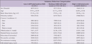
A total of 43,558 participants from the CLSA were included in our analysis (Table 1). Sixty percent of participants were middle aged (45-65 years), 48.8% were male, and more than 95% were White Caucasian. The average BMI was classified as overweight (BMI between 25 and 29.9), with 1/3 of participants reporting at least one chronic condition. The participants classified as having high sedentary behaviour (n= 16,555, 38.0%) were significantly older, heavier, reported more chronic conditions, did less exercise, and reported more fall-related injuries than those classified as low sedentary behaviour. At the baseline, 4813 (11%) participants reported a fall-related injury in the past 12 months, while only 2953 (6.8%) participants reported a fall-related injury at follow-up.

Table 1. Descriptive Characteristics of Participants (n=43,558)

Data presented as mean + SD or N (%); BMI = Body mass index; † = Only data from Tracking cohort (n=21,000).


At baseline, 22.3% of participants were classified as having a ‘low level’ of sedentary behaviour, with that number decreasing to 17.3% at follow-up (Figure 1). Inversely, 9.7% of participants classified as ‘high level’ of sedentary behaviour went from 38.0% to 47.7%.

Figure 1. Distribution of participants within sedentary behaviour groups (Low-Medium-High) n=43,558


Logistic regression investigated the association between injurious falls and sedentary behaviour at baseline. When the model was fully adjusted (age, sex, chronic conditions, moderate-to-vigorous physical activity, and resistance training), the odds of injurious falls increased by 1.10 (1.05 – 1.15) for each categorical increase in sedentary behaviour (Figure 2).

Figure 2. Association between injurious falls and sedentary behaviour at follow-up

Data are presented as RR (95% CI). Model 1 = Unadjusted; Model 2: Adjusted for age; Model 3: Adjusted for age and sex; Model 4: Adjusted for age, sex, chronic conditions, moderate to vigorous physical activity, and resistance training.


Findings from the logistic regression model investigating the association between injurious falls and an increase in sedentary behaviour between baseline and follow-up showed that the odds of injurious falls were not significant,1.00 (0.92-1.01). When the model was fully what? Adjusted? (age, sex, chronic conditions, moderate-to-vigorous physical activity, and resistance training) (Table 2). Additionally, when participants who reported injurious falls at baseline were removed, the odds of fall-related injuries were 1.05 (0.93-1.12) when adjusting for the same confounders.

Table 2. Association between change in sedentary behaviour and fall-related injuries

Model does not included people who reported a fall with injury at baseline


A subgroup analysis of middle-aged and older adults yielded similar results. Cross-sectionally, the logistic regression model showed a significant association between falls and sedentary behaviour for middle-aged 1.11 (1.05-1.17) and older adults 1.09 (1.02-1.17). However, a change in sedentary behaviour was not significantly associated with falls in any of he groups 1.02 (0.91-1.15) for middle-aged and 0.95 (0.84-1.09) for older adults.


Discussion

This study investigated the association between sedentary behaviour and fall-related injuries and whether sedentary behaviour changes impacted the risk of injurious falls. Our results revealed that sedentary behaviour was associated with fall-related injuries in the CLSA cohort of middle-aged and older adults independent of age, number of medical conditions, and physical activity. However, changes in sedentary behaviour between baseline and follow-up testing did not lead to a change in relative injury fall risk.
Considering the primary aim of this study, these results were consistent with those found in the systematic review by Semanik et al. (28), where sedentary behaviour was associated with increased falls. This study shows that this relationship exists independently of physical activity in both men and women. This could have significant implications for maintaining the health and well-being of middle-aged and older adults, as this study highlights the potential harms of sustained sedentary behaviour. High sedentary behaviour could also identify those most at risk of fall-related injuries in middle-aged and older adults.
Interestingly, our results are inconsistent with a similar study performed by Lustosa et al. (14), who found no association between sedentary time and fall-related injuries despite an association between sedentary time and self-reported falls. However, their study used a median split method, which only allowed for sedentary time to be classified as one of two (low or high) sedentary time categories. In contrast, the present study used a three-category model where participants were classified as having either low, moderate, or high levels of sedentary time. These findings reinforce the dose-response relationship between sedentary behaviour and health outcomes and highlight the necessity for a continuous measure of sedentary time rather than a categorical approach.
While the results of this paper confirmed the main hypothesis, exploring our secondary aim yielded surprising results, as categorical changes in sedentary behaviour (between baseline and follow-up) were not associated with a change in relative fall risk. This is incongruent with our primary finding that sedentary behaviour is associated with falls. In addition, our results challenge the work of Bea et al. (2017), who found that fall risk did increase with a reduction in sedentary behaviour in a female sample over six years (19). A longer period of sustained reduction in sedentary behaviour may be required to see the benefits. From this perspective, it is reasonable to conclude that short-term increases or decreases in sedentary behaviour are unlikely to harm or benefit this population’s risk of falls or fall-related injuries. It could be helpful to revisit this question with the CLSA data in the future to assess the impact of sedentary behaviour change over a longer duration.
This study has some limitations that must be noted. Firstly, in the PASE questionnaire, daily sedentary behaviour was capped at four hours. This creates an unbalanced weighting of participants in the ‘High’ sedentary condition and may have adversely affected the reporting of sedentary behaviour. The categories created to answer the research question are arbitrary and were directed by the tool and the exposure distribution. Future research would benefit from a more precise method of reporting sedentary behaviour (i.e., wearable device), particularly in aging adults, where it is more common (30). Secondly, the number of medical conditions and medications was reported and controlled. However, some medications will more substantially affect fall risk and fall-related injury than others. Without specific details of these medications, we cannot accurately determine their impact on the relationship between falls and sedentary behaviour. In addition, Fall-related injuries were collected through different questionnaires at baseline and follow-up. This may explain why fewer people recorded falling at follow-up.
Based on the results of this study, health practitioners should recommend limiting sedentary behaviour as young as possible to reduce the relative risk of falls and fall-related injuries. Future research in this area should focus on determining how much sedentary behaviour needs to be reduced and over what length to decrease the relative risk of injurious falls in middle—and older-aged adults.


Conclusion

In conclusion, higher levels of sedentary behaviour are associated with a higher risk of fall-related injury independent of confounders in people aged 45 to 85. Considering this, older middle and older adults or those at risk of falling may benefit from less sedentary time. However, we found that changes in sedentary behaviour were not associated with injurious falls. Despite the results, a more precise measure of sedentary behaviour is needed for epidemiology studies to capture changes over time better.


Acknowledgments: This research was made possible using the data/biospecimens collected by the Canadian Longitudinal Study on Aging (CLSA). Funding for the Canadian Longitudinal Study on Aging (CLSA) is provided by the Government of Canada through the Canadian Institutes of Health Research (CIHR) under grant reference: LSA 94473 and the Canada Foundation for Innovation. This research has been conducted using the CLSA dataset Baseline and Follow-Up 1 under Application Number 2002007. The CLSA is led by Drs. Parminder Raina, Christina Wolfson and Susan Kirkland. The opinions expressed in this manuscript are the author’s own and do not reflect the views of the Canadian Longitudinal Study on Aging.

Conflict of Interest Statement: All authors declare that they have no conflicts of interest.

Ethical standards: All subjects gave informed consent for inclusion before participating in the study. The study was conducted following the Declaration of Helsinki, and the protocol was approved by the Institution Ethics Committee of UNB 2023-019.

Open Access: This article is distributed under the terms of the Creative Commons Attribution 4.0 International License (http://creativecommons.org/licenses/by/4.0/), which permits use, duplication, adaptation, distribution and reproduction in any medium or format, as long as you give appropriate credit to the original author(s) and the source, provide a link to the Creative Commons license and indicate if changes were made.


References

1. Worl Health Organization. Falls. Published April 26, 2021. Accessed April 5, 2024. https://www.who.int/news-room/fact-sheets/detail/falls
2. Peel NM. Epidemiology of Falls in Older Age. Canadian Journal on Aging / La Revue canadienne du vieillissement. 2011;30(1):7-19. doi:10.1017/S071498081000070X
3. Davis JC, Robertson MC, Ashe MC, Liu-Ambrose T, Khan KM, Marra CA. International comparison of cost of falls in older adults living in the community: a systematic review. Osteoporosis International. 2010;21(8):1295-1306. doi:10.1007/s00198-009-1162-0
4. Vaishya R, Vaish A. Falls in Older Adults are Serious. Indian Journal of Orthopaedics. 2020;54(1):69-74. doi:10.1007/s43465-019-00037-x
5. Public Health Agency of Canada. Surveillance Report on Falls among Older Adults in Canada. Public Health Agency of Canada; 2022. https://www.canada.ca/content/dam/hc-sc/documents/reserch/surveillance/senior-falls-in-Canada-en.pdf
6. Peeters G, van Schoor NM, Cooper R, Tooth L, Kenny RA. Should prevention of falls start earlier? Co-ordinated analyses of harmonised data on falls in middle-aged adults across four population-based cohort studies. PLOS ONE. 2018;13(8):e0201989. doi:10.1371/journal.pone.0201989
7. Talbot LA, Musiol RJ, Witham EK, Metter EJ. Falls in young, middle-aged and older community dwelling adults: perceived cause, environmental factors and injury. BMC Public Health. 2005;5(1):86. doi:10.1186/1471-2458-5-86
8. World Health Organization. Global Status Report on Physical Activity 2022. World Health Organization; 2022. Accessed June 3, 2024. https://www.who.int/teams/health-promotion/physical-activity/global-status-report-on-physical-activity-2022
9. Tremblay MS, Aubert S, Barnes JD, et al. Sedentary Behavior Research Network (SBRN) – Terminology Consensus Project process and outcome. International Journal of Behavioral Nutrition and Physical Activity. 2017;14(1):75. doi:10.1186/s12966-017-0525-8
10. Dunstan DW, Howard B, Healy GN, Owen N. Too much sitting – A health hazard. Diabetes Research and Clinical Practice. 2012;97(3):368-376. doi:10.1016/j.diabres.2012.05.020
11. Patterson R, McNamara E, Tainio M, et al. Sedentary behaviour and risk of all-cause, cardiovascular and cancer mortality, and incident type 2 diabetes: a systematic review and dose response meta-analysis. Eur J Epidemiol. 2018;33(9):811-829. doi:10.1007/s10654-018-0380-1
12. Semanik PA, Lee J, Song J, et al. Accelerometer-Monitored Sedentary Behavior and Observed Physical Function Loss. Am J Public Health. 2015;105(3):560-566. doi:10.2105/AJPH.2014.302270
13. Blodgett J, Theou O, Kirkland S, Andreou P, Rockwood K. The association between sedentary behaviour, moderate–vigorous physical activity and frailty in NHANES cohorts. Maturitas. 2015;80(2):187-191. doi:10.1016/j.maturitas.2014.11.010
14. Zhao R, Bu W, Chen Y, Chen X. The Dose-Response Associations of Sedentary Time with Chronic Diseases and the Risk for All-Cause Mortality Affected by Different Health Status: A Systematic Review and Meta-Analysis. The Journal of nutrition, health and aging. 2020;24(1):63-70. doi:10.1007/s12603-019-1298-3
15. O’Flaherty EJ. Modeling Normal Aging Bone Loss, with Consideration of Bone Loss in Osteoporosis. Toxicological Sciences. 2000;55(1):171-188. doi:10.1093/toxsci/55.1.171
16. Mitchell WK, Williams J, Atherton P, Larvin M, Lund J, Narici M. Sarcopenia, dynapenia, and the impact of advancing age on human skeletal muscle size and strength; a quantitative review. Front Physiol. 2012;3:260. doi:10.3389/fphys.2012.00260
17. Jiang Y, Wang M, Liu S, Ya X, Duan G, Wang Z. The association between sedentary behavior and falls in older adults: A systematic review and meta-analysis. Frontiers in Public Health. 2022;10. doi:10.3389/fpubh.2022.1019551
18. Lustosa LG, Rudoler D, Theou O, Dogra S. Leisure Sedentary Time is Associated with Self-Reported Falls in Middle-aged and Older Females and Males: an Analysis of the CLSA. Canadian Geriatrics Journal. 2023;26(2):239-246. doi:10.5770/cgj.26.636
19. Bea JW, Thomson CA, Wallace RB, et al. Changes in physical activity, sedentary time, and risk of falling: The Women’s Health Initiative Observational Study. Preventive Medicine. 2017;95:103-109. doi:10.1016/j.ypmed.2016.11.025
20. Aunger J, Wagnild J. Objective and subjective measurement of sedentary behavior in human adults: A toolkit. American Journal of Human Biology. 2022;34(1):e23546. doi:10.1002/ajhb.23546
21. Lu Z, Lam FMH, Leung JCS, Kwok TCY. The U-Shaped Relationship Between Levels of Bouted Activity and Fall Incidence in Community-Dwelling Older Adults: A Prospective Cohort Study. The Journals of Gerontology: Series A. 2020;75(10):e145-e151. doi:10.1093/gerona/glaa058
22. Raina PS, Wolfson C, Kirkland SA, et al. The Canadian Longitudinal Study on Aging (CLSA). Canadian Journal on Aging / La Revue canadienne du vieillissement. 2009;28(3):221-229. doi:10.1017/S0714980809990055
23. Canadian Longitudinal Study on Aging. Data Collection. CLSA Canadian Longitudinal Study on Aging. Published n.d. Accessed April 5, 2024. https://www.clsa-elcv.ca/data-collection
24. Immonen M, Haapea M, Similä H, et al. Association between chronic diseases and falls among a sample of older people in Finland. BMC Geriatrics. 2020;20(1):225. doi:10.1186/s12877-020-01621-9
25. Yang X, Li L, Xie F, Wang Z. A prospective cohort study of the impact of chronic disease on fall injuries in middle-aged and older adults. 2023;18(1). doi:10.1515/med-2023-0748
26. Paliwal Y, Slattum PW, Ratliff SM. Chronic Health Conditions as a Risk Factor for Falls among the Community-Dwelling US Older Adults: A Zero-Inflated Regression Modeling Approach. Sosnoff JJ, ed. BioMed Research International. 2017;2017:5146378. doi:10.1155/2017/5146378
27. Appeadu MK, Bordoni B. Falls and Fall Prevention in Older Adults. StatPearls Publishing; 2023. https://pubmed.ncbi.nlm.nih.gov/32809596/
28. Semanik PA, Lee J, Song J, et al. Accelerometer-Monitored Sedentary Behavior and Observed Physical Function Loss. Am J Public Health. 2015;105(3):560-566. doi:10.2105/AJPH.2014.302270
29. Lustosa LG, Rudoler D, Theou O, Dogra S. Leisure Sedentary Time is Associated with Self-Reported Falls in Middle-aged and Older Females and Males: an Analysis of the CLSA. Canadian Geriatrics Journal. 2023;26(2):239-246. doi:10.5770/cgj.26.636
30. Copeland JL, Clarke J, Dogra S. Objectively measured and self-reported sedentary time in older Canadians. Preventive Medicine Reports. 2015;2:90-95. doi:10.1016/j.pmedr.2015.01.003

© The Authors 2024

EFFECT OF MODIFIABLE LIFESTYLE FACTORS ON BIOLOGICAL AGING


W.-H. Lu1,2


1. IHU HealthAge, Toulouse, France; 2. Institute on Aging, Toulouse University Hospital (CHU Toulouse), Toulouse, France.

Corresponding Author: Wan-Hsuan Lu, Postal address: Gérontopôle de Toulouse, Institut du Vieillissement, 37 Allée Jules Guesde, 31000 Toulouse, France, E-mail address: wanhsuanlu@gmail.com, Phone number: + 33- 561-145-691

J Aging Res & Lifestyle 2024;13:88-92
Published online June 5, 2024, http://dx.doi.org/10.14283/jarlife.2024.13


Abstract

Biological age is a concept that uses bio-physiological parameters to account for individual heterogeneity in the biological processes driving aging and aims to enhance the prediction of age-related clinical conditions compared to chronological age. Although engaging in healthy lifestyle behaviors has been linked to a lower mortality risk and a reduced incidence of chronic diseases, it remains unclear to what extent these health benefits result from slowing the pace of the biological aging process. This short review summarized how modifiable lifestyle factors — including diet, physical activity, smoking, alcohol consumption, and the aggregate of multiple healthy behaviors — were associated with established estimates of biological age based on clinical or cellular/molecular markers, including Klemera-Doubal Method biological age, homeostatic dysregulation, phenotypic age, DNA methylation age, and telomere length. In brief, the available studies tend to show a consistent association of lifestyle factors with physiological measures of biological age, while findings regarding molecular-based metrics vary. The limited evidence highlights the need for further research in this field, particularly with a life-course approach.

Key words: Healthy aging, healthspan, biomarker of aging, epigenetic age, age acceleration.


Aging is the time-related deterioration that occurs in an organism at all levels, from the molecular and cellular to the physiological and functional, ultimately increasing vulnerability to death (1). For decades, scientists and clinicians have observed that chronological age, representing the time since birth, is a significant predictor of various age-related health conditions; however, it may not accurately describe how an organism functions, especially in the later life stages (2, 3). Biological age seeks to quantify the bio-physiological processes driving aging. Generally, biomarkers or clinical metrics designed to forecast the remaining lifespan and healthspan (the absence of disability) are considered indicators of biological age (4, 5). The disparity between predicted biological and chronological age is defined as age acceleration, and the positive age acceleration implies that individuals may undergo age-related decline faster than their peers (6, 7).
Engaging in healthy lifestyle behaviors has been linked to a lower mortality risk (8) and decreased incidence of a myriad of medical conditions, including cardiovascular diseases (9), metabolic syndrome (10), cancer (11), neurodegenerative and psychiatric disorders (12), and geriatric syndromes (13, 14). Although the mechanisms connecting lifestyle factors to extended lifespan/ healthspan are not fully understood, it is plausible that the health benefits result, at least partly, from slowing down the biological aging process. This short review explored how modifiable lifestyle factors, such as diet, physical activity, smoking, and alcohol consumption, are associated with biological aging.


Biological age estimation

Articles published in English were searched from the Pubmed database for this review. Several papers investigating the relationship between modifiable lifestyle factors and biological age using various measures from physiological to molecular scales were identified. Only research that quantified biological age using validated algorithms, epigenetic clocks, or telomere length were included (see Table 1).

Table 1. Biological age estimation methods used in prior studies on modifiable lifestyle factors


Diet and biological aging

An unhealthy diet may accelerate biological aging due to its inflammatory and oxidative stress potentials. The cross-sectional study conducted by Wang and colleagues, which involved 8,839 participants from the National Health and Nutrition Examination Survey (NHANES) of the United States, showed a consistent association of consuming foods with higher Dietary Inflammatory Index (DII) and Dietary Oxidative Balance Score (DOBS) with accelerated biological aging. In this work, biological age was assessed through clinical biomarkers using established algorithms, including Klemera-Doubal Method biological age (KDM-BA), homeostatic dysregulation (HD), and phenotypic age (PA) (25). Another study of 10,191 Taiwanese aged ≥50 revealed that adopting a diet rich in plant foods was associated with a reduced likelihood of experiencing an acceleration in the multidimensional aging measure (MDAge) over 8 years, composed of selected clinical chemistry biomarkers (26). Kresovich et al.’s cross-sectional study demonstrated the beneficial impact of healthy eating approaches, including the diet designed for hypertension management and the Mediterranean diet (MED), on DNA methylation age (DNAmAge) acceleration among non-Hispanic white women (the Sister study); the most significant associations were observed in acceleration in PhenoAge and GrimAge (27). Conversely, an 18-month randomized controlled trial (RCT) in 294 adults with obesity or dyslipidemia observed no significant differences in the change of epigenetic ages between three dietary interventions, which included providing guidelines to promote a healthy diet and implementing a calorie-restricted MED and a plant-rich MED, respectively (28). In summary, the observational studies suggest that a healthy diet may decelerate biological aging, while further evidence is required to determine whether different dietary strategies are superior.


Physical activity and biological aging

Several non-interventional studies had reported that higher physical activity levels or lower sedentary time were associated with slower epigenetic aging (29–31). However, the association may be partially attributed to body mass index (BMI) and other confounders, with the associations tending to attenuate or disappear after adjusting for those confounders (29, 30). Further insights from Fox and colleagues revealed that cardiovascular health and immune function mediated the effect of physical activity on DNAm GrimAge acceleration (31). Physical activity also showed a favorable impact on telomere attrition. In their study recruiting 284,479 participants from the UK Biobank, Zhu et al. discovered that physical activities during leisure time, housework, and public transportation were associated with reduced leukocyte telomere length (LTL) deviation, which referred to the difference between genetically determined and observed LTL. Notably, engaging in job-related activities was linked to a greater LTL deviation (32). In short, engaging in physical activities outside of work could slow down the rate of biological age acceleration, as measured by cellular markers.


Smoking, alcohol consumption, and biological aging

As calculated by KDM-BA and PA, individuals who smoked and drank alcohol had a greater age acceleration than those who reported as non-smokers/non-drinkers, with evidence from 94,433 adults aged 30 to 70 in Taiwan (33). This finding is supported by a study investigating epigenetic age among 2,316 women from the Sister study, which indicated that both lifetime and recent alcohol consumption were associated with DNAm GrimAge acceleration (34). Furthermore, smoking and alcohol consumption were cross-sectionally associated with acceleration in several DNAmAge clocks in the GENOA study composed of 1,100 African Americans; however, only current smokers showed a significant association with increased PhenoAge acceleration over time (35). To summarize, tobacco and alcohol consumption have been correlated with accelerated biological aging, as demonstrated by cross-sectional studies, but longitudinal evidence supporting these associations remains insufficient.


Multiple lifestyle factors and biological aging

The effect of engaging in multiple healthy behaviors on deceleration in biological aging had also been evaluated, including nonsmoking, less alcohol consumption, daily fruit and vegetable intake, being physically active or regular exercise, good sleep habits, and maintaining normal BMI and waist-to-hip ratio (36–38). Overall, adherence to more health-promoting factors was associated with slower biological aging, either assessed via the phenotypic measure (frailty index) (36) or clinical biomarkers (KDM-BA and PA) (37, 38).
Despite limited sample sizes, data from RCTs suggested that lifestyle interventions may modify biological age. The pilot trial of Fitzgerald et al. performed an 8-week treatment program about diet, dietary supplements, sleep, exercise, and stress management for 43 men aged 50 to 72 without chronic diseases. Compared to the controls, the lifestyle intervention was associated with a decrease in Horvath DNAmAge of 3.23 years. Moreover, in the intervention group, Horvath DNAmAge decreased by an average of 1.96 years by the end of the program (not reaching statistical significance) (39). In a secondary analysis of a 24-month RCT that enrolled 219 healthy post-menopausal women, participants who received the healthy-dietary intervention had a lower GrimAge acceleration than their no-intervention counterparts. On the other hand, the physical activity intervention reduced the epigenetic mutation load (40), which reflects the age-related dysfunction of the epigenetic maintenance system (41). Finally, a secondary analysis of an RCT involving 93 obese older adults observed that a 12-month calorie-restricted diet, whether combined with exercise or not, was associated with decreased biological age as per three different algorithms. In contrast, the exercise intervention alone did not significantly alter biological age over time and showed no difference from controls (42). To sum up, multiple healthy behaviors may collectively slow biological aging.


Perspectives on the way forward

Individuals who engage in a healthy lifestyle may exhibit a slower pace of biological aging, as their DNA methylation profile and physiological biomarkers are in a healthier state that typically indicates lower risks of mortality and age-related diseases (Figure 1). However, most studies linking lifestyle factors and biological aging are cross-sectional designs, making it difficult to establish causation. Furthermore, it is worth noting that previous research investigating lifestyle factors and biological aging was commonly obtained from specific US cohorts, such as NHANES and the Sister study, probably due to the difficulty of having both biological age measures and comprehensive lifestyle data in other large cohorts. More evidence derived from diverse populations needs to be included. The impact of lifestyle factors on biological aging warrants investigation using a life-course approach. It is possible that alterations in biological mechanisms become evident only if these behaviors start at a younger age or are consistently adopted in the long term. Due to the difficulty of following individuals throughout their lifespans, initiatives such as the INSPIRE project (43) are crucial for contributing to this topic, as they enable the following of a large age range over a relatively long period. Finally, larger-sample RCTs are needed to validate observed effects.

Figure 1. Lifestyle factors showed an association with the deceleration of biological age suggested in prior studies

Note: This figure was created with BioRender.com.


Several measurement issues of biological age also remain in the field. For example, even if the same biological age algorithm is used, employing different biomarker selection strategies may result in the diverse compositions of the biomarkers and, thus, different performance of estimated biological age (44). Similarly, the lack of standardization in biomarker formulations and study design/performing procedures can lead to heterogeneous results when examining aging biomarkers across cohorts (45). Measuring and parameterizing biological age will continue to pose challenges in future observational studies or RCTs on lifestyle factors. Notably, recently proposed guidelines for validating biomarkers of aging offer a solution to harmonize future cross-population studies, which provide several recommendations for investigating omics-based aging biomarkers at different stages, from data maintenance and biomarker development to external validation (45). Lastly, using digital markers collected by wearable sensors for measuring biological age is a promising field that requires further exploration. A previous study showed that biological age acceleration estimated from step count data could distinguish morbidity and smoking status as effectively as blood-based markers (46). However, it remains to be investigated how this digital biomarker-based measure of biological age can help reveal the impact of modifiable lifestyle factors.
Some interventions demonstrated symptom-relieved effects without significantly altering the underlying pathology (47); the same question could be posed regarding the biological influence of lifestyle behaviors discussed in this article. The available evidence tends to show a consistent association of lifestyle factors with physiological measures of biological age, while findings regarding molecular-based metrics (especially epigenetic clocks) vary. This suggests that lifestyle factors have a greater impact on physiological health, reflecting the overall accumulation of cellular and molecular damage, rather than targeting a specific aging mechanism. Future research comparing multiple biological aging measures derived from different levels of organization within the body (physiological, cellular, and molecular) can provide insight into the topic. In addition, given that health-promoting factors have been shown to modify the association between disease pathologies and phenotypic outcomes — such as the role of physical activity in neurodegenerative diseases (48) — it is important to investigate whether these modifying effects result from the decelerated biological aging and the enhanced biological resilience.


Conflict of interest: The author has no conflicts of interest.

Open Access: This article is distributed under the terms of the Creative Commons Attribution 4.0 International License (http://creativecommons.org/licenses/by/4.0/), which permits use, duplication, adaptation, distribution and reproduction in any medium or format, as long as you give appropriate credit to the original author(s) and the source, provide a link to the Creative Commons license and indicate if changes were made.


References

1. Ferrucci L, Levine ME, Kuo PL, Simonsick EM. Time and the metrics of aging. Circ Res. 2018;123(7):740-744. doi:10.1161/CIRCRESAHA.118.312816
2. Baker GT, Sprott RL. Biomarkers of aging. Exp Gerontol. 1988;23(4-5):223-239. doi:10.1016/0531-5565(88)90025-3
3. Simm A, Nass N, Bartling B, Hofmann B, Silber RE, Navarrete Santos A. Potential biomarkers of ageing. Biol Chem. 2008;389(3):257-265. doi:10.1515/BC.2008.034
4. Jylhävä J, Pedersen NL, Hägg S. Biological Age Predictors. EBioMedicine. 2017;21:29-36. doi:10.1016/j.ebiom.2017.03.046
5. Lohman T, Bains G, Berk L, Lohman E. Predictors of Biological Age: The Implications for Wellness and Aging Research. Gerontol Geriatr Med. 2021;7. doi:10.1177/23337214211046419
6. Zhang Q. An interpretable biological age. Lancet Heal Longev. 2023;4(12):e662-e663. doi:10.1016/S2666-7568(23)00213-1
7. Elliott ML, Caspi A, Houts RM, et al. Disparities in the pace of biological aging among midlife adults of the same chronological age have implications for future frailty risk and policy. Nat Aging. 2021;1(3):295-308. doi:10.1038/s43587-021-00044-4
8. Loef M, Walach H. The combined effects of healthy lifestyle behaviors on all cause mortality: A systematic review and meta-analysis. Prev Med (Baltim). 2012;55(3):163-170. doi:10.1016/j.ypmed.2012.06.017
9. Barbaresko J, Rienks J, Nöthlings U. Lifestyle Indices and Cardiovascular Disease Risk: A Meta-analysis. Am J Prev Med. 2018;55(4):555-564. doi:10.1016/j.amepre.2018.04.046
10. Garralda-Del-Villar M, Carlos-Chillerón S, Diaz-Gutierrez J, et al. Healthy lifestyle and incidence of metabolic syndrome in the SUN cohort. Nutrients. 2019;11(1):65. doi:10.3390/nu11010065
11. Zhang YB, Pan XF, Chen J, et al. Combined lifestyle factors, incident cancer, and cancer mortality: a systematic review and meta-analysis of prospective cohort studies. Br J Cancer. 2020;122(7):1085-1093. doi:10.1038/s41416-020-0741-x
12. Kip E, Parr-Brownlie LC. Healthy lifestyles and wellbeing reduce neuroinflammation and prevent neurodegenerative and psychiatric disorders. Front Neurosci. 2023;17:1092537. doi:10.3389/fnins.2023.1092537
13. Abe T, Nofuji Y, Seino S, et al. Healthy lifestyle behaviors and transitions in frailty status among independent community-dwelling older adults: The Yabu cohort study. Maturitas. 2020;136:54-59. doi:10.1016/j.maturitas.2020.04.007
14. Bruyère O, Reginster JY, Beaudart C. Lifestyle approaches to prevent and retard sarcopenia: A narrative review. Maturitas. 2022;161:44-48. doi:10.1016/j.maturitas.2022.02.004
15. Klemera P, Doubal S. A new approach to the concept and computation of biological age. Mech Ageing Dev. 2006;127(3):240-248. doi:10.1016/j.mad.2005.10.004
16. Graf GH, Crowe CL, Kothari M, et al. Testing Black-White Disparities in Biological Aging Among Older Adults in the United States: Analysis of DNA-Methylation and Blood-Chemistry Methods. Am J Epidemiol. 2022;191(4):613-625. doi:10.1093/aje/kwab281
17. Cohen AA, Milot E, Yong J, et al. A novel statistical approach shows evidence for multi-system physiological dysregulation during aging. Mech Ageing Dev. 2013;134(3-4):110-117. doi:10.1016/j.mad.2013.01.004
18. Levine ME, Lu AT, Quach A, et al. An epigenetic biomarker of aging for lifespan and healthspan. Aging (Albany NY). 2018;10(4):573-591. doi:10.18632/aging.101414
19. Wang S, Wen CP, Li W, et al. Development of a Novel Multidimensional Measure of Aging to Predict Mortality and Morbidity in the Prospective MJ Cohort. J Gerontol A Biol Sci Med Sci. 2023;78(4):690-697. doi:10.1093/gerona/glac161
20. Horvath S, Raj K. DNA methylation-based biomarkers and the epigenetic clock theory of ageing. Nat Rev Genet. 2018;19(6):371-384. doi:10.1038/s41576-018-0004-3
21. Ye Q, Apsley AT, Etzel L, et al. Telomere length and chronological age across the human lifespan: A systematic review and meta-analysis of 414 study samples including 743,019 individuals. Ageing Res Rev. 2023;90(July):102031. doi:10.1016/j.arr.2023.102031
22. Blasco MA. Telomeres and human disease: Ageing, cancer and beyond. Nat Rev Genet. 2005;6(8):611-622. doi:10.1038/nrg1656
23. Rockwood K, Song X, MacKnight C, et al. A global clinical measure of fitness and frailty in elderly people. CMAJ. 2005;173(5):489-495. doi:10.1503/cmaj.050051
24. Clegg A, Young J, Iliffe S, Rikkert MO, Rockwood K. Frailty in elderly people. Lancet. 2013;381(9868):752-762. doi:10.1016/S0140-6736(12)62167-9
25. Wang X, Sarker S kumar, Cheng L, et al. Association of dietary inflammatory potential, dietary oxidative balance score and biological aging. Clin Nutr. 2024;43(1):1-10. doi:10.1016/j.clnu.2023.11.007
26. Wang S, Li W, Li S, et al. Association between plant-based dietary pattern and biological aging trajectory in a large prospective cohort. BMC Med. 2023;21(1):310. doi:10.1186/s12916-023-02974-9
27. Kresovich JK, Park YMM, Keller JA, Sandler DP, Taylor JA. Healthy eating patterns and epigenetic measures of biological age. Am J Clin Nutr. 2022;115(1):171-179. doi:10.1093/ajcn/nqab307
28. Yaskolka Meir A, Keller M, Hoffmann A, et al. The effect of polyphenols on DNA methylation-assessed biological age attenuation: the DIRECT PLUS randomized controlled trial. BMC Med. 2023;21(1). doi:10.1186/s12916-023-03067-3
29. Kresovich JK, Garval EL, Martinez Lopez AM, et al. Associations of Body Composition and Physical Activity Level with Multiple Measures of Epigenetic Age Acceleration. Am J Epidemiol. 2021;190(6):984-993. doi:10.1093/aje/kwaa251
30. Spartano NL, Wang R, Yang Q, et al. Association of Accelerometer-Measured Physical Activity and Sedentary Time with Epigenetic Markers of Aging. Med Sci Sports Exerc. 2023;55(2):264-272. doi:10.1249/MSS.0000000000003041
31. Fox FAU, Liu D, Breteler MMB, Aziz NA. Physical activity is associated with slower epigenetic ageing—Findings from the Rhineland study. Aging Cell. 2023;22(6). doi:10.1111/acel.13828
32. Zhu J, Yang Y, Zeng Y, et al. The Association of Physical Activity Behaviors and Patterns With Aging Acceleration: Evidence From the UK Biobank. Journals Gerontol – Ser A Biol Sci Med Sci. 2023;78(5):753-761. doi:10.1093/gerona/glad064
33. Lin WY. Lifestyle Factors and Genetic Variants on 2 Biological Age Measures: Evidence from 94 443 Taiwan Biobank Participants. Journals Gerontol – Ser A Biol Sci Med Sci. 2022;77(6):1189-1198. doi:10.1093/gerona/glab251
34. Kresovich JK, Martinez Lopez AM, Garval EL, et al. Alcohol Consumption and Methylation-Based Measures of Biological Age. Journals Gerontol – Ser A Biol Sci Med Sci. 2021;76(12):2107-2111. doi:10.1093/gerona/glab149
35. Zhao W, Ammous F, Ratliff S, et al. Education and lifestyle factors are associated with dna methylation clocks in older African Americans. Int J Environ Res Public Health. 2019;16(17):3141. doi:10.3390/ijerph16173141
36. Fan J, Yu C, Pang Y, et al. Adherence to Healthy Lifestyle and Attenuation of Biological Aging in Middle-Aged and Older Chinese Adults. Journals Gerontol – Ser A Biol Sci Med Sci. 2021;76(12):2232-2241. doi:10.1093/gerona/glab213
37. Ng TP, Zhong X, Gao Q, Gwee X, Chua DQL, Larbi A. Socio-Environmental, Lifestyle, Behavioural, and Psychological Determinants of Biological Ageing: The Singapore Longitudinal Ageing Study. Gerontology. 2020;66(6):603-613. doi:10.1159/000511211
38. Zhang R, Wu M, Zhang W, et al. Association between life’s essential 8 and biological ageing among US adults. J Transl Med. 2023;21(1). doi:10.1186/s12967-023-04495-8
39. Fitzgerald KN, Hodges R, Hanes D, et al. Potential reversal of epigenetic age using a diet and lifestyle intervention: a pilot randomized clinical trial. Aging (Albany NY). 2021;13(7):9419-9432. doi:10.18632/aging.202913
40. Fiorito G, Caini S, Palli D, et al. DNA methylation-based biomarkers of aging were slowed down in a two-year diet and physical activity intervention trial: the DAMA study. Aging Cell. 2021;20(10). doi:10.1111/acel.13439
41. Yan Q, Paul KC, Lu AT, et al. Epigenetic mutation load is weakly correlated with epigenetic age acceleration. Aging (Albany NY). 2020;12(18):17863. doi:10.18632/AGING.103950
42. Ho E, Qualls C, Villareal DT. Effect of Diet, Exercise, or Both on Biological Age and Healthy Aging in Older Adults with Obesity: Secondary Analysis of a Randomized Controlled Trial. J Nutr Heal Aging. 2022;26(6):552-557. doi:10.1007/s12603-022-1812-x
43. de Souto Barreto P, GUYONNET S, Ader I, et al. The INSPIRE research initiative: a program for GeroScience and healthy aging research going from animal models to humans and the healthcare system. J Frailty Aging. 2021;10(2):86-93. doi:10.14283/jfa.2020.18
44. Wei K, Peng S, Liu N, et al. All-Subset Analysis Improves the Predictive Accuracy of Biological Age for All-Cause Mortality in Chinese and U.S. Populations. Journals Gerontol – Ser A Biol Sci Med Sci. 2022;77(11):2288-2297. doi:10.1093/gerona/glac081
45. Moqri M, Herzog C, Poganik JR, et al. Validation of biomarkers of aging. Nat Med. 2024;30(2):360-372. doi:10.1038/s41591-023-02784-9
46. Pyrkov T V., Sokolov IS, Fedichev PO. Deep longitudinal phenotyping of wearable sensor data reveals independent markers of longevity, stress, and resilience. Aging (Albany NY). 2021;13(6):7900-7913. doi:10.18632/aging.202816
47. Coley N, Zetterberg H, Cantet C, et al. Plasma p-tau181 as an outcome and predictor of multidomain intervention effects: a secondary analysis of a randomised, controlled, dementia prevention trial. Lancet Heal Longev. 2024;5(2):e120-e130. doi:10.1016/S2666-7568(23)00255-6
48. Raffin J, Rolland Y, Aggarwal G, et al. Associations Between Physical Activity, Blood-Based Biomarkers of Neurodegeneration, and Cognition in Healthy Older Adults: The MAPT Study. Journals Gerontol Ser A. Published online April 17, 2021. doi:10.1093/gerona/glab094

© The Authors 2024


METABOLIC SYNDROME AND POSITIVE FRAILTY SCREENING: A CROSS-SECTIONAL STUDY WITH COMMUNITY-DWELLING OLDER ADULTS


M.C.B. de Souza1, G. da Silva Rocha2, E. de Souza Sampaio3, P.C. de Oliveira Garcia Rodrigues4, R.A. Vieira5, A.F. Souza Gomes6, T.R. Pereira de Brito7


1. Institute of Motricity Sciences, Federal University of Alfenas, Alfenas, Brazil; 2. Health and Sport Sciences Center, Federal University of Acre, Rio Branco, Brazil; 3. Faculty of Nutrition, Federal University of Alfenas, Alfenas, Brazil; 4. Faculty of Nutrition, Federal University of Alfenas, Alfenas, Brazil; 5. Nursing school, Federal University of Alfenas, Alfenas, Brazil; 6. Postgraduate Program in Health and Nutrition, Nutrition School, Federal University of Ouro Preto; Ouro Preto, MG, Brazil 7. Faculty of Nutrition, Federal University of Alfenas, Alfenas, Brazil

Corresponding Author: Tábatta Renata Pereira de Brito, PhD, Faculty of Nutrition, Federal University of Alfenas, Alfenas, Brazil, Rua Gabriel Monteiro da Silva, 700, Centro, Alfenas, Minas Gerais, CEP: 37130-000, Faculty of Nutrition. Telephone Number: +55 35 3701 9742. E-mail address: tabatta.brito@unifal-mg.edu.br, ORCID: https://orcid.org/0000-0001-9466-2993

J Aging Res & Lifestyle 2024;13:82-87
Published online May 27, 2024, http://dx.doi.org/10.14283/jarlife.2024.12


Abstract

BACKGROUND: Metabolic Syndrome is a set of disorders that characterized by the association of three or more risk factors, like the obesity central, dyslipidemia, borderline blood pressure, hyperglycemia, and the increase of triglycerides. However, these factors also can be associated with pathophysiology of frailty.
OBJECTIVES: verifying whether the metabolic syndrome is associated to the positive frailty screening in the older people.
DESIGN: Cross-sectional study. Participants: 443 older people living in Rio Branco, Brazil.
SETTING: Data collection was carried out in two stages: a personal interview and blood collection.
MEASUREMENTS: The diagnosis of metabolic syndrome was based on the criteria of the Third Report of the National Cholesterol Education Program Expert Panel on Detection, Evaluation and Treatment of High Blood Cholesterol in Adults. The frailty screening was performed using subjective questions validated in a previous study. Descriptive statistics and multinomial logistic regression were used for data analyses.
RESULTS: There was a predominance of female older people (69.07%), aged between 60 and 79 years (87.13%), with an income greater than or equal to one minimum wage (72.09%), no cognitive decline (75.94%) and depressive symptoms (63.31%), independent for BADL (86.46%) and dependent for IADL (51.69%). From the total sample, 56.88% of the older people were identified as frail, 34.09% pre-frail and 9.03% non frail. The prevalence of metabolic syndrome was 51.69%. After adjusting by the independent variables, an association between metabolic syndrome and pre-frailty was observed, and older people with metabolic syndrome were more likely to be prefrail (RRR=2.36; 95%CI=1.08-5.18).
CONCLUSION: The metabolic syndrome was associated to the increase chance of screening for prefrailty in the older people evaluated, which reinforces the needy to establish preventive measures in relation to the metabolic syndrome to avoid frailty in the older people.

Key words: Cross-sectional studies, frail older people, frailty, metabolic syndrome, older adults.


Introduction

According to the World Health Organization, the combination between common aspects of modernity, such as globalization, urbanization, and changes in the lifestyle, makes chronic non-communicable diseases one of the main causes of mortality (1). In this context, metabolic syndrome stands out, as it is a health condition strongly associated with behavioral issues, increasing the incidence of cardiovascular diseases and mortality (2).
Metabolic Syndrome (MetS) is a set of disorders that affect the cardiovascular system, characterized, according to the National Cholesterol Education Program’s Adult Treatment Panel III (NCEP-ATP III), by the association of three or more risk factors, like the obesity central, dyslipidemia, borderline blood pressure, hyperglycemia, and the increase of triglycerides (3, 4). Once the circulatory system is affected in the metabolic syndrome, some chronic diseases are commonly associated, such as atherosclerosis and coronary artery disease (5).
The prevalence of metabolic syndrome varies according to ethnic differences, sex and the criteria used to define the syndrome (4). For example, the World Health Organization (WHO) criteria consider the presence of type 2 Diabetes to be necessary, so when these criteria are used, the prevalence tends to be lower compared to the criteria of the International Diabetes Federation (IDF) or the NCEP-ATPIII (6).
However, the associated factors with MetsS are not limited only to cardiometabolic complications, and frailty may occur, for example, since characteristics of the metabolic syndrome, such as obesity and insulin resistance, may be present in the pathophysiology of frailty (7). Frailty is a geriatric syndrome with multiple causes, characterized by the decrease of strength, of the resistance and the physiological function, leading to increased vulnerability, functional loss, institutionalization, falls, and high risk for mortality (8, 9).
Due to the different definitions of frailty proposed, there are several ways to identify the syndrome, and the identification of phenotypes that require objective measures, such as grip strength, making this difficult to apply in clinical practice, especially in developing countries, where human resources may be scarce. Thus, alternative frailty assessment strategies have been elaborated to be applied in the clinical context, basing mainly on self-reported measures. These alternatives aim to capture the central aspects of frailty, maintaining predictive validity for adverse results, and may be useful for frailty screening (10, 11).
Despite the high prevalence of MetS and frailty, and the vast literature about these conditions, studies about the association between the two syndromes, especially among older people, are still low. Studies utilizing self-reported measures for frailty screening can be valuable for initiating early preventive actions targeting metabolic syndrome-associated frailty. Especially in developing countries, human resources for conducting assessments of older individuals, including physical measurements, may be scarce. Therefore, this study aims to investigate the association between frailty, measured through self-reported screening instruments, and metabolic syndrome in older individuals.


Materials and Methods

Study design and participants

This is a cross-sectional study, carried out in the municipality from Rio Branco, Acre, Brazil. Rio Branco is the capital of Acre, situated in the northern region of Brazil. It stands as the most populous and developed city in the state. We followed the Strengthening the Reporting of Observational Studies in Epidemiology (STROBE) reporting guidelines for cross-sectional studies (12).
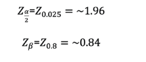
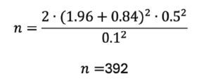
The calculation of the sample size was obtained considering the estimation of proportions in the order of 0.50, a confidence interval of 95%, a design effect (deff) of 1.17, and a population of 24.043 older people, resulting in a sample of 443 older people. The design effect is determined as the ratio between the variance of an estimate obtained through a specific sampling strategy and the variance of the identical estimate derived from a simple random sample comprising the same number of observational units (13). The deff of 1.17 was adopted based on previous study (14) (Figure 1).

Figure 1. Sample definition


The random selection of the sample was carried out from the records of the older people in the G-MUS System – Municipal Health Management, using Microsoft Office Excel. To perform the draw, the list of older people registered in November 2018 was used, which added up to 22.370 older people, that is, 89.3% of the population aged 60 years or older used to the sample size calculation.
Inclusion criteria were being aged 60 years or older, presenting neurological and/or cognitive conditions that enabled answering of the questionnaire (perceived by the interviewer during the presentation of the research and invitation to participate), and the absence of permanent or temporary inability to walk, except with the use of a walking aid device. Exclusion criteria were refusing to donate biological material (blood).
Data were collected from July to December 2019 at two different times. First, a home interview was carried out and, up to seven days after the interview, blood was collected at the older people’s home.

Ethical aspects

All procedures were performed in accordance with the ethical standards of the Helsinki Declaration (as revised in Brazil 2013) and based on resolution 466/2012 of the National Research Ethics Committee in Brazil. This research was submitted to the Research Ethics Committee of Federal University of Acre, being approved in October 2017 under opinion No. 2.319.053. All participants signed an informed consent form and were able to clarify any possible doubts regarding their participation in this study.

Study variables

Dependent variable

The dependent variable of this study was the self-reported frailty obtained through questions related to the components of this syndrome: unintentional weight loss, strength reduction, slowness (walking speed reduction), low physical activity and fatigue. The questions used were validated in a study carried out in Brazil (15). They were considered “frail” older people who scored for three or more components, “prefrail” those who scored positively for one or two, and “non frail” those who didn’t score in any of the components described.

Independent variable of interest

The independent variable of interest in the study was the metabolic syndrome identified as recommended by the NCEP ATPIII, which considers the combination of at least three components: abdominal obesity (measured by waist circumference, >102cm for men and >88cm for women), triglycerides ≥150mg/dL or the use of lipid-lowering drugs, HDL cholesterol <40mg/dL for men and <50mg/dL for women, blood pressure ≥130mmHg or ≥85mmHg or the use of antihypertensive drugs, fasting glucose ≥100mg/dL or previous diagnosis of Type 2 diabetes mellitus or the use of hypoglycemic agents (16).

Descriptive and adjustment variables

Descriptive and adjustment variables were sex (male; female); age group (60 – 79 years; 80 years and over); cognitive decline (no decline; with decline); depressive symptoms (no; yes); performance in Basic Activities of Daily Living (BADLs) (independent; dependent); and performance in Instrumental Activities of Daily Living (IADLs) (independent; dependent).

Instruments used to collect information

To evaluate cognitive decline, depressive symptoms, and performance in BADLs and IADLs, the validated instruments described below were used.
Cognitive Abilities Screening Instrument – Short Form (CASI-S): an instrument designed to identify cognitive alterations in older people. The maximum score is 33 points and the cut-off point adopted for screening for cognitive decline is 23 (17, 18).
Geriatric Depression Scale (GDS): identifies the presence of depressive symptoms in older adults through 15 questions with yes/no answers. Positive screening for depressive symptoms is considered a score ≥ 6 (19, 20).
Katz scale: evaluates performance in BADLs. The BADLs consist of self-care tasks, including six functions: going to the bathroom, dressing, taking a shower, moving around, being continent (keeping control over eliminations), and eating (21). Older adults who performed all BADLs without assistance were considered independent.
Lawton & Brody scale: the scale evaluates the performance of the older adults in IADLs (22), which are adaptive tasks developed together with the community in an independent life and which include tasks such as using transport, doing household chores (taking care of the house and preparing meals), shopping, making phone calls, managing their own finances, and taking medication. Older people who performed all IADLs without assistance were considered independent.

Data treatment and statistical analysis

The database was built in Microsoft Office Excel, version 2019 (16.0), with double data entry being performed in order to correct possible typing errors. Statistical analyses were performed using Stata software, version 13.0. In the descriptive analysis of the data, the proportions were estimated and the differences between the groups were identified using the Pearson’s χ2 test. For the association analysis, multinomial logistic regression was used. All independent variables were kept in the final model for adjustment. In all analyses, a significance index of 5% was used.


Results

There was a predominance of female older people (69.07%), aged between 60 and 79 years (87.13%), with an income greater than or equal to one minimum wage (72.09%), with no cognitive decline (75.94%) and depressive symptoms (63.31%), independent for BADL (86.46%) and dependent for IADL (51.69%) (Table 1).

Table 1. Percentage distribution of the older people according to the socioeconomic, health and metabolic syndrome characteristics. Rio Branco, Acre, Brazil, 2019. (n=443)

a. BADL (Basic Activities of Daily Living); b. IADL (Instrumental Activities of Daily Living).

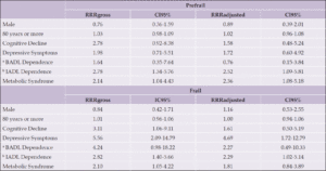
Table 2. Association between metabolic syndrome and frailty syndrome in the older people. Rio Branco, Acre, Brazil, 2019. (n=443)

a. BADL (Basic Activities of Daily Living); b. IADL (Instrumental Activities of Daily Living).


Positive frailty screening was identified in 56.88% of the older people, prefrail represented 34.09% and only 9.03% were classified as non-frail. The prevalence of metabolic syndrome was 51.69%.
Despite the proportion of older people with metabolic syndrome being higher among prefrail and frail, the test of difference in proportions wasn’t statistically significant. In the univariate multinomial regression analysis, it was observed that the older people with metabolic syndrome were more likely to be prefrail and frail (Table 2).
After adjusting by the independent variables, there was an association between metabolic syndrome and prefrailty, and the older people with metabolic syndrome were more likely to be prefrail (RRR=2.36; 95%CI=1.08-5.18). The dependence for IADL was associated with both prefrailty condition (RRR=2.52; 95%CI=1.09-5.81) and frailty (RRR=2.29; 95%CI=1.02-5.14). Depressive symptoms were associated only with the frailty condition (RRR=4.69; 95%CI=1.72-12.79) (Table 2).


Discussion

This study aimed to investigate the association between frailty, measured through a self-reported screening tool, and metabolic syndrome in older people. The results indicated that metabolic syndrome was associated with an increased likelihood of pre-frailty in the evaluated older population, consistent with other studies that used objective measures to identify the frailty phenotype (7, 23, 24). Considering that the association between the two conditions was identified using a self-reported frailty screening tool, these findings are important in demonstrating that a simple and easily applicable tool can be used to screen frail older people in Brazil, which may help prevent metabolic syndrome.
A meta-analysis determined in the adult population in Brazil estimated, a prevalence of 42% when using the NCEP-ATPIII among the oldest (age ≥ 45 years) (25). As in Brazil, studies carried out in other countries with samples only of the older people are low. Similar results of prevalence were found in a study with 1099 Australians aged between 50 and 80 years, which estimated the prevalence of metabolic syndrome at 32% using the NCEP ATPIII criteria (26). On another hand, the prevalence of frailty in community-dwelling older people in Latin America and the Caribbean was 19.6% (95% CI: 15.4–24.3%) with a range of 7.7% to 42.6% in the studies reviewed, depending on the definition adopted (27).
A divergent result was found in a study that used data from the US National Health and Nutrition Examination Survey (NHANES) which found that younger people had a higher prevalence of metabolic syndrome and higher frailty index compared with the older people. This divergence of results can be explained by the fact that in the study with data from NHANES, different criteria were used to define MetS and frailty, and that the correlation analyzes were not adjusted for other variables (28).
Metabolic syndrome and frailty may share common pathophysiological mechanisms that put the older people at risk due to cardiovascular risk factors, coagulopathies, and metabolic deregulation (29). It is observed that the increase in blood pressure, a risk factor in MetS, can be correlated with a sedentary lifestyle, which in turn is associated with a decrease in functional capacity, and so can lead to a decrease in walking performance, making it slower. This decline in walking speed is present in the cycle of clinical manifestations of frailty (30).
Metabolic, immunological, and endocrine changes characteristic of MetS in older people may be related to the mechanisms of the frailty syndrome, since the chances of being prefrail or frail increased by about 50% with the presence of the MetS (7). The literature presents limited studies regarding the relationship between frailty and metabolic syndrome. The absence of similar research conducted with older people Brazilians complicates result comparisons. Nevertheless, it is noteworthy to mention a study conducted in Spain, revealing that. older people with MetS and compared with individuals without MetS presents an increase in the risk of frailty over a period from 3.5 years (23).
Regard to a possible explanation for the effect of metabolic syndrome about the frailty, didn’t find any association between the isolated components of MetS and frailty, suggesting that MetS as a set of disorders, rather than the sum of its parts, can increase the chances of frailty (31). The presence of hyperglycemia and hypercholesterolemia, as well as the use of drugs to treat these conditions, chronic inflammatory processes, and the catabolic state in an individual’s body, when associated, can cause clinical manifestations (31, 32).
These changes can lead to a loss of weight, muscle mass, energy, and walking speed, which are components of the Frailty Phenotype (9, 33). In addition, MetS is followed by peripheral insulin resistance, chronic microinflammation, activation of oxidative and prothrombotic mechanisms, and deregulation of the renin-angiotensin axis (31). All these mechanisms can have a detrimental effect on nutrition, as well as on the neuromuscular system and cognition of the older people. Furthermore, MetS has been associated to the higher occurrence and severity of microvascular brain damage, a condition that can accelerate cognitive and functional decline, leading individuals to the frailty (24).
The limitations of this study include the cross-sectional outline, which makes impossible the accomplishing causal inference and the difficulty in generalizing the results, since it is a sample of older people living in the Amazon region of Brazil. Furthermore, unfortunately, even with so much information available on the main physiopathological mechanisms of both syndromes, it was not possible to analyze, for example, blood biomarkers such as pro-inflammatory cytokines, immunological or hormonal profiles that could add more information to our study.
A strong point of this study is using a frailty tracking instrument that can be easily used by any professional in different health services. In addition to easy application and accessibility due to low cost, some evidence already indicates that the use of these tools can reliably contribute to frailty screening, which will make their use more common in the field of research and clinical practice (10, 11). Future studies may benefit from the use of these instruments and thus add to the body of existing evidence for their improvement. In the long term, this may enable the creation of guidelines that will add new elements to objective diagnostic methods.


Conclusion

In conclusion, the metabolic syndrome was associated to the increase chance of screening for prefrailty in the older people evaluated. The results found from the use of this instrument in the present study indicate that frailty screening can be crucial among the older people with MetS, since frailty is a reversible process and that early interventions can prevent adverse outcomes potentiated by MetS. In addition, the diagnosis of MetS is easy and accessible, so that the identification of this condition in the older people can alert health professionals to the need for a more specific investigation of the health status of the older people, using instruments to identify frailty. Thus, the diagnosis of MetS in the older people can lead to a treatment that already considers the prevention of frailty.


Acknowledgements: The present study had resources obtained through research funding by the Research Program for SUS: Shared Health Management – PPSUS/AC. Call FAPAC-SESACRE-Decit/SCTIE/MS-CNPq under process No. 33376.512.21332.21092017. The authors wish to thank all participants for being involved in this study.

Data Availability Statement: The data that support the findings of this study are available from the corresponding author upon reasonable request.

Conflict of interest: The authors report no conflicts of interest.

Open Access: This article is distributed under the terms of the Creative Commons Attribution 4.0 International License (http://creativecommons.org/licenses/by/4.0/), which permits use, duplication, adaptation, distribution and reproduction in any medium or format, as long as you give appropriate credit to the original author(s) and the source, provide a link to the Creative Commons license and indicate if changes were made.


References

1. Pan-Americana Da Saúde O. Fatores de risco para doenças crônicas não transmissíveis nas Américas: Considerações sobre o fortalecimento da capacidade regulatória Documento de Referência Técnica REGULA. 2016
2. Shi TH, Wang B, Natarajan S. The Influence of Metabolic Syndrome in Predicting Mortality Risk Among US Adults: Importance of Metabolic Syndrome Even in Adults With Normal Weight. Prev Chronic Dis 2020; 17:200020. https://doi:10.5888/pcd17.200020
3. Decker JJ, Norby FL, Rooney MR, et al. Metabolic Syndrome and Risk of Ischemic Stroke in Atrial Fibrillation. Stroke 2019; 50:3045–3050. https://doi:10.1161/STROKEAHA.119.025376
4. Baygi F, Herttua K, Sheidaei A, et al. Association of Serum Uric Acid with cardio-metabolic risk factors and metabolic syndrome in seafarers working on tankers. BMC Public Health 2020; 20:442. https://doi:10.1186/s12889-020-08466-2
5. Barcelos ALV, de Oliveira EA, Haute GV, et al. Association of IL-10 to coronary disease severity in patients with metabolic syndrome. Clinica Chimica Acta 2019; 495:394–398. https://10.1016/j.cca.2019.05.006
6. Eckstein N, Buchmann N, Demuth I, et al. Association between Metabolic Syndrome and Bone Mineral Density – Data from the Berlin Aging Study II (BASE-II). Gerontology 2016; 62:337–344. https://doi:10.1159/000434678
7. Buchmann N, Spira D, König M, et al. FRAILTY AND THE METABOLIC SYNDROME – RESULTS OF THE BERLIN AGING STUDY II (BASE-II). J Frailty Aging 2019;1–7. https://doi:10.14283/jfa.2019.15
8. Morley JE, Vellas B, Abellan van Kan G, et al. Frailty Consensus: A Call to Action. J Am Med Dir Assoc 2013; 14:392–397. https://doi:10.1016/j.jamda.2013.03.022
9. Fried LP, Tangen CM, Walston J, et al. Frailty in Older Adults: Evidence for a Phenotype. 2001. https://doi:10.1093/gerona/56.3.m146
10. Aprahamian I, Lin SM, Suemoto CK, et al. Feasibility and Factor Structure of the FRAIL Scale in Older Adults. J Am Med Dir Assoc 2017; 18:367.e11-367.e18. https://10.1016/j.jamda.2016.12.067
11. Pialoux T, Goyard J , Lesourd B. Screening tools for frailty in primary healthcare: A systematic review. 2012; 12(2):189-97. https://doi:10.1111/j.1447-0594.2011.00797.x
12. von Elm E, Altman DG, Egger M, Pocock SJ, Gøtzsche PC, Vandenbroucke JP. The strengthening the reporting of observational studies in epidemiology (STROBE) statement: Guidelines for reporting observational studies. International Journal of Surgery 2014;12.https://doi.org/10.1016/j.ijsu.2014.07.013.
13. Suchindran CM. Sample Size. Encyclopedia of Social Measurement, 2005; 437-441. https://doi.org/10.1016/B0-12-369398-5/00057-8
14. Alves MCGP, Escuder MML, Goldbaum M, Barros MB de A, Fisberg RM, Cesar CLG. Sampling plan in health surveys, city of São Paulo, Brazil, 2015. Rev Saúde Pública. 2018;52:81. https://doi:10.11606/S1518-8787.2018052000471
15. Nunes DP, Duarte YA de O, Santos JLF, et al. Screening for frailty in older adults using a self-reported instrument. Rev Saude Publica 2015; 49. https://doi:10.1590/S0034-8910.2015049005516
16. Alberti KGMM, Eckel RH, Grundy SM, et al. Harmonizing the Metabolic Syndrome. Circulation 2009; 120:1640–1645. https://doi:10.1161/CIRCULATIONAHA.109.192644
17. Damasceno A, Delicio AM, Mazo DFC, et al. Validation of the Brazilian version of mini-test CASI-S. Arq Neuropsiquiatr 2005; 63:416–421. https://doi:10.1590/S0004-282X2005000300010
18. de Oliveira GM, Yokomizo JE, e Silva L dos SV, et al. The applicability of the cognitive abilities screening instrument–short (CASI-S) in primary care in Brazil. Int Psychogeriatr 2016; 28:93–99. https://doi:10.1017/S1041610215000642
19. Yesavage JA, Sheikh JI. Geriatric Depression Scale (GDS). Clin Gerontol 1986; 5:165–173. https://doi:10.1300/J018v05n0109
20. Paradela EMP, Lourenço RA, Veras RP. Validação da escala de depressão geriátrica em um ambulatório geral. Rev Saude Publica 2005; 39:918–923. https://doi:10.1590/S0034-89102005000600008
21. Katz S. Studies of Illness in the Aged. JAMA 1963; 185:914. https://doi:10.1001/jama.1963.03060120024016
22. Lawton MP, Brody EM. Assessment of older people: self-maintaining and instrumental activities of daily living. Gerontologist 1969; 9:179–86.
23. Pérez-Tasigchana RF, León-Muñoz LM, Lopez-Garcia E, et al. Metabolic syndrome and insulin resistance are associated with frailty in older adults: a prospective cohort study. Age Ageing 2017; 46:807–812. https://doi:10.1093/ageing/afx023
24. Viscogliosi G. The Metabolic Syndrome: A Risk Factor for the Frailty Syndrome? J Am Med Dir Assoc 2016; 17:364–366. https://10.1016/j.jamda.2016.01.005
25. de Siqueira Valadares LT, de Souza LSB, Salgado Júnior VA, et al. Prevalence of metabolic syndrome in Brazilian adults in the last 10 years: a systematic review and meta-analysis. BMC Public Health 2022; 22:327. https://doi:10.1186/s12889-022-12753-5
26. Pan F, Tian J, Mattap SM, et al. Association between metabolic syndrome and knee structural change on MRI. Rheumatology. 2019. https://doi:10.1093/rheumatology/kez266
27. Da Mata FAF, Pereira PP da S, Andrade KRC de, et al. Prevalence of Frailty in Latin America and the Caribbean: A Systematic Review and Meta-Analysis. PLoS One. 2016; 11:e0160019. https://doi:10.1371/journal.pone.0160019
28. Kane AE, Gregson E, Theou O, et al. The association between frailty, the metabolic syndrome, and mortality over the lifespan. Geroscience 2017; 39:221–229. https://doi:10.1007/s11357-017-9967-9
29. Damluji AA, Chung S-E, Xue Q-L, et al. Frailty and cardiovascular outcomes in the National Health and Aging Trends Study. Eur Heart J 2021; 42:3856–3865. https://doi:10.1093/eurheartj/ehab468
30. Neves T, Silva LM, Carolina A, et al. Aprovado em: 13 dez 2019 Conscientiae saúde, jul./set. 2019; 18:338–351. https://doi.org/10.5585/ConsSaude.v18n
31. Rodríguez-Mañas L, Angulo J, Carnicero JA, et al. Dual effects of insulin resistance on mortality and function in non-diabetic older adults: findings from the Toledo Study of Healthy Aging. Geroscience 2022; 44:1095–1108. https://doi:10.1007/s11357-021-00384-4
32. Taylor JA, Greenhaff PL, Bartlett DB, et al. Multisystem physiological perspective of human frailty and its modulation by physical activity. Physiol Rev. 2023. 103:1137–1191. https://10.1152/physrev.00037.2021
33. Fried LP. Frailty. In: Hazzard’s Geriatric Medicine and Gerontology, 6th ed. 2010.

© The Authors 2024
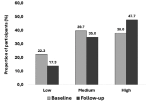
DOES PHYSICAL EXERCISE MODIFY THE PATHOPHYSIOLOGY OF ALZHEIMER’S DISEASE IN OLDER PERSONS?


J. Raffin1,2


1. Institut Hospitalo-Universitaire (IHU) HealthAge, Toulouse, France; 2. Institut du Vieillissement, Gérontopôle de Toulouse, Centre Hospitalo-Universitaire de Toulouse, 37 allées Jules Guesde, 31000 Toulouse, France

Corresponding Author: Jérémy Raffin, PhD, Gérontopôle de Toulouse, Institut du Vieillissement, Bâtiment B, 37 Allées Jules Guesde, 31000, Toulouse, France,+ 33 5 61 14 56 28, E-mail: jeremy.raffin@live.fr

J Aging Res & Lifestyle 2024;13:77-81
Published online May 22, 2024, http://dx.doi.org/10.14283/jarlife.2024.11


Abstract

Physical exercise is well known for its benefits on brain health. However, the mechanisms through which these benefits occur remain discussed, especially in the context of cognitive conditions such as Alzheimer’s disease. The present short review summarizes the findings of interventional studies that examined the effects of exercise training on the specific and non-specific biomarkers of Alzheimer’s disease. Controlled exercise intervention studies published in the English language were selected if they assessed the effects of a physical exercise intervention of at least 2 weeks in middle-aged or older adults on one of the following biomarkers measured either in the brain, the cerebrospinal fluid or the blood: beta-amyloid, tau, neurofilament light chain, and glial fibrillary acidic protein. Overall, there was no strong evidence of significant effects of exercise interventions on any of the selected biomarkers. However, in specific populations, such as women with obesity, pre-diabetes, or depression, favorable changes in blood beta-amyloid concentrations were reported. Further benefits on cerebrospinal fluid beta-amyloid were also demonstrated in APOE-ε4 allele carriers with Alzheimer’s disease. In conclusion, the current evidence suggests that physical exercise does not modulate the pathophysiology of Alzheimer’s disease in the overall population of middle-aged and older adults. Nonetheless, some specific populations, such as women with metabolic disorders and Alzheimer’s disease patients with APOE-ε4 genotype, seem to be favorably affected. Further studies, including long follow-ups, large sample sizes, and concomitantly assessing the effects of other factors such as sedentary behavior and diet, are required to bring further evidence to the field.

Key words: Alzheimer’s disease, physical exercise, biomarkers, amyloid, tau.


Introduction

Cognitive impairment and dementia are major causes of disability during aging (1). The current number of people living with dementia has been estimated at about 50 million, and this number will triple by 2050 (1, 2). Hence, preventing cognitive decline and dementia represents a major goal in aging societies, given the economic and social impact they induce (3). Alzheimer’s disease (AD) is the leading cause of dementia (4) and is notably characterized by an abnormal accumulation of dysfunctional beta-amyloid (Aβ) and phosphorylated tau proteins in the brain (5). In addition, the pathophysiology of AD also involves neuronal damages and neuro-inflammation that are respectively mirrored by an increased production of neurofilament light chain (NFL) and glial fibrillary acidic protein (GFAP) (6). Hence, all together, Aβ, phosphorylated tau, NFL and GFAP have been defined as main biomarkers for AD, the former two being core biomarkers and the latter two being non-specific biomarkers (6).
Various strategies to prevent AD have been developed, including drug therapy trials (7) as well as lifestyle interventions comprising cognitive stimulation, diet regulation, and physical exercise (8). While the positive effects of physical exercise on cognitive function are well demonstrated (9, 10), the pathway through which physical exercise induces beneficial effects remains unclear (11, 12). More specifically, the question of whether chronic physical exercise modulates the physiopathology of AD has not been established, and only a few studies have been conducted in humans (11). The present narrative review addresses this question by summarizing the effects of the published controlled interventions conducted in middle-aged and older adults that investigated the effect of regular physical exercise on the main biomarkers of AD (13).


Methods

The present work is a short non-systematic narrative review on the effects of regular exercise on the main biomarkers of AD. We selected the interventional controlled studies that examined whether physical exercise intervention of any type (eg, aerobic, resistance, balance exercises, or multicomponent exercise training), conducted for at least 2 weeks, had an effect on AD biomarkers. AD biomarkers may have been measured either in the brain, the cerebrospinal fluid (CSF) or in the blood. Four biomarkers of AD were selected based on the latest version of the Revised Criteria for Diagnosis and Staging of Alzheimer’s Disease (6): Aβ (including Aβ38, Aβ40, Aβ42, and Aβ42/40 species), tau proteins (including total and phosphorylated species), NFL and GFAP. Only human studies conducted in adults were chosen with no restriction regarding age, sex, chronic diseases or cognitive status. Studies not published in the English language were not included.


Results

Studies conducted on Aβ species, including middle-aged and older men and women with normal cognitive status, mild cognitive impairment or AD indicated no significant effect of exercise interventions, lasting from 8 weeks to 1 year and including 3 to 5 sessions of 45 to 60 min weekly, on either blood (14–18), CSF(19) or brain (20–22) amyloid levels, compared to control groups. However, subgroup analyses demonstrated that in APOE-ε4 allele carriers with AD, an increase in CSF Aβ40 was observed in inactive controls after 16 weeks of follow-up while the carriers from the exercise group maintained their baseline concentrations (19). Other interventions specifically conducted in older women reported that 12 to 16 weeks of resistance exercise training induced a significant reduction in blood Aβ42 concentrations in obese (23) or pre-diabetic (24) individuals, compared to inactive groups. Such changes were accompanied by significant reductions in glycated haemoglobin (24). Likewise, 12 weeks of Taekwondo exercise administrated in older women with depression significantly reduced the blood levels of Aβ42 (25) compared to no exercise. Exercise interventions performed in older women with no specific health condition reported mixed findings with both significant changes and no change reported after 12 (26) and 16 weeks of aerobic training (27).
Regarding the effects of exercise on tau proteins, studies are scarce but it has been shown that in non-demented middle-aged individuals, 2 weeks of resistance exercise concomitant to a bed-rest intervention did not modulate the blood levels of total tau compared to bed rest alone (28). Likewise, 6 months of cycling exercise had no impact on the blood levels of phosphorylated tau 181 in cognitively healthy older adults (18). Furthermore, in older adults with AD, neither 16 (19) or 24 weeks (17) of aerobic exercise impacted the concentrations of total and phosphorylated tau proteins measured in the CSF or in the blood.
Similar negative findings have been reported on the non-specific markers of AD. In non-demented middle-aged and older adults, 2 weeks of resistance exercise conducted in parallel to a bed rest protocol did not impact NFL and GFAP blood concentrations compared to bed rest without exercise (28). Similarly, the blood concentrations in GFAP and NFL were not affected by 6 months of aerobic training in older adults with no cognitive impairment (18). A long-term intervention of 2 years of combined resistance and aerobic exercise reported no effects on blood concentrations in NFL in comparison to a control group (29). In patients with AD, it was reported that, compared to a no-exercise group, 16 weeks of aerobic training did not produce any significant changes in CSF NFL concentrations (30), even in subjects that were classified as amyloid positive (CSF concentrations Aβ42 < 550 pg/ml) (19).


Discussion

Overall, the studies included in this short review demonstrated no effect of exercise interventions on the main biomarkers of AD, which is in line with previous reviews on the topic (31, 32). Nonetheless, some factors such as sex, APOE genotype or health status, seem to modify the effect of exercise training, as favourable and significant findings regarding Aβ levels were reported in women with obesity (23), pre-diabetes (24), or depression (25) and in APOE-ε4 carriers individuals with AD (19). Hence, it is possible that exercise still has an effect on specific populations that display risks factors for AD such as women (33), APOE-ε4 genotype carriers (34), individuals with metabolic disorders (35) or depression (36). Notably, the favourable effects found in AD patients with APOE-ε4 genotype is also in accordance with previous work demonstrating that APOE-ε4 carriers display greater improvements in cognitive functions in response to exercise compared to non-carriers (37). Regarding the lack of effects in the overall population, because most of trials were performed in older adults, it is possible that an advanced age might counteract the benefits of physical training, although some authors have reported greater effects of exercise on cognition in healthy adults older than 60 (38) compared to younger counterparts. Our findings also diverge from the observational studies and meta-analyses that demonstrated beneficial associations between PA and amyloid (39, 40), NFL (41–43) and GFAP (43) levels, although the relationships with tau remain contrasted (39, 44–48).
Even though interventional studies reported little effects on the specific biomarkers, PA may still improve cognition through other pathways. Recent reviews published on this topic have reported that chronic exercise has a positive effect on brain glucose metabolism, vascular function, and BDNF concentrations, along with providing benefits on cognition (11, 32, 38). This indicates a pleiotropic effect of exercise that is consistent with its positive impact on the biological hallmarks of aging (49), which are thought to be the common roots of most of age-related diseases (50). Future studies should thus focus on long term interventions not only in middle-aged and older adults, but also in young adults as abnormal proteins deposition may start decades before the disease onset (51, 52). Studies examining the factors that may moderate the effect of exercise, such as sex, genotype, or health status are also required, as well as studies with large sample sizes, given that the subject samples of the studies selected for this review were relatively small, ranging from 156 to 14 individuals. We also recommend that cognition should be assessed along with the measurement of the neurodegeneration biomarkers in order to determine whether changes in the latter could mediate changes in the former. In addition, most of the studies included herein focused on tau, amyloid, and NFL proteins, but there is a lack of evidence regarding the impact of exercise on GFAP. Yet, the research on the non-specific markers of AD, namely NFL and GFAP, remains important. Indeed, GFAP is an indicator of astrocyte activation (6) and has been shown to be an early predictor of Aβ production (53) while NFL reflect axonal damages (6). Yet, higher blood levels of both NFL and GFAP have been associated with reduced cognitive capacity (54, 55) and greater prospective cognitive decline (54). Importantly also, the biomarkers examined herein may interact with each other, such that the benefits of exercise on one biomarker may depend on the levels of other biomarkers. Interventional studies simultaneously measuring the effects of several biomarkers and examining their interactions may thus provide significant contributions to the field.
In conclusion, physical exercise interventions do not demonstrate favorable AD-modifying effects, except in women with impaired metabolic health or depression and APOE-ε4 carriers patients with AD. While more studies are needed given the paucity of available evidence, other important factors such as diet (56, 57), cognitive stimulation (58), or sleep quality (59) may also modulate AD pathophysiology and should be explored collectively. Importantly, sedentary behaviour, which demonstrated significant association with incident dementia (60), may have deleterious independent and/or exercise-counteracting effects on brain physiopathology. All of these factors may act synergistically and potentiate the single effects of each individually, emphasizing the need for holistic approach interventions to prevent dementia (61).


Conflict of interest: The author declares no conflict of interest.

Open Access: This article is distributed under the terms of the Creative Commons Attribution 4.0 International License (http://creativecommons.org/licenses/by/4.0/), which permits use, duplication, adaptation, distribution and reproduction in any medium or format, as long as you give appropriate credit to the original author(s) and the source, provide a link to the Creative Commons license and indicate if changes were made.


References

1. GBD 2016 Dementia Collaborators. Global, regional, and national burden of Alzheimer’s disease and other dementias, 1990-2016: a systematic analysis for the Global Burden of Disease Study 2016. Lancet Neurol. 2019;18:88–106. doi: 10.1016/S1474-4422(18)30403-4. Cited: in: : PMID: 30497964.
2. World Health Organization. Risk Reduction of Cognitive Decline and Dementia: WHO Guidelines [Internet]. Geneva: World Health Organization; 2019 [cited 2024 Apr 2]. Available from: http://www.ncbi.nlm.nih.gov/books/NBK542796/.
3. Meijer E, Casanova M, Kim H, Llena-Nozal A, Lee J. Economic costs of dementia in 11 countries in Europe: Estimates from nationally representative cohorts of a panel study. Lancet Reg Health Eur. 2022;20:100445. doi: 10.1016/j.lanepe.2022.100445. Cited: in: : PMID: 35781926.
4. Prince M, Wimo A, Guerchet M, Ali G-C, Wu Y-T, Prina M. World Alzheimer Report 2015, The Global Impact of Dementia: An analysis of prevalence, incidence, cost and trends. 2015;
5. Mangialasche F, Solomon A, Winblad B, Mecocci P, Kivipelto M. Alzheimer’s disease: clinical trials and drug development. The Lancet Neurology. 2010;9:702–716. doi: 10.1016/S1474-4422(10)70119-8. Cited: in: : PMID: 20610346.
6. Revised Criteria for Diagnosis and Staging of Alzheimer’s | AAIC [Internet]. Revised Criteria for Diagnosis and Staging of Alzheimer’s | AAIC. [cited 2024 Apr 2]. Available from: https://aaic.alz.org/diagnostic-criteria.asp.
7. Huang L-K, Kuan Y-C, Lin H-W, Hu C-J. Clinical trials of new drugs for Alzheimer disease: a 2020–2023 update. J Biomed Sci. 2023;30:83. doi: 10.1186/s12929-023-00976-6. Cited: in: : PMID: 37784171.
8. Ko Y, Chye SM. Lifestyle intervention to prevent Alzheimer’s disease. Rev Neurosci. 2020;/j/revneuro.ahead-of-print/revneuro-2020-0072/revneuro-2020-0072.xml. doi: 10.1515/revneuro-2020-0072. Cited: in: : PMID: 32804681.
9. Brini S, Sohrabi HR, Peiffer JJ, Karrasch M, Hämäläinen H, Martins RN, Fairchild TJ. Physical Activity in Preventing Alzheimer’s Disease and Cognitive Decline: A Narrative Review. Sports Med. 2018;48:29–44. doi: 10.1007/s40279-017-0787-y.
10. Tyndall AV, Clark CM, Anderson TJ, Hogan DB, Hill MD, Longman RS, Poulin MJ. Protective Effects of Exercise on Cognition and Brain Health in Older Adults. Exercise and Sport Sciences Reviews. 2018;46:215–223. doi: 10.1249/JES.0000000000000161.
11. Abdullahi A, Wong TW, Ng SS. Understanding the mechanisms of disease modifying effects of aerobic exercise in people with Alzheimer’s disease. Ageing Res Rev. 2024;94:102202. doi: 10.1016/j.arr.2024.102202. Cited: in: : PMID: 38272266.
12. Xu L, Liu R, Qin Y, Wang T. Brain metabolism in Alzheimer’s disease: biological mechanisms of exercise. Transl Neurodegener. 2023;12:33. doi: 10.1186/s40035-023-00364-y. Cited: in: : PMID: 37365651.
13. Hansson O. Biomarkers for neurodegenerative diseases. Nat Med. 2021;27:954–963. doi: 10.1038/s41591-021-01382-x.
14. Baker LD, Frank LL, Foster-Schubert K, Green PS, Wilkinson CW, McTiernan A, Cholerton BA, Plymate SR, Fishel MA, Watson GS, et al. Aerobic exercise improves cognition for older adults with glucose intolerance, a risk factor for Alzheimer’s disease. J Alzheimers Dis. 2010;22:569–579. doi: 10.3233/JAD-2010-100768. Cited: in: : PMID: 20847403.
15. Ruiz JR, Gil-Bea F, Bustamante-Ara N, Rodríguez-Romo G, Fiuza-Luces C, Serra-Rexach JA, Cedazo-Minguez A, Lucia A. Resistance training does not have an effect on cognition or related serum biomarkers in nonagenarians: a randomized controlled trial. Int J Sports Med. 2015;36:54–60. doi: 10.1055/s-0034-1375693. Cited: in: : PMID: 25329433.
16. Yokoyama H, Okazaki K, Imai D, Yamashina Y, Takeda R, Naghavi N, Ota A, Hirasawa Y, Miyagawa T. The effect of cognitive-motor dual-task training on cognitive function and plasma amyloid β peptide 42/40 ratio in healthy elderly persons: a randomized controlled trial. BMC Geriatr. 2015;15:60. doi: 10.1186/s12877-015-0058-4. Cited: in: : PMID: 26018225.
17. Yu F, Han SY, Salisbury D, Pruzin JJ, Geda Y, Caselli RJ, Li D. Feasibility and preliminary effects of exercise interventions on plasma biomarkers of Alzheimer’s disease in the FIT-AD trial: a randomized pilot study in older adults with Alzheimer’s dementia. Pilot Feasibility Stud. 2022;8:243. doi: 10.1186/s40814-022-01200-2. Cited: in: : PMID: 36461134.
18. Sewell KR, Rainey-Smith SR, Pedrini S, Peiffer JJ, Sohrabi HR, Taddei K, Markovic SJ, Martins RN, Brown BM. The impact of exercise on blood-based biomarkers of Alzheimer’s disease in cognitively unimpaired older adults. GeroScience [Internet]. 2024 [cited 2024 May 2]; doi: 10.1007/s11357-024-01130-2.
19. Steen Jensen C, Portelius E, Siersma V, Høgh P, Wermuth L, Blennow K, Zetterberg H, Waldemar G, Gregers Hasselbalch S, Hviid Simonsen A. Cerebrospinal Fluid Amyloid Beta and Tau Concentrations Are Not Modulated by 16 Weeks of Moderate- to High-Intensity Physical Exercise in Patients with Alzheimer Disease. Dement Geriatr Cogn Disord. 2016;42:146–158. doi: 10.1159/000449408. Cited: in: : PMID: 27643858.
20. Frederiksen KS, Madsen K, Andersen BB, Beyer N, Garde E, Høgh P, Waldemar G, Hasselbalch SG, Law I. Moderate- to high-intensity exercise does not modify cortical β-amyloid in Alzheimer’s disease. Alzheimers Dement (N Y). 2019;5:208–215. doi: 10.1016/j.trci.2019.04.006. Cited: in: : PMID: 31198839.
21. Tarumi T, Rossetti H, Thomas BP, Harris T, Tseng BY, Turner M, Wang C, German Z, Martin-Cook K, Stowe AM, et al. Exercise Training in Amnestic Mild Cognitive Impairment: A One-Year Randomized Controlled Trial. J Alzheimers Dis. 2019;71:421–433. doi: 10.3233/JAD-181175. Cited: in: : PMID: 31403944.
22. Vidoni ED, Morris JK, Watts A, Perry M, Clutton J, Van Sciver A, Kamat AS, Mahnken J, Hunt SL, Townley R, et al. Effect of aerobic exercise on amyloid accumulation in preclinical Alzheimer’s: A 1-year randomized controlled trial. PLoS One. 2021;16:e0244893. doi: 10.1371/journal.pone.0244893. Cited: in: : PMID: 33444359.
23. Kang D, Koh S, Kim T, Bressel E, Kim D. Circuit Training Improves the Levels of β-Amyloid and Brain-Derived Neurotrophic Factor Related to Cognitive Impairment Risk Factors in Obese Elderly Korean Women. J Clin Med. 2024;13:799. doi: 10.3390/jcm13030799. Cited: in: : PMID: 38337492.
24. Kim B-R, Lim S-T. Effects of Leisure-Time Physical Activity on Cognitive Reserve Biomarkers and Leisure Motivation in the Pre-Diabetes Elderly. Healthcare (Basel). 2022;10:737. doi: 10.3390/healthcare10040737. Cited: in: : PMID: 35455914.
25. Baek S-H, Hong G-R, Min D-K, Kim E-H, Park S-K. Effects of Functional Fitness Enhancement through Taekwondo Training on Physical Characteristics and Risk Factors of Dementia in Elderly Women with Depression. International Journal of Environmental Research and Public Health. 2021;18:7961. doi: 10.3390/ijerph18157961.
26. Kwon Y, Park S, Kim E, Park H. Effects of combined exercise on β-Amyloid and DHEAs in elderly women. Japanese Journal of Physical Fitness and Sports Medicine. 2007;56:149–156. doi: 10.7600/jspfsm.56.149.
27. Kim J-H, Jung Y-S, Kim J-W, Ha M-S, Ha S-M, Kim D-Y. Effects of aquatic and land-based exercises on amyloid beta, heat shock protein 27, and pulse wave velocity in elderly women. Exp Gerontol. 2018;108:62–68. doi: 10.1016/j.exger.2018.03.024. Cited: in: : PMID: 29604402.
28. Blaber AP, Sadeghian F, Naz Divsalar D, Scarisbrick IA. Elevated biomarkers of neural injury in older adults following head-down bed rest: links to cardio-postural deconditioning with spaceflight and aging. Front Hum Neurosci. 2023;17:1208273. doi: 10.3389/fnhum.2023.1208273. Cited: in: : PMID: 37822710.
29. Monti E, Tagliaferri S, Zampieri S, Sarto F, Sirago G, Franchi MV, Ticinesi A, Longobucco Y, Adorni E, Lauretani F, et al. Effects of a 2-year exercise training on neuromuscular system health in older individuals with low muscle function. J Cachexia Sarcopenia Muscle. 2023;14:794–804. doi: 10.1002/jcsm.13173. Cited: in: : PMID: 36708273.
30. Jensen CS, Portelius E, Høgh P, Wermuth L, Blennow K, Zetterberg H, Hasselbalch SG, Simonsen AH. Effect of physical exercise on markers of neuronal dysfunction in cerebrospinal fluid in patients with Alzheimer’s disease. Alzheimers Dement (N Y). 2017;3:284–290. doi: 10.1016/j.trci.2017.03.007. Cited: in: : PMID: 29067334.
31. Frederiksen KS, Gjerum L, Waldemar G, Hasselbalch SG. Effects of Physical Exercise on Alzheimer’s Disease Biomarkers: A Systematic Review of Intervention Studies. Burns J, editor. JAD. 2017;61:359–372. doi: 10.3233/JAD-170567.
32. Moniruzzaman M, Kadota A, Akash MS, Pruitt PJ, Miura K, Albin R, Dodge HH. Effects of physical activities on dementia-related biomarkers: A systematic review of randomized controlled trials. Alzheimers Dement (N Y). 2020;6:e12109. doi: 10.1002/trc2.12109. Cited: in: : PMID: 33521235.
33. Beam CR, Kaneshiro C, Jang JY, Reynolds CA, Pedersen NL, Gatz M. Differences Between Women and Men in Incidence Rates of Dementia and Alzheimer’s Disease. J Alzheimers Dis. 2018;64:1077–1083. doi: 10.3233/JAD-180141. Cited: in: : PMID: 30010124.
34. Liu C-C, Kanekiyo T, Xu H, Bu G. Apolipoprotein E and Alzheimer disease: risk, mechanisms, and therapy. Nat Rev Neurol. 2013;9:106–118. doi: 10.1038/nrneurol.2012.263. Cited: in: : PMID: 23296339.
35. Craft S. The Role of Metabolic Disorders in Alzheimer Disease and Vascular Dementia: Two Roads Converged. Archives of Neurology. 2009;66:300–305. doi: 10.1001/archneurol.2009.27.
36. Sáiz-Vázquez O, Gracia-García P, Ubillos-Landa S, Puente-Martínez A, Casado-Yusta S, Olaya B, Santabárbara J. Depression as a Risk Factor for Alzheimer’s Disease: A Systematic Review of Longitudinal Meta-Analyses. J Clin Med. 2021;10:1809. doi: 10.3390/jcm10091809. Cited: in: : PMID: 33919227.
37. Colovati MES, Novais IP, Zampol M, Mendes GD, Cernach MCS, Zanesco A. Interaction between physical exercise and APOE gene polymorphism on cognitive function in older people. Braz J Med Biol Res. 2020;54:e10098. doi: 10.1590/1414-431X202010098. Cited: in: : PMID: 33331535.
38. Zhang M, Jia J, Yang Y, Zhang L, Wang X. Effects of exercise interventions on cognitive functions in healthy populations: A systematic review and meta-analysis. Ageing Research Reviews. 2023;92:102116. doi: 10.1016/j.arr.2023.102116.
39. Brown BM, Peiffer J, Rainey-Smith SR. Exploring the relationship between physical activity, beta-amyloid and tau: A narrative review. Ageing Res Rev. 2019;50:9–18. doi: 10.1016/j.arr.2019.01.003. Cited: in: : PMID: 30615936.
40. Rodriguez-Ayllon M, Solis-Urra P, Arroyo-Ávila C, Álvarez-Ortega M, Molina-García P, Molina-Hidalgo C, Gómez-Río M, Brown B, Erickson KI, Esteban-Cornejo I. Physical activity and amyloid beta in middle-aged and older adults: A systematic review and meta-analysis. Journal of Sport and Health Science. 2024;13:133–144. doi: 10.1016/j.jshs.2023.08.001.
41. Casaletto KB, Kornack J, Paolillo EW, Rojas JC, VandeBunte A, Staffaroni AS, Lee S, Heuer H, Forsberg L, Ramos EM, et al. Association of Physical Activity With Neurofilament Light Chain Trajectories in Autosomal Dominant Frontotemporal Lobar Degeneration Variant Carriers. JAMA Neurol. 2023;80:82–90. doi: 10.1001/jamaneurol.2022.4178. Cited: in: : PMID: 36374516.
42. Raffin J, Rolland Y, Aggarwal G, Nguyen AD, Morley JE, Li Y, Bateman RJ, Vellas B, Barreto P de S, MAPT/DSA Group. Associations Between Physical Activity, Blood-Based Biomarkers of Neurodegeneration, and Cognition in Healthy Older Adults: The MAPT Study. J Gerontol A Biol Sci Med Sci. 2021;76:1382–1390. doi: 10.1093/gerona/glab094. Cited: in: : PMID: 33864068.
43. VandeBunte AM, Lee SY, Paolillo EW, Rojas JC, Chan B, Lago AL, Kramer JH, Casaletto KB. Physical Activity Relates to Lower Astrocytic Activation and Axonal Breakdown in Clinically Normal Older Adults. Alzheimer’s & Dementia. 2022;18:e063455. doi: 10.1002/alz.063455.
44. Brown BM, Rainey-Smith SR, Dore V, Peiffer JJ, Burnham SC, Laws SM, Taddei K, Ames D, Masters CL, Rowe CC, et al. Self-Reported Physical Activity is Associated with Tau Burden Measured by Positron Emission Tomography. J Alzheimers Dis. 2018;63:1299–1305. doi: 10.3233/JAD-170998. Cited: in: : PMID: 29758940.
45. Frederiksen KS, Gjerum L, Waldemar G, Hasselbalch SG. Physical Activity as a Moderator of Alzheimer Pathology: A Systematic Review of Observational Studies. Curr Alzheimer Res. 2019;16:362–378. doi: 10.2174/1567205016666190315095151. Cited: in: : PMID: 30873924.
46. Hou X-H, Xu W, Bi Y-L, Shen X-N, Ma Y-H, Dong Q, Tan L, Yu J-T. Associations of healthy lifestyles with cerebrospinal fluid biomarkers of Alzheimer’s disease pathology in cognitively intact older adults: the CABLE study. Alzheimers Res Ther. 2021;13:81. doi: 10.1186/s13195-021-00822-7. Cited: in: : PMID: 33875016.
47. Law LL, Rol RN, Schultz SA, Dougherty RJ, Edwards DF, Koscik RL, Gallagher CL, Carlsson CM, Bendlin BB, Zetterberg H, et al. Moderate intensity physical activity associates with CSF biomarkers in a cohort at risk for Alzheimer’s disease. Alzheimers Dement (Amst). 2018;10:188–195. doi: 10.1016/j.dadm.2018.01.001. Cited: in: : PMID: 29527551.
48. Zhong S, Zhao B, Ma Y-H, Sun Y, Zhao Y-L, Liu W-H, Ou Y-N, Dong Q, Tan L, Yu J-T. Associations of Physical Activity with Alzheimer’s Disease Pathologies and Cognition: The CABLE Study. J Alzheimers Dis. 2022;89:483–492. doi: 10.3233/JAD-220389. Cited: in: : PMID: 35871345.
49. Garatachea N, Pareja-Galeano H, Sanchis-Gomar F, Santos-Lozano A, Fiuza-Luces C, Morán M, Emanuele E, Joyner MJ, Lucia A. Exercise Attenuates the Major Hallmarks of Aging. Rejuvenation Research. 2015;18:57–89. doi: 10.1089/rej.2014.1623.
50. López-Otín C, Blasco MA, Partridge L, Serrano M, Kroemer G. Hallmarks of aging: An expanding universe. Cell. 2023;186:243–278. doi: 10.1016/j.cell.2022.11.001. Cited: in: : PMID: 36599349.
51. Gordon BA, Blazey TM, Su Y, Hari-Raj A, Dincer A, Flores S, Christensen J, McDade E, Wang G, Xiong C, et al. Spatial patterns of neuroimaging biomarker change in individuals from families with autosomal dominant Alzheimer’s disease: a longitudinal study. Lancet Neurol. 2018;17:241–250. doi: 10.1016/S1474-4422(18)30028-0. Cited: in: : PMID: 29397305.
52. Villemagne VL, Burnham S, Bourgeat P, Brown B, Ellis KA, Salvado O, Szoeke C, Macaulay SL, Martins R, Maruff P, et al. Amyloid β deposition, neurodegeneration, and cognitive decline in sporadic Alzheimer’s disease: a prospective cohort study. The Lancet Neurology. 2013;12:357–367. doi: 10.1016/S1474-4422(13)70044-9. Cited: in: : PMID: 23477989.
53. Pereira JB, Janelidze S, Smith R, Mattsson-Carlgren N, Palmqvist S, Teunissen CE, Zetterberg H, Stomrud E, Ashton NJ, Blennow K, et al. Plasma GFAP is an early marker of amyloid-β but not tau pathology in Alzheimer’s disease. Brain. 2021;144:3505–3516. doi: 10.1093/brain/awab223. Cited: in: : PMID: 34259835.
54. Chatterjee P, Pedrini S, Doecke JD, Thota R, Villemagne VL, Doré V, Singh AK, Wang P, Rainey-Smith S, Fowler C, et al. Plasma Aβ42/40 ratio, p-tau181, GFAP, and NfL across the Alzheimer’s disease continuum: A cross-sectional and longitudinal study in the AIBL cohort. Alzheimers Dement. 2023;19:1117–1134. doi: 10.1002/alz.12724. Cited: in: : PMID: 36574591.
55. Snellman A, Ekblad LL, Ashton NJ, Karikari TK, Lantero-Rodriguez J, Pietilä E, Koivumäki M, Helin S, Karrasch M, Zetterberg H, et al. Head-to-head comparison of plasma p-tau181, p-tau231 and glial fibrillary acidic protein in clinically unimpaired elderly with three levels of APOE4-related risk for Alzheimer’s disease. Neurobiology of Disease. 2023;183:106175. doi: 10.1016/j.nbd.2023.106175.
56. Bartochowski Z, Conway J, Wallach Y, Chakkamparambil B, Alakkassery S, Grossberg GT. Dietary Interventions to Prevent or Delay Alzheimer’s Disease: What the Evidence Shows. Curr Nutr Rep. 2020;9:210–225. doi: 10.1007/s13668-020-00333-1.
57. van Soest AP, Beers S, van de Rest O, de Groot LC. The Mediterranean-Dietary Approaches to Stop Hypertension Intervention for Neurodegenerative Delay (MIND) Diet for the Aging Brain: A Systematic Review. Advances in Nutrition. 2024;15:100184. doi: 10.1016/j.advnut.2024.100184.
58. Sabbagh MN, Perez A, Holland TM, Boustani M, Peabody SR, Yaffe K, Bruno M, Paulsen R, O’Brien K, Wahid N, et al. Primary prevention recommendations to reduce the risk of cognitive decline. Alzheimer’s & Dementia. 2022;18:1569–1579. doi: 10.1002/alz.12535.
59. Cordone S, Scarpelli S, Alfonsi V, De Gennaro L, Gorgoni M. Sleep-Based Interventions in Alzheimer’s Disease: Promising Approaches from Prevention to Treatment along the Disease Trajectory. Pharmaceuticals (Basel). 2021;14:383. doi: 10.3390/ph14040383. Cited: in: : PMID: 33921870.
60. Raichlen DA, Aslan DH, Sayre MK, Bharadwaj PK, Ally M, Maltagliati S, Lai MHC, Wilcox RR, Klimentidis YC, Alexander GE. Sedentary Behavior and Incident Dementia Among Older Adults. JAMA. 2023;330:934–940. doi: 10.1001/jama.2023.15231.
61. Zheng S, Edney SM, Goh CH, Tai BC, Mair JL, Castro O, Salamanca-Sanabria A, Kowatsch T, van Dam RM, Müller-Riemenschneider F. Effectiveness of holistic mobile health interventions on diet, and physical, and mental health outcomes: a systematic review and meta-analysis. EClinicalMedicine. 2023;66:102309. doi: 10.1016/j.eclinm.2023.102309. Cited: in: : PMID: 38053536.

© The Authors 2024
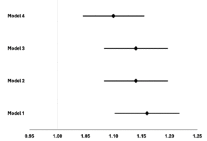
DOES NUTRITIONAL SUPPLEMENTATION HAVE A DISEASEMODIFYING EFFECT ON THE ALZHEIMER’S DISEASE NEURODEGENERATIVE PROCESS?


K.V. Giudici1


1. Institute of Aging, Gerontopole of Toulouse, Toulouse University Hospital, Université Toulouse III Paul Sabatier, Toulouse, France.

Corresponding Author: Kelly Virecoulon Giudici, Institute of Aging, Gérontopôle of Toulouse, Toulouse University Hospital, Université Toulouse III Paul Sabatier, 37 Allée Jules Guesde, 31000 Toulouse, France, E-mail: kellygiudici@gmail.com

J Aging Res & Lifestyle 2024;13:73-76
Published online May 22, 2024, http://dx.doi.org/10.14283/jarlife.2024.10


Abstract

Because nutrition is one of the main factors related to Alzheimer’s disease (AD), questions arise about how taking nutrients as supplements can affect its pathophysiological process. In the present study, an overview of the potential effects of nutritional supplementation on the main biomarkers related to the AD pathophysiology (i.e., amyloid-β and tau) is explored. Trials testing the supplementation of single or combined nutrients versus placebo identified effects on some AD biomarkers, but changes were not always accompanied by positive effects on cognitive function. Differences in characteristics of studied populations (cognitive status, age, educational level), choice of nutrient combinations and doses, duration of intervention, and adjustments for potential confounders are some factors that may explain discrepancies in findings.

Key words: Alzheimer’s, supplementation, amyloid, tau, cognitive decline, aging.

Abbreviations: Aβ: amyloid-β; AD: Alzheimer’s disease; ADAS-cog: Alzheimer disease assessment scale-cognitive subscale; ALA: α-lipoic acid; APOE: apolipoprotein E; CSF: cerebrospinal fluid; DHA: docosahexaenoic acid; EPA: eicosapentaenoic acid; IQ: intelligence quotient; MAPT: Multidomain Alzheimer Preventive Trial; MCI: mild cognitive impairment; MMSE: Mini Mental State Examination; PET: positron emission tomography; p-tau181: phosphorylated tau at threonine 181; PUFA: polyunsaturated fatty acids; RCT: randomized controlled trial; t-tau: total tau; VISP: Vitamin Intervention for Stroke Prevention.


Introduction

The pathophysiological process leading to the characterization of Alzheimer’s disease (AD) as a unique neurodegenerative disorder, among other types of dementia, consists of the accumulation of amyloid-β (Aβ) plaques and pathologic tau deposits in the brain (1). The neurodegeneration influenced by these processes, coupled with dementia, results in gradual cognitive decline that may reach advanced stages in which quality of life is severely affected (2).
Although there is a strong genetic risk factor for the development of AD (the presence of the APOE ε4 allele) (3), many other factors such as diet, physical activity level, stress management and sleep quality are known to affect the probability of accumulating Aβ and contributing to tau phosphorylation and aggregation in the brain, and consequently increasing the risk of AD (4).
The detection and diagnosis of AD has been classically based on the evaluation of Aβ and tau biomarkers in the brain (by positron emission tomography – PET) or in the cerebrospinal fluid (CSF) (1, 2), which are either expensive or invasive methods. More recently, blood-based biomarkers have emerged as less complex alternatives, but with compatible reliability (5, 6). These measures have been used as outcomes in trials testing what can be done, in terms of lifestyle changes, to prevent or fight this neurodegenerative disease. Since nutrition is one of the main factors related to AD (7), questions arise if taking nutrients as supplements is able to affect its pathophysiological process. In the present study, an overview on the potential effects of nutritional supplementation on the main biomarkers related to the AD pathophysiology (i.e., Aβ and tau) is explored.


Effects of nutritional supplementation on Aβ and tau biomarkers

The main omega-3 polyunsaturated fatty acids (PUFA) (eicosapentaenoic acid – EPA and docosahexaenoic acid – DHA) are known by their substantial anti-inflammatory and antioxidant properties (8). DHA is especially important to brain function: besides its essential structural properties, it regulates synaptic function, modulates gene expression, acts as an indirect antioxidant and contributes to neuroprotection (9). The Multidomain Alzheimer Preventive Trial (MAPT) explored the effects of a 3-year supplementation with omega-3 PUFA (800mg DHA and 225mg EPA/day), alone or combined to physical activity and cognitive training, on AD biomarkers and clinical tests among 1,680 community-dwelling older adults living in France and Monaco (10, 11). At the end of the 3-year follow-up, no effects of interventions were observed in cognitive function (evaluated with a composite cognitive score) (11), nor in plasma phosphorylated tau at threonine 181 (p-tau181) when a subsample of 527 participants with this measure was analyzed (12). Interestingly, in another secondary analysis of MAPT among a subsample of 483 participants with plasma Aβ42/40 ratio assessments, the combined intervention showed a positive effect on cognitive function in the per-protocol positive amyloid group (i.e., Aβ42/40≤0.0107; n=154), after 1 year and 3 years. However, no differences were found between intervention and placebo groups after two additional years of observational follow-up (13).
In the OmegAD Study, a 6-month omega-3 PUFA supplementation (2.3g/day) or placebo was offered to 35 patients diagnosed with AD. Compared to placebo, intervention did not affect CSF Aβ38, Aβ40, Aβ42, total tau (t-tau) and p-tau (14, 15). A secondary analysis with 33 participants revealed that changes in CSF levels of DHA due to supplementation were inversely correlated with CSF levels of t-tau and p-tau, indicating that the more DHA increased in CSF, greater was the change in CSF tau biomarkers (16). Another trial offered 2g/day of DHA or placebo for 240 individuals with mild cognitive impairment (MCI) living in China over 2 years, and found decreases in blood Aβ42 levels and expression of Aβ protein precursor mRNA, which were accompanied by increases in scores of full-scale intelligence quotient (IQ), verbal IQ and subdomains of information and digit span, among those taking the DHA supplement (17).
Vitamin D is another nutrient believed to contribute to the development of cognition and its maintenance over time (18). Acting as a hormone with multiple actions in metabolism, it impacts neurocognition by inducing neuroprotection, modulating oxidative stress, regulating calcium homeostasis and inhibition inflammation (19). A trial with 210 participants testing a 1-year vitamin D supplementation (800IU/day) in older adults with AD observed a decrease in plasma Aβ42 and improvements in cognitive assessments (information, arithmetic, digit span, vocabulary, block design and picture arrange scores) among the intervention group (20). Another trial testing the effect of a high-dose short-term vitamin D supplementation (50,000IU/week for 8 weeks) versus placebo on plasma Aβ40 of 24 vitamin D insufficient adults observed a greater plasma Aβ40 increase among the intervention group, what authors suggested to be an indicative of decreased brain Aβ (21).
Excessive homocysteine (Hcy) has direct neurotoxic effects, due to inducing oxidative stress, causing DNA damage and apoptosis, and stimulating Aβ deposition in the brain (22). Some B-vitamins are known by their role in Hcy remethylation, thus contributing to decrease Hcy levels and to potentially avoid its neurotoxicity (23). Chen et al. (24) explored the effects of a 6-month folate supplementation (1.25mg/day) on inflammatory biomarkers and cognitive function among patients with AD. They found that plasma Aβ40 levels were lower, Aβ42/40 ratio was higher and mean Mini Mental State Examination (MMSE) score was slightly increased in the intervention group compared to the control group at the end of follow-up. In the Vitamin Intervention for Stroke Prevention (VISP) trial, 300 adults with ischemic stroke and high total Hcy (tHcy) levels (a risk factor for AD) were treated with either a high-dose supplement (composed of 25mg of pyridoxine, 0.4mg of cobalamin, and 2.5mg of folic acid) or a low-dose supplement (200mcg of pyridoxine, 6mcg of cobalamin, and 20mcg of folic acid) for 2 years (25). At the end of follow-up, no alterations were observed for Aβ40, Aβ42 or Aβ42/40 ratio. On the other hand, tHcy levels significantly decreased in both groups (more among participants taking the high dose) and were strongly correlated with plasma Aβ40, but not with Aβ42 concentrations (25).
Important copper concentrations and localization changes have been identified in AD cerebral regions, raising questions whether therapeutic approaches for regulating its levels could affect AD pathophysiology (26). Kessler et al. (27) offered a 12-month supplementation of 8mg/day of this nutrient or placebo to a sample of patients with mild AD. They found no effect on the progression of CSF tau and p-tau levels nor in MMSE and Alzheimer disease assessment scale-cognitive subscale (ADAS-cog) scores. Still, a lower decline in CSF Aβ42 was observed in the intervention group (a positive finding, since decreased CSF Aβ42 is a diagnostic marker for AD).
The Nolan Study, in turn, tested the effect of a 1-year multi-nutrient supplementation (including omega-3 PUFA, vitamin C, vitamin D, vitamin E, thiamin, riboflavin, niacin, pantothenic acid, pyridoxine, folic acid, biotin, cobalamin, selenium, choline and citrulline) on clinical tests, imaging and blood biomarkers related to the AD pathology among a sample of 362 community-dwelling older adults living in France (28, 29). At the end of the follow-up, supplementation could not postpone the increase in plasma p-tau181 (observed in both intervention and placebo groups) (29), and neither showed an effect on cognitive function (28). Another randomized controlled trial (RCT) found no benefits of a 16-week supplementation of combined vitamin E (800IU/day), vitamin C (500mg/day) and α-lipoic acid (ALA) (900mg/day), or coenzyme Q alone (1200mg/day) on CSF Aβ42, t-tau or p-tau181 in a sample of 66 subjects with mild to moderate AD (30). Surprisingly, a faster decline in MMSE score was identified among the group receiving vitamin E, vitamin C and ALA (30).


Conclusions and perspectives

Despite the well-established influence of diet in the development of the AD neurodegenerative process (7), it is still not clear how nutritional supplementation may contribute to preventing or postponing it and, consequently, to protect cognitive function. Trials currently show that some biomarkers related to the AD development can be modified with supplementation protocols varying from months to years. Still, changes are not always accompanied by positive effects on cognitive function. Differences in characteristics of studied populations (cognitively normal participants or subjects with MCI or AD, age ranges, educational level), choice of nutrients’ combinations and doses, duration of interventions and adjustments for potential confounders (such as APOE ε4 status) are some factors that may explain discrepancies in findings.
It is known that AD slowly develops for decades before cognitive decline is perceptible and starts negatively affecting a person’s life (2). It is thus comprehensible that nutritional supplementation alone in advanced age seem to be not able to neutralize the decades of metabolic processes that have been slowly acting on a person’s body and brain (and triggered not only by inadequate diet, but also by other lifestyle factors as stress, bad sleep quality and sedentary behavior (31), leading to the development of AD.
In spite of increasing costs and complexity, future research on the topic might benefit of enhanced sample sizes and/or duration of interventions (covering a higher percentage of average lifespan) in study protocols. Another point to consider is that not every person may benefit from supplementation. In this sense, scanning for nutritional deficiencies related to cognitive performance may help identify individuals for whom supplementation would be more probably effective. Moreover, genetic variants are able to affect the way nutrients act on metabolism. In AD, the APOE ε4 allele is recognized as the major genetic risk factor in late onset Alzheimer’s (3), partly due to impairing lipid transport from neurons to astrocytes (32), altering microglia function (33), impairing neuronal insulin signaling (34), favoring blood-brain barrier dysfunction (35) and increasing DHA β-oxidation (36). Thus, studies taking into account major polymorphisms related to AD physiopathology take a step forward in this investigation.
Current and growing knowledge on the theme must be used to support a careful choice of nutrients in future studies. Since evidence points towards oxidative stress as an early event leading to Aβ deposition and dimerization of tau protein and its subsequent hyperphosphorylation (37, 38), therapeutic approaches focusing on antioxidants (as vitamin C, vitamin E and selenium) might be considered. Additionally, inflammation (39) and impaired glucose metabolism (40) are both related to the development of AD, and might be the target of nutritional interventions as well – by offering, for example, nutrients with anti-inflammatory abilities (as omega-3 PUFA (8) and folate (24)), and nutrients known to improve glucose homeostasis (as vitamin D (41)).
Finally, it should be kept in mind that nutritional supplementation does not aim to overlap the importance of following balanced dietary patterns over the life course in order to prevent AD and other diseases – even because, to date, nutrients synergy as experienced with food intake cannot be replicated with supplements. Notwithstanding, identifying specific nutrients or bioactive compounds for which a high dose (incompatible with usual food intake) would be effective in fighting the AD pathophysiological process is another factor that may justify supplementation. Altogether, the state of art points towards this as a hot topic in research, for which further important discoveries are yet to be achieved.


Conflict of interest: None.

Open Access: This article is distributed under the terms of the Creative Commons Attribution 4.0 International License (http://creativecommons.org/licenses/by/4.0/), which permits use, duplication, adaptation, distribution and reproduction in any medium or format, as long as you give appropriate credit to the original author(s) and the source, provide a link to the Creative Commons license and indicate if changes were made.


References

1. Jack CR, Bennett DA, Blennow K, Carrillo MC, Dunn B, Haeberlein SB, et al. NIA-AA Research Framework: Toward a biological definition of Alzheimer’s disease. Alzheimers Dement J Alzheimers Assoc. 2018;14(4):535–62. https://doi.org/10.1016/j.jalz.2018.02.018.
2. Lloret, Esteve, Lloret, Cervera-Ferri, Lopez, Nepomuceno, et al. When Does Alzheimer′s Disease Really Start? The Role of Biomarkers. Int J Mol Sci. 2019 Nov 6;20(22):5536. https://doi.org/ 10.1176/appi.focus.19305
3. Michaelson DM. APOE ε4: The most prevalent yet understudied risk factor for Alzheimer’s disease. Alzheimers Dement. 2014 Nov;10(6):861–8. https://doi.org/ 10.1016/j.jalz.2014.06.015
4. Pacholko AG, Wotton CA, Bekar LK. Poor Diet, Stress, and Inactivity Converge to Form a “Perfect Storm” That Drives Alzheimer’s Disease Pathogenesis. Neurodegener Dis. 2019;19(2):60–77. https://doi.org/ 10.1159/000503451
5. Nakamura A, Kaneko N, Villemagne VL, Kato T, Doecke J, Doré V, et al. High performance plasma amyloid-β biomarkers for Alzheimer’s disease. Nature. 2018 08;554(7691):249–54. https://doi.org/ 10.1038/nature25456
6. Karikari TK, Pascoal TA, Ashton NJ, Janelidze S, Benedet AL, Rodriguez JL, et al. Blood phosphorylated tau 181 as a biomarker for Alzheimer’s disease: a diagnostic performance and prediction modelling study using data from four prospective cohorts. Lancet Neurol. 2020;19(5):422–33. https://doi.org/ 10.1016/S1474-4422(20)30071-5
7. Xu Lou I, Ali K, Chen Q. Effect of nutrition in Alzheimer’s disease: A systematic review. Front Neurosci. 2023 May 4;17:1147177. https://doi.org/ 10.3389/fnins.2023.1147177
8. Ajith TA. A Recent Update on the Effects of Omega-3 Fatty Acids in Alzheimer’s Disease. Curr Clin Pharmacol. 2019 Jan 14;13(4):252–60. https://doi.org/ 10.2174/1574884713666180807145648
9. Díaz M, Mesa-Herrera F, Marín R. DHA and Its Elaborated Modulation of Antioxidant Defenses of the Brain: Implications in Aging and AD Neurodegeneration. Antioxidants. 2021 Jun 3;10(6):907. https://doi.org/ 10.3390/antiox10060907
10. Vellas B, Carrie I, Gillette-Guyonnet S, Touchon J, Dantoine T, Dartigues JF, et al. MAPT study: a multidomain approach for preventing Alzheimer’s disease: design and baseline data. J Prev Alzheimers Dis. 2014 Jun;1(1):13–22.
11. Andrieu S, Guyonnet S, Coley N, Cantet C, Bonnefoy M, Bordes S, et al. Effect of long-term omega 3 polyunsaturated fatty acid supplementation with or without multidomain intervention on cognitive function in elderly adults with memory complaints (MAPT): a randomised, placebo-controlled trial. Lancet Neurol. 2017 May;16(5):377–89. https://doi.org/ 10.1016/S1474-4422(17)30040-6
12. Coley N, Zetterberg H, Cantet C, Guyonnet S, Ashton NJ, Vellas B, et al. Plasma p-tau181 as an outcome and predictor of multidomain intervention effects: a secondary analysis of a randomised, controlled, dementia prevention trial. Lancet Healthy Longev. 2024 Feb;5(2):e120–30. https://doi.org/ 10.1016/S2666-7568(23)00255-6
13. Delrieu J, Vellas B, Guyonnet S, Cantet C, Ovod V, Li Y, et al. Cognitive impact of multidomain intervention and omega 3 according to blood Aβ42/40 ratio: a subgroup analysis from the randomized MAPT trial. Alzheimers Res Ther. 2023 Oct 23;15(1):183. https://doi.org/ 10.1186/s13195-023-01325-3
14. Freund-Levi Y, Hjorth E, Lindberg C, Cederholm T, Faxen-Irving G, Vedin I, et al. Effects of omega-3 fatty acids on inflammatory markers in cerebrospinal fluid and plasma in Alzheimer’s disease: the OmegAD study. Dement Geriatr Cogn Disord. 2009;27(5):481–90. https://doi.org/ 10.1159/000218081
15. Tofiq A, Zetterberg H, Blennow K, Basun H, Cederholm T, Eriksdotter M, et al. Effects of Peroral Omega-3 Fatty Acid Supplementation on Cerebrospinal Fluid Biomarkers in Patients with Alzheimer’s Disease: A Randomized Controlled Trial-The OmegAD Study. J Alzheimers Dis JAD. 2021;83(3):1291–301. https://doi.org/ 10.3233/JAD-210007
16. Freund Levi Y, Vedin I, Cederholm T, Basun H, Faxén Irving G, Eriksdotter M, et al. Transfer of omega-3 fatty acids across the blood–brain barrier after dietary supplementation with a docosahexaenoic acid-rich omega-3 fatty acid preparation in patients with Alzheimer’s disease: the OmegAD study. J Intern Med. 2014 Apr;275(4):428–36. https://doi.org/ 10.1111/joim.12166
17. Zhang YP, Lou Y, Hu J, Miao R, Ma F. DHA supplementation improves cognitive function via enhancing Aβ-mediated autophagy in Chinese elderly with mild cognitive impairment: a randomised placebo-controlled trial. J Neurol Neurosurg Psychiatry. 2018 Apr;89(4):382–8. https://doi.org/ 10.1136/jnnp-2017-316176
18. Gáll Z, Székely O. Role of Vitamin D in Cognitive Dysfunction: New Molecular Concepts and Discrepancies between Animal and Human Findings. Nutrients. 2021 Oct 20;13(11):3672. https://doi.org/ 10.3390/nu13113672
19. Bivona G, Gambino CM, Iacolino G, Ciaccio M. Vitamin D and the nervous system. Neurol Res. 2019 Sep 2;41(9):827–35. https://doi.org/ 10.1080/01616412.2019.1622872
20. Jia J, Hu J, Huo X, Miao R, Zhang Y, Ma F. Effects of vitamin D supplementation on cognitive function and blood Aβ-related biomarkers in older adults with Alzheimer’s disease: a randomised, double-blind, placebo-controlled trial. J Neurol Neurosurg Psychiatry. 2019 Jul 11;jnnp-2018-320199. https://doi.org/ 10.1136/jnnp-2018-320199
21. Miller BJ, Whisner CM, Johnston CS. Vitamin D Supplementation Appears to Increase Plasma Aβ40 in Vitamin D Insufficient Older Adults: A Pilot Randomized Controlled Trial. Fiala M, editor. J Alzheimers Dis. 2016 May 23;52(3):843–7. https://doi.org/ 10.3233/JAD-150901
22. Obeid R, Herrmann W. Mechanisms of homocysteine neurotoxicity in neurodegenerative diseases with special reference to dementia. FEBS Lett. 2006 May 29;580(13):2994–3005. https://doi.org/ 10.1016/j.febslet.2006.04.088
23. Morris MS. The Role of B Vitamins in Preventing and Treating Cognitive Impairment and Decline. Adv Nutr. 2012 Nov;3(6):801–12. https://doi.org/ 10.3945/an.112.002535
24. Chen H, Liu S, Ji L, Wu T, Ji Y, Zhou Y, et al. Folic Acid Supplementation Mitigates Alzheimer’s Disease by Reducing Inflammation: A Randomized Controlled Trial. Mediators Inflamm. 2016;2016:1–10. https://doi.org/ 10.1155/2016/5912146
25. Viswanathan A, Raj S, Greenberg SM, Stampfer M, Campbell S, Hyman BT, et al. Plasma Aβ, homocysteine, and cognition: The Vitamin Intervention for Stroke Prevention (VISP) trial. Neurology. 2009 Jan 20;72(3):268–72. https://doi.org/10.1212/01.wnl.0000339486.63862.db
26. Mathys ZK, White AR. Copper and Alzheimer’s Disease. In: Aschner M, Costa LG, editors. Neurotoxicity of Metals. Advances in Neurobiology, vol 18. Springer, Cham. https://doi.org/10.1007/978-3-319-60189-2_10
27. Kessler H, Pajonk FG, Bach D, Schneider-Axmann T, Falkai P, Herrmann W, et al. Effect of copper intake on CSF parameters in patients with mild Alzheimer’s disease: a pilot phase 2 clinical trial. J Neural Transm. 2008 Dec;115(12):1651–9. https://doi.org/ 10.1007/s00702-008-0136-2
28. Giudici KV, Guyonnet S, Cantet C, de Souto Barreto P, Weiner MW, Tosun D, et al. A 1-year randomized controlled trial of a nutritional blend to improve nutritional biomarkers and prevent cognitive decline among community-dwelling older adults: The Nolan Study. Alzheimers Dement N Y N. 2022;8(1):e12314. https://doi.org/ 10.1002/trc2.12314
29. Giudici KV, de Souto Barreto P, Guyonnet S, Cantet C, Zetterberg H, Boschat C, et al. Effect of a 1-Year Nutritional Blend Supplementation on Plasma p-tau181 and GFAP Levels among Community-Dwelling Older Adults: A Secondary Analysis of the Nolan Trial. JAR Life. 2023;12:25–34. https://doi.org/ 10.14283/jarlife.2023.7
30. Galasko DR, Peskind E, Clark CM, Quinn JF, Ringman JM, Jicha GA, et al. Antioxidants for Alzheimer disease: a randomized clinical trial with cerebrospinal fluid biomarker measures. Arch Neurol. 2012 Jul;69(7):836–41. https://doi.org/ 10.1001/archneurol.2012.85
31. Yu JT, Xu W, Tan CC, Andrieu S, Suckling J, Evangelou E, et al. Evidence-based prevention of Alzheimer’s disease: systematic review and meta-analysis of 243 observational prospective studies and 153 randomised controlled trials. J Neurol Neurosurg Psychiatry. 2020 Nov;91(11):1201–9. https://doi.org/ 10.1136/jnnp-2019-321913
32. Martens YA, Zhao N, Liu CC, Kanekiyo T, Yang AJ, Goate AM, et al. ApoE Cascade Hypothesis in the pathogenesis of Alzheimer’s disease and related dementias. Neuron. 2022 Apr;110(8):1304–17. https://doi.org/ 10.1016/j.neuron.2022.03.004
33. Maezawa I, Nivison M, Montine KS, Maeda N, Montine TJ. Neurotoxicity from innate immune response is greatest with targeted replacement of ε4 allele of apolipoprotein E gene and is mediated by microglial p38MAPK. FASEB J. 2006 Apr;20(6):797–9. https://doi.org/ 10.1096/fj.05-5423fje
34. Zhao N, Liu CC, Van Ingelgom AJ, Martens YA, Linares C, Knight JA, et al. Apolipoprotein E4 Impairs Neuronal Insulin Signaling by Trapping Insulin Receptor in the Endosomes. Neuron. 2017 Sep;96(1):115–129.e5. https://doi.org/ 10.1016/j.neuron.2017.09.003
35. Montagne A, Nation DA, Sagare AP, Barisano G, Sweeney MD, Chakhoyan A, et al. APOE4 leads to blood–brain barrier dysfunction predicting cognitive decline. Nature. 2020 May 7;581(7806):71–6. https://doi.org/ 10.1038/s41586-020-2247-3
36. Chouinard-Watkins R, Rioux-Perreault C, Fortier M, Tremblay-Mercier J, Zhang Y, Lawrence P, et al. Disturbance in uniformly 13 C-labelled DHA metabolism in elderly human subjects carrying the apoE ε4 allele. Br J Nutr. 2013 Nov 28;110(10):1751–9. https://doi.org/ 10.1017/S0007114513001268
37. Da Cunha Germano BC, De Morais LCC, Idalina Neta F, Fernandes ACL, Pinheiro FI, Do Rego ACM, et al. Vitamin E and Its Molecular Effects in Experimental Models of Neurodegenerative Diseases. Int J Mol Sci. 2023 Jul 7;24(13):11191. https://doi.org/10.3390/ijms241311191
38. Roy RG, Mandal PK, Maroon JC. Oxidative Stress Occurs Prior to Amyloid Aβ Plaque Formation and Tau Phosphorylation in Alzheimer’s Disease: Role of Glutathione and Metal Ions. ACS Chem Neurosci. 2023 Sep 6;14(17):2944–54. https://doi.org/ 10.1021/acschemneuro.3c00486
39. Shen XN, Niu LD, Wang YJ, Cao XP, Liu Q, Tan L, et al. Inflammatory markers in Alzheimer’s disease and mild cognitive impairment: a meta-analysis and systematic review of 170 studies. J Neurol Neurosurg Psychiatry. 2019;90(5):590–8. https://doi.org/ 10.1136/jnnp-2018-319148
40. González A, Calfío C, Churruca M, Maccioni RB. Glucose metabolism and AD: evidence for a potential diabetes type 3. Alzheimers Res Ther. 2022 Dec;14(1):56. https://doi.org/ 10.1186/s13195-022-00996-8
41. Argano C, Mirarchi L, Amodeo S, Orlando V, Torres A, Corrao S. The Role of Vitamin D and Its Molecular Bases in Insulin Resistance, Diabetes, Metabolic Syndrome, and Cardiovascular Disease: State of the Art. Int J Mol Sci. 2023 Oct 23;24(20):15485. https://doi.org/ 10.3390/ijms242015485

© The Authors 2024

EFFECTS OF MULTIDOMAIN INTERVENTIONS ON SARCOPENIA


M. Nunes-Pinto1,2, R.G. Bandeira de Mello2,3


1. Gerontopôle de Toulouse, Institut du Vieillissement, Centre Hospitalo-Universitaire de Toulouse, France; 2. Postgraduate Program in Medical Sciences (Endocrinology), Federal University of Rio Grande do Sul (UFRGS), Porto Alegre, Brazil; 3. Master of Public Health Program, Bloomberg School of Public Health, Johns Hopkins University, Baltimore, USA

Corresponding Author: Mariá Nunes-Pinto, Gerontopôle de Toulouse, Institut du Vieillissement, Centre Hospitalo-Universitaire de Toulouse, France, nunespintomaria@gmail.com, +33744749342

J Aging Res & Lifestyle 2024;13:65-72
Published online May 22, 2024, http://dx.doi.org/10.14283/jarlife.2024.9


Abstract

Sarcopenia, a complex muscular condition driven by multi-systemic dysregulation and its interactions with lifestyle, physical attributes, and mental health, lacks effective drug treatments, relying primarily on non-pharmacological interventions. Fragmented approaches may prove suboptimal due to its complexity, underscoring the potential for multidomain interventions—a combination of two or more strategies to improve individual health—as a promising treatment option. This review examines the possible roles of multidomain interventions in sarcopenia, specifically addressing their effects on muscle mass and quality, muscle strength, and physical performance in older adults. While the updated literature highlights the beneficial consequences of multidomain interventions in enhancing physical performance outcomes, gaps persist in understanding their influence on the biological aspects of sarcopenia. Promising initial findings suggest changes in plasma inflammatory markers or muscle turnover networks, but further research is necessary to clarify the disease-modifying effects of multidomain intervention in sarcopenic patients.

Key words: Sarcopenia, multidomain interventions, non-pharmacological treatments, muscle health, disease-modifying effects.


Introduction

The Multidomain intervention is a broad term encompassing strategies involving at least two interventions, typically associating physical exercise, nutrition, cognitive training, or psychosocial components (1). Due to its simultaneous multiple targets, multidomain interventions are believed to promote comprehensive improvement in an individual’s health and prevent disability (2). Sarcopenia is a complex geriatric syndrome characterized by the progressive loss of muscle mass or quality, reduced strength, and declining physical performance (3). It is an age-related disease with systemic impacts, contributing to disability and mortality (4), thus making it a potential candidate for multidomain interventions. Although multidomain interventions may apply as a therapeutic option for sarcopenia, the extent to which they intervene on the pathophysiology of the disease (i.e., disease-modifying effect), on sarcopenia symptoms, or on both is unknown. The aim of this short review is to gather current data on the potential roles of non-pharmacological multidomain interventions in the treatment of sarcopenia.


Methods and Results

A comprehensive and sensitive search strategy was conducted in the PubMed database, combining multidomain and sarcopenia terms to identify relevant evidence in this field. We looked for randomized controlled trials (RCT) that operationalized a multidomain intervention in older people. Very limited research was uncovered regarding sarcopenic populations. Considering established connections between sarcopenia and frailty (5), we included studies enrolling participants with sarcopenia, pre-frailty or frailty, and mobility impairment (low short physical performance battery (SPPB) scores).
All RCTs evaluated older adults aged 65 years or older. The interventions, lasting from 12 weeks to 3 years, always comprised physical exercise, usually along with nutritional counseling or supplementation. Only two studies did not involve any nutritional strategy (6, 7). Additional components like cognitive training, psychosocial support, or comorbidities management were sometimes aggregated. Further details about included studies are summarized in Table 1.

Table 1. Summarized information on studies included in the review

Legend: IG Intervention group; CG Control group; n Number of participants; F female participants; mo Month; wk Week; x/ times per; HR Hazard ratio; ¹ 95% confidence interval; BGD Between-group difference; HGS Handgrip strength (kilogram); GS gait speed (meter per seconds); SPPB Short physical performance battery scores (points); 5CST Five chair stand test (seconds); TUG Timed Up and Go test (seconds); 400mt 400m walk test; KES Knee extension test (kilograms); ALM Appendicular lean mass (kilograms); ASMI Appendicular skeletal muscle mass index (ASM/height². Kilograms per meters²); CHS Cardiovascular Health Study; FNIH Foundation for the National Institutes of Health; PUFA Polyunsaturated fatty acid; IADL Instrumental Activities of Daily Living; IU International units; TNFα Tumor necrosis factor alpha (pg/mL); CSA Cross sectional area (cm²), RA radiological attenuation (Hounsfield units). Please refer to the original articles for further information.


The collected data refers to the effects of multidomain interventions in each of the components of sarcopenia (muscle mass/quality, muscle strength, or physical performance). For organizational clarity, the findings are here categorized into these three components in alignment with the consensus of the European Working Group on Sarcopenia in Older People (3). Finally, information related to the biological influences of multidomain interventions (disease-modifying effects) will be presented.

Physical performance

Regarding multidomain interventions in participants diagnosed with sarcopenia, two studies identified greater improvement in the SPPB total score in the multidomain intervention group compared to control group (8, 9). While one study found no differences between groups in gait speed (GS) after 12 weeks of intervention (10), Lu et al. identified that among the components of sarcopenia, GS exhibited the greatest change associated with multidomain intervention, with 22 out of 30 participants becoming free of low GS after 6 months (11). Moreover, in a multicenter European study involving 1519 older adults, a subgroup analysis of participants with lower SPPB score (3 to 7) revealed that the multidomain intervention was associated with a reduced risk of mobility disability (inability to perform the 400-meter walk test) during the 36-month follow-up (8).
Similar findings were seen among pre-frail/frail or mobility-impaired samples. Several studies identified greater improvement in the SPPB total score (6, 7, 12–15), GS (6, 12, 14–17) and timed-up-and-go test (TUG) (16, 17) in the multidomain intervention group compared to control groups. Only three studies found no difference between groups in GS (18, 19) and TUG (20).

Muscle strength

Examining samples with diagnosed sarcopenia, three studies evaluated muscle strength. Two of them found improved completion time for the 5-times chair sit-to-stand test (5CST) after the intervention compared to the control group (9, 10). Additionally, knee extension strength (KES) was greater in the intervention group than in the control group (10). While one study found no difference between groups in handgrip strength (HGS) after a two-year intervention (9), Bernabei et al. identified a smaller decline in HGS over the same period in women assigned to the multidomain intervention than those in the control group; however, no difference was seen among men (8).
In the two other populations, several studies also identified improvements in the 5CST (6, 12, 16, 20, 21) and lower limb strength (19, 20, 22) with the intervention compared to the control group, except for one study that did not find differences in KES between the groups (17). Casals et al. found a decrease in the prevalence of individuals with low HGS in the intervention group, with no significant changes observed in the control group (12). However, three other studies found no significant difference in HGS between groups (7, 17, 18).

Muscle mass and quality

Regarding individuals with sarcopenia, Zhu et al. identified increased appendicular skeletal muscle mass (ASM) index (ASM/height²) in the combined exercise-program-and-nutrition-supplement group over 12 weeks, while it decreased among controls (10). Bernabei et al. also found that women that received the multidomain intervention experienced less loss of appendicular lean mass (ALM) compared to the control group at 36 months. However, no significant differences were observed in men (8). Interestingly, in a sample of 92 sarcopenic individuals, among the three components of sarcopenia, muscle mass exhibited the least change, with only 7.6% achieving a normal ALM index (ALM/height²) after 6 months (11). In the other populations, one study observed a smaller decline in ASM index among the intervention group compared to controls. (6). Two other studies found no group differences concerning ALM (17, 22), although in one of them, the control group also received physical exercise (22).
More recently, studies have analyzed muscle quality parameters using image assessment techniques such as computed tomography scans (CT) or ultrasound (US). Performing US measurements on the vastus lateralis muscle of sarcopenic individuals, Monti et al. demonstrated preserved muscle architecture (pennation angle and fascicle length) despite a reduction in cross-sectional area (CSA) after the intervention. Conversely, in the control group, these architectural parameters declined along with a further decrease in CSA (9). Two studies assessed thigh measurements in mobility-impaired individuals. Within the intervention group, Englund et al. verified CT improvements in thigh composition (higher thigh muscle CSA, lower subcutaneous adipose tissue, lower intermuscular fat) (22). Skoglund et al. also observed significant increases in CT CSA and enhanced radiological attenuation, a parameter of muscle density, in knee extensors and hip adductors following the intervention. However, despite improvements in physical performance within this sample, multivariate analysis revealed that these changes were not directly associated with the alterations in muscle CSA and density post-intervention (15).

Biological evaluations

Ongoing research into the pathophysiology of sarcopenia reveals a complex multisystemic dysregulation indicative of altered muscle anabolic and catabolic responses, chronic inflammation, mitochondrial dysfunction, and neuromuscular changes (23). Two of the included studies assessed biological parameters after multidomain interventions in sarcopenic individuals. Through the analysis of blood T cells, Ma et al. discovered significant differences in the expression of seven genes associated with T cell regulation and inflammation (RASGRP1, BIN1, LEF1, ANXA6, IL-7R, LRRN3, and PRKCQ) between pre- and post-intervention measurements. These differences were found to correlate with leg extension strength (24). Moreover, Monti et al. found that C-terminal agrin fragment (CAF) levels, a marker of neuro-muscular junction (NMJ) instability, remained unchanged in the intervention group. At the same time, they increased in the control group after a two-year follow-up. They also observed a correlation between better SPPB scores and lower CAF concentration. Conversely, no difference was found in plasma levels of neurofilament light chain, another marker of the motoneuron health (9).
Other authors assessed biomarkers following multidomain interventions in pre-frail/frail populations. Concerning inflammation, Caldo-Silva et al. verified that plasma tumor necrosis factor alpha (TNF-α) levels decreased in the group receiving physical exercise and branched-chain amino acids supplementation, but only after a detraining period followed by a second wave of 16 weeks of intervention (21). Tan et al. also identified a reduction of TNF-α levels after 12 weeks of intervention compared to controls (6). No differences were seen among groups for other inflammatory indicators such as interleukin (IL) 6 (6), IL-10 (6, 21), and myeloperoxidase (21), nor in the hematological profile (hemoglobin, erythrocyte, white blood cell, or platelet counts) (25). Markers related to nutrient sensing and muscle turnover were also assessed. One study verified lower insulin-like growth factor (IGF) Binding Protein-3 to IGF-1 plasma ratios in the multidomain group compared to placebo, alongside reduced myostatin and increased brain-derived neurotrophic factor serum levels within the group (17). After the intervention, no difference was seen concerning growth differentiation factor 15 (6), Beta-2 microglobulin (17), albumin (21), and growth hormone (17).

Final considerations and future perspectives

Sarcopenia is a multifaceted geriatric syndrome with significant implications for both individuals and society, as it is associated with quality of life, independence, morbidity, and mortality (3). As of today, there is currently no evidence supporting drug treatments for sarcopenia; strength exercise training and nutritional support remain the main management options (26). Therefore, multidomain interventions are an interesting approach, especially when combining these last two aspects.
Findings from this review, summarized in figure 1, indicate beneficial impacts of multidomain interventions on the muscular components of sarcopenia among older adults. Muscle performance appears to be the more consistently improved aspect. Greater lower limb strength measured by CST or KES was also observed. Muscle mass seems to ameliorate with multidomain intervention, probably in a smaller degree compared to the other components. Conversely, the variability in improvements in HGS suggests a less uniform response. This could arise from several factors, including larger muscle mass volume, better response to interventions (27), or more pronounced age-related declines in lower limb muscle compared to the upper body (28). Moreover, lower limbs are extensively involved in weight-bearing and functional activities, and balance and mobility exercises often prioritize the lower body (29). Overall, responses seemed to be influenced by age, sex, degree of baseline impairment, and the type of intervention. Although not within this review’s scope, it’s important to recognize that since all studies combined physical exercise with another modality, and typically compared the intervention to a control group, the exclusive effect of physical exercise cannot be ruled out.

Figure 1. A summarized framework of multidomain interventions on sarcopenia and its components, including modifications in physical manifestations and potential biological effects, according to the main findings of our review


While multidomain interventions demonstrate a more consistent impact on the physical manifestations of sarcopenia, there is still limited understanding of their pathophysiology effects. Recent research delves into qualitative changes in the muscle, with modifications in architectural parameters and intramuscular fat being seen even without major changes in muscle mass. Furthermore, there may be primary effects concerning some inflammatory markers, nutrient sensing, and muscle turnover networks, or measurements of the neuromuscular junction. Still, current data is derived from a limited number of studies with small sample sizes, often lacking comprehensive analysis of biological parameters and their correlation with physical outcomes. Future research addressing larger populations aimed specifically at sarcopenic patients is needed, with a special focus on the biological primary pathways and disease-modifying effects of multidomain interventions.


Conflict of Interest: All authors declare no conflict of interest.

Open Access: This article is distributed under the terms of the Creative Commons Attribution 4.0 International License (http://creativecommons.org/licenses/by/4.0/), which permits use, duplication, adaptation, distribution and reproduction in any medium or format, as long as you give appropriate credit to the original author(s) and the source, provide a link to the Creative Commons license and indicate if changes were made.


References

1. Bevilacqua R, Soraci L, Stara V, et al. A systematic review of multidomain and lifestyle interventions to support the intrinsic capacity of the older population. Front Med (Lausanne). 2022;9. doi:10.3389/FMED.2022.929261
2. Chen LK, Hwang AC, Lee WJ, et al. Efficacy of multidomain interventions to improve physical frailty, depression and cognition: data from cluster-randomized controlled trials. J Cachexia Sarcopenia Muscle. 2020;11(3):650-662. doi:10.1002/JCSM.12534
3. Cruz-Jentoft AJ, Bahat G, Bauer J, et al. Sarcopenia: revised European consensus on definition and diagnosis. Age Ageing. 2019;48(1):16. doi:10.1093/AGEING/AFY169
4. Chen LK, Woo J, Assantachai P, et al. Asian Working Group for Sarcopenia: 2019 Consensus Update on Sarcopenia Diagnosis and Treatment. J Am Med Dir Assoc. 2020;21(3):300-307.e2. doi:10.1016/J.JAMDA.2019.12.012
5. Landi F, Cherubini A, Cesari M, et al. Sarcopenia and frailty: From theoretical approach into clinical practice. Eur Geriatr Med. 2016;7(3):197-200. doi:10.1016/j.eurger.2015.12.015
6. Tan LF, Chan YH, Seetharaman S, et al. Impact of Exercise and Cognitive Stimulation Therapy on Physical Function, Cognition and Muscle Mass in Pre-Frail Older Adults in the Primary Care Setting: A Cluster Randomized Controlled Trial. Journal of Nutrition, Health and Aging. 2023;27(6):438-447. doi:10.1007/S12603-023-1928-7
7. Stathi A, Greaves CJ, Thompson JL, et al. Effect of a physical activity and behaviour maintenance programme on functional mobility decline in older adults: the REACT (Retirement in Action) randomised controlled trial. Lancet Public Health. 2022;7(4):e316-e326. doi:10.1016/S2468-2667(22)00004-4
8. Bernabei R, Landi F, Calvani R, et al. Multicomponent intervention to prevent mobility disability in frail older adults: randomised controlled trial (SPRINTT project). BMJ. 2022;377. doi:10.1136/BMJ-2021-068788
9. Monti E, Tagliaferri S, Zampieri S, et al. Effects of a 2-year exercise training on neuromuscular system health in older individuals with low muscle function. J Cachexia Sarcopenia Muscle. 2023;14(2):794-804. doi:10.1002/JCSM.13173
10. Zhu LY, Chan R, Kwok T, Cheng KCC, Ha A, Woo J. Effects of exercise and nutrition supplementation in community-dwelling older Chinese people with sarcopenia: A randomized controlled trial. Age Ageing. 2019;48(2):220-228. doi:10.1093/ageing/afy179
11. Lu Y, Niti M, Yap KB, et al. Assessment of Sarcopenia Among Community-Dwelling At-Risk Frail Adults Aged 65 Years and Older Who Received Multidomain Lifestyle Interventions: A Secondary Analysis of a Randomized Clinical Trial. JAMA Netw Open. 2019;2(10). doi:10.1001/JAMANETWORKOPEN.2019.13346
12. Casals C, Ávila-Cabeza-de-Vaca L, González-Mariscal A, et al. Effects of an educational intervention on frailty status, physical function, physical activity, sleep patterns, and nutritional status of older adults with frailty or pre-frailty: the FRAGSALUD study. Front Public Health. 2023;11. doi:10.3389/FPUBH.2023.1267666
13. Cameron ID, Fairhall N, Langron C, et al. A multifactorial interdisciplinary intervention reduces frailty in older people: Randomized trial. BMC Med. 2013;11(1):1-10. doi:10.1186/1741-7015-11-65/TABLES/5
14. Fairhall N, Sherrington C, Lord SR, et al. Effect of a multifactorial, interdisciplinary intervention on risk factors for falls and fall rate in frail older people: a randomised controlled trial. Age Ageing. 2014;43(5):616-622. doi:10.1093/AGEING/AFT204
15. Skoglund E, Lundberg TR, Rullman E, et al. Functional improvements to 6 months of physical activity are not related to changes in size or density of multiple lower-extremity muscles in mobility-limited older individuals. Exp Gerontol. 2022;157. doi:10.1016/J.EXGER.2021.111631
16. Gené Huguet L, Navarro González M, Kostov B, et al. Pre Frail 80: Multifactorial Intervention to Prevent Progression of Pre-Frailty to Frailty in the Elderly. J Nutr Health Aging. 2018;22(10):1266-1274. doi:10.1007/S12603-018-1089-2
17. Kim H, Suzuki T, Kim M, et al. Effects of exercise and milk fat globule membrane (MFGM) supplementation on body composition, physical function, and hematological parameters in community-dwelling frail Japanese women: a randomized double blind, placebo-controlled, follow-up trial. PLoS One. 2015;10(2). doi:10.1371/JOURNAL.PONE.0116256
18. Kwon J, Yoshida Y, Yoshida H, Kim H, Suzuki T, Lee Y. Effects of a combined physical training and nutrition intervention on physical performance and health-related quality of life in prefrail older women living in the community: a randomized controlled trial. J Am Med Dir Assoc. 2015;16(3):263.e1-263.e8. doi:10.1016/J.JAMDA.2014.12.005
19. Ng TP, Feng L, Nyunt MSZ, et al. Nutritional, Physical, Cognitive, and Combination Interventions and Frailty Reversal among Older Adults: A Randomized Controlled Trial. American Journal of Medicine. 2015;128(11):1225-1236.e1. doi:10.1016/j.amjmed.2015.06.017
20. Rydwik E, Lammes E, Frändin K, Akner G. Effects of a physical and nutritional intervention program for frail elderly people over age 75. A randomized controlled pilot treatment trial. Aging Clin Exp Res. 2008;20(2):159-170. doi:10.1007/BF03324763
21. Caldo-Silva A, Furtado GE, Chupel MU, et al. Effect of training-detraining phases of multicomponent exercises and BCAA supplementation on inflammatory markers and albumin levels in frail older persons. Nutrients. 2021;13(4). doi:10.3390/NU13041106
22. Englund DA, Kirn DR, Koochek A, et al. Nutritional supplementation with physical activity improves muscle composition in mobility-limited older adults, the VIVE2 study: A randomized, double-blind, placebo-controlled trial. Journals of Gerontology – Series A Biological Sciences and Medical Sciences. 2018;73(1):95-101. doi:10.1093/gerona/glx141
23. Wiedmer P, Jung T, Castro JP, et al. Sarcopenia – Molecular mechanisms and open questions. Ageing Res Rev. 2021;65:101200. doi:10.1016/J.ARR.2020.101200
24. Ma SL, Wu J, Zhu L, et al. Peripheral blood T cell gene expression responses to exercise and HMB in sarcopenia. Nutrients. 2021;13(7). doi:10.3390/NU13072313
25. Caldo-Silva A, Furtado GE, Chupel MU, et al. Empowering frail older adults: multicomponent elastic-band exercises and BCAA supplementation unleash physical health and preserve haematological biomarkers. Front Sports Act Living. 2023;5. doi:10.3389/FSPOR.2023.1171220
26. Rolland Y, Dray C, Vellas B, Barreto PDS. Current and investigational medications for the treatment of sarcopenia. Metabolism. 2023;149. doi:10.1016/J.METABOL.2023.155597
27. Jung R, Gehlert S, Geisler S, Isenmann E, Eyre J, Zinner C. Muscle strength gains per week are higher in the lower-body than the upper-body in resistance training experienced healthy young women-A systematic review with meta-analysis. PLoS One. 2023;18(4). doi:10.1371/JOURNAL.PONE.0284216
28. Bullo V, Roma E, Gobbo S, et al. Lower Limb Strength Profile in Elderly with Different Pathologies: Comparisons with Healthy Subjects. Geriatrics. 2020;5(4):1-15. doi:10.3390/GERIATRICS5040083
29. De Labra C, Guimaraes-Pinheiro C, Maseda A, Lorenzo T, Millán-Calenti JC. Effects of physical exercise interventions in frail older adults: A systematic review of randomized controlled trials Physical functioning, physical health and activity. BMC Geriatr. 2015;15(1):1-16. doi:10.1186/S12877-015-0155-4/TABLES/2

© The Authors 2024
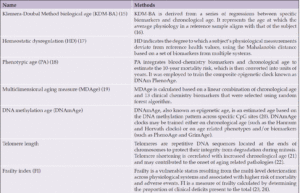
COGNITIVE INTERVENTIONS: SYMPTOMATIC OR DISEASEMODIFYING TREATMENTS IN THE BRAIN?


F. Bellelli1,2


1. Fellowship in Geriatric and Gerontology, University of Milan, Milan, Italy; 2. Gérontopôle de Toulouse, Institut du Vieillissement, Centre Hospitalo-Universitaire de Toulouse, Toulouse, France.

Corresponding Author: Federico Bellelli, MD, Fellowship in Geriatric and Gerontology, University of Milan, Milan, Italy, + 39 3319709061, federico.bellelli@unimi.it

J Aging Res & Lifestyle 2024;13:60-64
Published online May 22, 2024, http://dx.doi.org/10.14283/jarlife.2024.8


Abstract

Recent findings suggest that brain-stimulating activities may have beneficial effects on both Mild Cognitive Impairment (MCI) and Alzheimer’s Disease (AD). However, whether cognitive interventions merely enhance cognitive reserve or truly attenuate, or even reverse, the disease’s pathophysiology is still controversial. The aim of the present article is to discuss the potential for brain-stimulating activities, including cognitive stimulation (CS), cognitive rehabilitation (CR), and cognitive training (CT), to be symptomatic or disease-modifying interventions in the context of cognitive decline. While emerging evidence indicates that CT can enhance synaptic plasticity, suggesting a potential role in augmenting cognitive reserve, its impact on AD pathology remains uncertain. Small-scale studies suggest that CT and CS may slow down neurodegeneration in MCI patients and that multidomain interventions combining physical activity with CT may benefit Aβ pathology. However, the considerable heterogeneity across studies limits the comparability of findings. It underscores the necessity for a more standardized approach to cognitive interventions in future guidelines for preventing and managing cognitive decline.

Key words: Cognitive stimulation, cognitive rehabilitation, cognitive training, Alzheimer disease, disease-modifying treatments.


The World Health Organization (WHO) and the National Institute for Health and Care Excellence (NICE) recommend social interactions and other brain-stimulating activities as non-pharmacological treatments for dementia (1, 2). However, the extent to which such interventions merely alleviate signs and symptoms of cognitive decline, reduce the pathophysiological burden of Alzheimer’s disease (AD – most prevalent type of dementia), benefit both aspects or have no discernible effect remains unclear and deserve further debate. The present article aims to discuss the potential for brain-stimulating activities to be symptomatic or disease-modifying interventions in the context of cognitive decline during aging.


Brain stimulating activities

Among various brain-stimulating interventions, most recommendations endorse cognitive stimulation (CS) (1, 3), cognitive rehabilitation (CR)(1), or cognitive training (CT) (4). CS consists of various activities and discussions aimed at improving social and cognitive functioning (1). Similarly, CR works to achieve goals relevant to the person living with dementia and his family, trying to enhance and maintain functioning in everyday life (1). On the other hand, CT is a more specific approach that works on a set of standardized tasks designed to reflect singular cognitive functions (i.e., episodic memory) (1) and is, therefore, particularly suitable for individuals with Mild Cognitive Impairment (MCI) (5).
Multiple systematic reviews have suggested in the last two decades that individuals with MCI may experience slightly to moderate improvements following cognitive interventions. Still, the heterogeneity of the studies made it challenging to distinguish which method had the strongest impact (6, 7). Recently, a systematic review and meta-analysis extended the evaluation of cognitive interventions to individuals with AD dementia. Analyzing 25 studies involving 2012 participants, the review concluded that while further research on CR and CS is warranted, there is some indication of temporary benefits on global cognitive function following CT (8).
However, the mechanisms by which these interventions may improve cognitive functions are still debated. Indeed, according to the cognitive reserve hypothesis, individuals with a greater cognitive reserve can withstand a higher AD pathological burden before developing dementia by employing mental processing approaches or compensatory brain networks (synaptic plasticity) (9). Given that, do cognitive interventions enhance cognitive reserve, or do they genuinely attenuate or reverse the disease’s pathophysiology?


Tackling cognitive decline symptoms through improved cognitive reserve

Recent findings suggest that cognitive interventions might have a beneficial impact on synaptic plasticity and, consequently, on cognitive reserve. Indeed, several studies have demonstrated that CT can enhance regional activity in functional Magnetic Resonance Imaging (fMRI) scans of patients with MCI following an intervention ranging from 2 weeks to 12 months (10–13). In particular, Hampstead et al. proposed that the most robust training-specific increases occur within areas of the default network that are abnormal in MCI and AD (medial frontal and parietal cortices and around the temporoparietal junction) (12). Accordingly, two studies found that compared to standard care, 8-week CR and 7-week CS programs could improve fMRI even in individuals with mild dementia (14, 15). Interestingly, Bentham et al. observed that individuals with high vascular burden had a lower functional connectivity response to CS than those with low burden, suggesting that vascular pathology could limit the potential for a neuroplastic response to cognitive interventions (16).


Brain stimulation and the biomarkers of AD

Regarding the core biomarkers of AD (Aβ and Tau), evidence on cognitive interventions remains scarce and inconclusive. A small study employed a questionnaire to retrospectively evaluate the engagement in cognitively stimulating activities (e.g., reading, writing, playing games) across the lifespan of 65 healthy older adults and 10 AD patients. The study observed greater participation in cognitively stimulating activities, particularly during early and middle adulthood, was associated with decreased amyloid PET burden compared to individuals with limited involvement in such activities (17). Accordingly, secondary analyses of a subset of the Multidomain Alzheimer Preventive Trial (MAPT), an extensive study on community-dwelling older adults at risk of cognitive decline, suggested that a three-year multidomain intervention, including CT and physical activity (PA) advice, was associated with lower Aβ burden on amyloid PET compared to controls (18). In contrast, no significant difference was observed in plasma phosphorylated-tau levels (19). However, a small study on community-dwelling older adults (n=27) suggested that PA rather than CT may primarily drive the effects on Aβ levels. Indeed, the study reported reduced plasma Aβ levels after a 12-week program in both single-task (PA) and dual-task training groups (PA + CT), with no between-group difference (20).
On the other hand, a similar study on individuals with AD (n=34) observed that an 8-week dual-task training significantly lowered plasma Aβ levels. In contrast, no significant difference was reported for the group doing only PA, suggesting that CT might influence AD pathophysiology (21). Notably, a study on individuals with MCI assessing the effects of a 9-month cognitive intervention alone (mindful awareness practice; MAP) on Aβ-42 levels (salivary sample) found no significant differences between the treatment arm (MAP) and the active control group (22). Even in preclinical AD models, evidence remains unclear, with some studies suggesting decreased (23-25), no change (26, 27), or increased (28) Aβ load in transgenic mice exposed to cognitive stimulation (i.e., enriched environment or spatial training).


Brain stimulation and the biomarkers of neurodegeneration

According to the Revised Criteria for Diagnosis and Staging of Alzheimer’s Disease, anatomic MRI and Fluorodeoxyglucose Positron Emission Tomography (FDG-PET) are biomarkers of non-specific degeneration in AD pathophysiology (29). However, cognitive interventions’ effects on structural and metabolic changes in the brain are still unclear. A retrospective study involving 329 cognitively unimpaired middle-aged adults revealed that individuals engaging in cognitively stimulating activities, such as playing card games, exhibited larger gray matter volumes in brain regions susceptible to AD pathology (30). In contrast, secondary analysis of a subset (n= 244) of the Finnish Geriatric Intervention Study to Prevent Cognitive Impairment and Disability (FINGER), a large-scale trial involving older adults at risk for dementia, found no changes in regional brain volumes or cortical thickness after two years of a multidomain intervention comprising PA, CT, diet, and vascular risk monitoring (31). The study suggests that multidomain intervention has no beneficial effect on neurodegeneration among cognitively unimpaired individuals at risk of AD. However, the design of the FINGER study did not permit the evaluation of the impact of interventions in individuals already experiencing some level of cognitive impairment. Three small-scale studies suggested that cognitive interventions may benefit MRI changes in individuals with MCI. Specifically, a small study allocated individuals with mild dementia to either a 7-week CS program (n=16) or standard care (n=13). The study observed that individuals in the treatment group maintained their total brain volume, as assessed by MRI, while those in the standard care group experienced a decrease (15). Accordingly, another study suggested that a 6-month multi-intervention program based on aerobic exercises and CS could decelerate atrophy in AD-related brain regions among MCI patients (32). Furthermore, the last study observed that undergoing 24 sessions of computerized CT led to focal increases in cortical thickness among individuals with MCI (33), even hinting at a potential for reversibility of neurodegeneration.
Regarding brain metabolism, some studies showed a trend toward regional metabolic changes following cognitive interventions among cognitively unimpaired individuals at risk for dementia. Secondary analyses of a subset population (n=67) of the MAPT study suggested that combined treatment with omega-3 supplementation and multidomain intervention (CT and physical activity; PA) did not significantly increase overall brain metabolism after six or twelve months of treatment. However, exploratory analyses employing voxel-wise approaches suggested that multidomain intervention could enhance metabolism in specific brain regions, including the right hippocampus, right posterior cingulate, left posterior para-hippocampal gyrus, and right insular cortex (34). Likewise, a small study on community-dwelling older adults (n=45) showed that a 16-week computerized CT had a trend toward a metabolic increase in the right inferior frontal gyrus without reaching the statistical significance (35). However, evidence in individuals with cognitive decline is limited and contradicting. A small study suggested that a six-month cognitive intervention might decelerate the widespread cortical metabolic decline in AD and MCI patients, with a more pronounced effect observed in the latter (36). On the contrary, another study observed that a 12-week home-based CT program did not yield significant improvements in brain metabolism in individuals with MCI (37).


Conclusions

In conclusion, evidence indicates a rise in synaptic plasticity following CT, suggesting that the beneficial effects of cognitive interventions may be partially attributed to the enhancement of cognitive reserve. However, whether cognitive interventions can attenuate or reverse AD pathophysiology (amyloid beta, tau, and neurodegeneration) remains subject to debate, and further studies are required (Figure 1). The need for robust evidence in this field could be attributed to various factors, including small study sample sizes, the substantial methodological heterogeneity across trials (rendering comparability across studies a problematic exercise), and combined interventions. Indeed, most studies evaluated the combined effects of physical activity and CT, making it challenging to discern the effects of one intervention. Additionally, the high cost of neuroimaging and limited accessibility to AD core biomarkers in cerebrospinal fluid (CSF) have posed significant constraints on large-scale population studies in previous years. However, the recent validation of more cost-effective and less invasive plasma-based biomarkers (29) may offer a valuable opportunity further to investigate cognitive interventions’ effects on AD pathogenesis.

Figure 1. Effects of cognitive interventions on the brain

Legend: CS, Cognitive Stimulation; CT, Cognitive Training; CR, Cognitive Rehabilitation; fMRI, functional Magnetic Resonance Imaging; FDG-PET, Fluorodeoxyglucose Positron Emission Tomography; MCI, Mild Cognitive Impairment; PA, Physical Activity


Moreover, considering that cognitive interventions may enhance cognitive reserve, it is reasonable to expect different results based on participants’ baseline cognitive reserve. Therefore, further studies must consider at least the subjects’ educational level. Lastly, the primary reason for more evidence in the field might be the substantial heterogeneity of the studies. Notably, the duration (ranging from a few days to 3 years) and the methodologies employed in cognitive interventions were quite different between various trials. Indeed, although most guidelines endorse non-pharmacological approaches as first-line treatment for dementia (4) and MCI (38), official recommendations regarding the specifics of these interventions have often been lacking in previous years. Recently, the WHO ICOPE guidelines recommended cognitive stimulation for older adults with cognitive impairment, suggesting a standard group approach involving up to 14 themed sessions lasting approximately 45 minutes each, held twice a week (3). Continuing this path, future guidelines on the prevention of cognitive decline, as well as on the treatment and management of dementia, should aim to standardize at least the duration and the modalities of cognitive interventions while preserving the person-centered care approach that is a cornerstone of geriatric medicine.


Conflict of Interest: The author declares no conflicts of interest.

Open Access: This article is distributed under the terms of the Creative Commons Attribution 4.0 International License (http://creativecommons.org/licenses/by/4.0/), which permits use, duplication, adaptation, distribution and reproduction in any medium or format, as long as you give appropriate credit to the original author(s) and the source, provide a link to the Creative Commons license and indicate if changes were made.


References

1. Dementia: assessment, management and support for people living with dementia and their carers. NICE Guideline. 2018. https://www.nice.org.uk/guidance/ng97. Accessed 21 Feb 2024.
2. World Health Organization. Risk reduction of cognitive decline and dementia: WHO guidelines. Geneva: World Health Organization. 2019. https://iris.who.int/handle/10665/312180. Accessed 21 Feb 2024.
3. Integrated care for older people (ICOPE): guidance for person-centred assessment and pathways in primary care. 2019. https://www.who.int/publications-detail-redirect/WHO-FWC-ALC-19.1. Accessed 21 Feb 2024.
4. Kane RL, Butler M, Fink HA, et al. Interventions to Prevent Age-Related Cognitive Decline, Mild Cognitive Impairment, and Clinical Alzheimer’s-Type Dementia. Rockville (MD): Agency for Healthcare Research and Quality (US); 2017. http://www.ncbi.nlm.nih.gov/books/NBK442425/ Accessed 22 Feb 2024.
5. Zhang H, Huntley J, Bhome R, et al. Effect of computerised cognitive training on cognitive outcomes in mild cognitive impairment: a systematic review and meta-analysis. BMJ Open. 2019 Aug 1;9(8):e027062. DOI: 10.1136/bmjopen-2018-027062
6. Reijnders J, van Heugten C, van Boxtel M. Cognitive interventions in healthy older adults and people with mild cognitive impairment: A systematic review. Ageing Res Rev. 2013 Jan 1;12(1):263–75. DOI: 10.1016/j.arr.2012.07.003
7. Simon SS, Yokomizo JE, Bottino CMC. Cognitive intervention in amnestic Mild Cognitive Impairment: A systematic review. Neurosci Biobehav Rev. 2012 Apr 1;36(4):1163–78. DOI: 0.1016/j.neubiorev.2012.01.007
8. Wang YY, Yang L, Zhang J, Zeng XT, Wang Y, Jin YH. The Effect of Cognitive Intervention on Cognitive Function in Older Adults With Alzheimer’s Disease: A Systematic Review and Meta-Analysis. Neuropsychol Rev. 2022 Jun 1;32(2):247–73. DOI: 10.1007/s11065-021-09486-4
9. Roe CM, Mintun MA, D’Angelo G, Xiong C, Grant EA, Morris JC. Alzheimer Disease and Cognitive Reserve: Variation of Education Effect With Carbon 11–Labeled Pittsburgh Compound B Uptake. Arch Neurol. 2008 Nov 10;65(11):1467–71. DOI: 10.1001/archneur.65.11.1467
10. Rosen AC, Sugiura L, Kramer JH, Whitfield-Gabrieli S, Gabrieli JD. Cognitive Training Changes Hippocampal Function in Mild Cognitive Impairment: A Pilot Study. J Alzheimers Dis. 2011;26(Suppl 3):349–57. DOI: 10.3233/JAD-2011-0009
11. Li B, Tang H, He G, et al. Tai Chi enhances cognitive training effects on delaying cognitive decline in mild cognitive impairment. Alzheimers Dement. 2023;19(1):136–49. DOI: 10.1002/alz.12658
12. Hampstead BM, Stringer AY, Stilla RF, et al. ACTIVATION AND EFFECTIVE CONNECTIVITY CHANGES FOLLOWING EXPLICIT MEMORY TRAINING FOR FACE-NAME PAIRS IN PATIENTS WITH MILD COGNITIVE IMPAIRMENT: A PILOT STUDY. Neurorehabil Neural Repair. 2011;25(3):210–22. DOI: 10.1177/1545968310382424
13. Belleville S, Clément F, Mellah S, Gilbert B, Fontaine F, Gauthier S. Training-related brain plasticity in subjects at risk of developing Alzheimer’s disease. Brain J Neurol. 2011 Jun;134(Pt 6):1623–34. DOI: 10.1093/brain/awr037
14. van Paasschen J, Clare L, Yuen KSL, Woods RT, Evans SJ, Parkinson CH, et al. Cognitive rehabilitation changes memory-related brain activity in people with Alzheimer disease. Neurorehabil Neural Repair. 2013 Jun;27(5):448–59. DOI: 10.1177/1545968312471902
15. Liu T, Spector A, Mograbi DC, Cheung G, Wong GHY. Changes in Default Mode Network Connectivity in Resting-State fMRI in People with Mild Dementia Receiving Cognitive Stimulation Therapy. Brain Sci. 2021 Sep;11(9):1137. DOI: 10.3390/brainsci11091137
16. Bentham C, De Marco M, Venneri A. The Modulatory Effect of Cerebrovascular Burden in Response to Cognitive Stimulation in Healthy Ageing and Mild Cognitive Impairment. Neural Plast. 2019 Aug 6;2019:2305318. DOI: 10.1155/2019/2305318
17. Landau SM, Marks SM, Mormino EC, et al. Association of Lifetime Cognitive Engagement and Low β-Amyloid Deposition. Arch Neurol. 2012 May 1;69(5):623–9. DOI: 10.1001/archneurol.2011.2748
18. Hooper C, Coley N, De Souto Barreto P, et al. Cortical β-Amyloid in Older Adults Is Associated with Multidomain Interventions with and without Omega 3 Polyunsaturated Fatty Acid Supplementation. J Prev Alzheimers Dis. 2020 Mar 1;7(2):128–34. DOI: 10.14283/jpad.2020.4
19. Coley N, Zetterberg H, Cantet C, et al. Plasma p-tau181 as an outcome and predictor of multidomain intervention effects: a secondary analysis of a randomised, controlled, dementia prevention trial. Lancet Healthy Longev. 2024 Feb;5(2):e120–30. DOI: 10.1016/S2666-7568(23)00255-6
20. Yokoyama H, Okazaki K, Imai D, et al. The effect of cognitive-motor dual-task training on cognitive function and plasma amyloid β peptide 42/40 ratio in healthy elderly persons: a randomized controlled trial. BMC Geriatr. 2015 May 28;15:60. DOI: 10.1186/s12877-015-0058-4
21. Nam SM, Kim S gil. Dual-Task Training Effect on Cognitive and Body Function, β-amyloid Levels in Alzheimer’s Dementia Patients: A Randomized Controlled Trial. J Korean Phys Ther. 2021 Jun 30;33(3):136–41. DOI: 10.18857/jkpt.2021.33.3.136
22. Ng TKS, Slowey PD, Beltran D, Ho RCM, Kua EH, Mahendran R. Effect of mindfulness intervention versus health education program on salivary Aβ-42 levels in community-dwelling older adults with mild cognitive impairment: A randomized controlled trial. J Psychiatr Res. 2021 Apr 1;136:619–25. DOI: 10.1016/j.jpsychires.2020.10.038
23. Herring A, Lewejohann L, Panzer AL, Donath A, Kröll O, Sachser N, et al. Preventive and therapeutic types of environmental enrichment counteract beta amyloid pathology by different molecular mechanisms. Neurobiol Dis. 2011 Jun;42(3):530–8. DOI: 10.1016/j.nbd.2011.03.007
24. Gerenu G, Dobarro M, Ramirez MJ, Gil-Bea FJ. Early cognitive stimulation compensates for memory and pathological changes in Tg2576 mice. Biochim Biophys Acta BBA – Mol Basis Dis. 2013 Jun 1;1832(6):837–47. DOI: 10.1016/j.bbadis.2013.02.018
25. Mehla J, Deibel SH, Karem H, et al. Repeated multi-domain cognitive training prevents cognitive decline, anxiety and amyloid pathology found in a mouse model of Alzheimer disease. Commun Biol. 2023 Nov 10;6:1145. DOI: 10.1038/s42003-023-05506-6
26. Arendash GW, Garcia MF, Costa DA, Cracchiolo JR, Wefes IM, Potter H. Environmental enrichment improves cognition in aged Alzheimer’s transgenic mice despite stable beta-amyloid deposition. Neuroreport. 2004 Aug 6;15(11):1751–4. DOI: 10.1097/01.wnr.0000137183.68847.4e
27. Costa DA, Cracchiolo JR, Bachstetter AD, Hughes TF, Bales KR, Paul SM, et al. Enrichment improves cognition in AD mice by amyloid-related and unrelated mechanisms. Neurobiol Aging. 2007 Jun;28(6):831–44. DOI: 10.1016/j.neurobiolaging.2006.04.009
28. Stuart KE, King AE, Fernandez-Martos CM, Summers MJ, Vickers JC. Environmental novelty exacerbates stress hormones and Aβ pathology in an Alzheimer’s model. Sci Rep. 2017 Jun 5;7:2764. DOI: 10.1038/s41598-017-03016-0
29. AAIC. Revised Criteria for Diagnosis and Staging of Alzheimer’s. 2023. http: //aaic.alz.org/diagnostic-criteria.asp. Accessed: 24 Feb 2024.
30. Schultz S, Larson J, Oh J, et al. Participation in cognitively-stimulating activities is associated with brain structure and cognitive function in preclinical Alzheimer’s disease. Brain Imaging Behav. 2015 Dec;9(4):729–36. DOI: 10.1007/s11682-014-9329-5
31. Stephen R, Liu Y, Ngandu T, et al. Brain volumes and cortical thickness on MRI in the Finnish Geriatric Intervention Study to Prevent Cognitive Impairment and Disability (FINGER). Alzheimers Res Ther. 2019 Jun 4;11(1):53. DOI: 10.1186/s13195-019-0506-z
32. Köbe T, Witte AV, Schnelle A, et al. Combined omega-3 fatty acids, aerobic exercise and cognitive stimulation prevents decline in gray matter volume of the frontal, parietal and cingulate cortex in patients with mild cognitive impairment. NeuroImage. 2016 May 1;131:226–38. DOI: 10.1016/j.neuroimage.2015.09.050
33. Na HR, Lim JS, Kim WJ, et al. Multimodal Assessment of Neural Substrates in Computerized Cognitive Training: A Preliminary Study. J Clin Neurol Seoul Korea. 2018 Oct;14(4):454–63. DOI: 10.3988/jcn.2018.14.4.454
34. Delrieu J, Voisin T, Saint-Aubert L, et al. The impact of a multi-domain intervention on cerebral glucose metabolism: analysis from the randomized ancillary FDG PET MAPT trial. Alzheimers Res Ther. 2020 Oct 19;12(1):134. DOI: 10.1186/s13195-020-00683-6
35. Shah T, Verdile G, Sohrabi H, et al. A combination of physical activity and computerized brain training improves verbal memory and increases cerebral glucose metabolism in the elderly. Transl Psychiatry. 2014 Dec 2;4(12):e487. DOI: 10.1038/tp.2014.122
36. Förster S, Buschert VC, Teipel SJ, et al. Effects of a 6-month cognitive intervention on brain metabolism in patients with amnestic MCI and mild Alzheimer’s disease. J Alzheimers Dis JAD. 2011;26 Suppl 3:337–48. DOI: 10.3233/JAD-2011-0025
37. Park J, Kim SE, Kim EJ, et al. Effect of 12-week home-based cognitive training on cognitive function and brain metabolism in patients with amnestic mild cognitive impairment. Clin Interv Aging. 2019 Jun 28;14:1167–75. DOI: 10.2147/CIA.S200269
38. Petersen RC, Lopez O, Armstrong MJ, et al. Practice guideline update summary: Mild cognitive impairment. Neurology. 2018 Jan 16;90(3):126–35. DOI: 10.1212/WNL.0000000000004826

© The Authors 2024
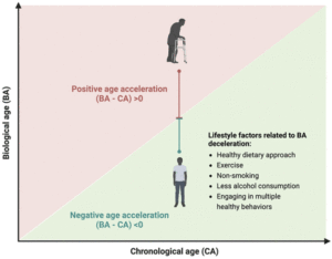
MEDICAL-GRADE HONEY IS A VERSATILE WOUND CARE PRODUCT FOR THE ELDERLY


D. Chrysostomou1-3, A. Pokorná2,4, N.A.J. Cremers5,6, L.J.F. Peters5


1. Wound Clinic Health@45, Linksfield Road 45, Dowerglen, Johannesburg 1612, South Africa; 2. Department of Health Sciences, Faculty of Medicine, Masaryk University, Brno, Czech Republic; 3. Department of Public Health, Faculty of Medicine, Masaryk University, Brno, Czech Republic; 4. College of Polytechnics Jihlava, Jihlava, Czech Republic; 5. Triticum Exploitatie BV, Sleperweg 44, 6222NK Maastricht, The Netherlands; 6. Department of Gynecology and Obstetrics, Maastricht University Medical Center, 6202 AZ Maastricht, The Netherlands.

Corresponding Author: Linsey J.F. Peters, Triticum Exploitatie BV, Sleperweg 44, 6222NK Maastricht, The Netherlands, research@mesitran.com, +31 (0)43 325 1773.

J Aging Res & Lifestyle 2024;13:51-59
Published online May 17, 2024, http://dx.doi.org/10.14283/jarlife.2024.7


Abstract

INTRODUCTION: Ageing of the global population has led to an increase in the demand for the treatment of wounds, especially considering the challenges of managing wounds in the elderly. Therefore, more effective treatment strategies need to be explored. In this article, we aimed to compare medical-grade honey (MGH) products with other wound care products and to provide guidelines on using MGH in wounds commonly found in the elderly.
METHODS: Based on literature research and expert opinion, an overview of commonly used wound care products and their wound healing characteristics is provided. In addition, literature-based classification of wounds in the elderly and the recommendations for treatments are provided.
RESULTS: Frequently used wound care products include povidone-iodine, enzymatic products, absorbing dressings, larvae, silver dressings, and MGH dressings. Supported by systematic reviews and meta-analyses, MGH dressings were identified as the most potent and all-round wound care product compared to the others. Next, we provided basic guidelines for managing the most common wounds in the elderly, both acute and chronic, and specified how and which MGH products can be used in these wounds.
CONCLUSION: MGH is a widely applicable, safe, easy-to-use, and cost-effective product to manage wounds in the elderly. In case of doubt, refer to a trained wound care specialist who can support the treatment of difficult-to-heal wounds.

Key words: Wound care, medical-grade honey, geriatric patients, chronic wounds, acute wounds.


Background

The standard of wound care has increased tremendously in the last few years. Scientific research combined with expert opinion led to the creation of guidelines for wound management. All medical practitioners will come across wounds in their daily practice, especially regarding elderly patients. Looking ahead to the year 2050, the population of older adults will rise from 1 billion to 2.1 billion (1). Population ageing has major consequences for healthcare, including rising demand, economic effects, and personal expenditures (2). Moreover, elderly will encounter a variety of health issues, including chronic wounds. These wounds fail to heal in a predictable timeframe (4 weeks to 3 months) or in a typical set of stages without responding to standard therapies (3).
Chronic wounds, also known as hard-to-heal or non-healing wounds, are associated with considerable morbidity and mortality in elderly (4). These wounds are common among elderly due to various factors such as comorbidities, e.g. vascular insufficiency or diabetes (4, 5). Chronic wounds are characterized by a disrupted healing process, which normally exists in four overlapping phases (6, 7): hemostasis, inflammation, proliferation, and maturation. Wounds turn chronic often due to bacterial infections, but also comorbidities and/or higher fragility and slower healing of the skin consequent to aging (7-9). Thus, as the elderly population continues to grow, the burden of managing wounds becomes even more critical. Therefore, exploring effective strategies to improve wound healing and prevent acute wounds from becoming chronic in older adults is crucial. Special attention must be paid to the effective management of infection as it is known historically that in the elderly, the symptomatology of infection is altered in their clinical presentation due to age-related alterations in immunology (10).
Honey has been used to treat wounds throughout history but became forgotten as a wound care product because of the discovery of antibiotics (11-14). With the advent of antibiotic-resistant bacteria, there has been increasing interest in honey as a wound care product (14-18). The wound care properties of honey are based on two main aspects: its antimicrobial and pro-healing effects (15, 19, 20). Because honey targets the bacteria in a multifaceted manner, it works against a broad spectrum of bacteria and lacks the risk of resistance. Commercially available honey has lower antibacterial activities due to production processes and adulteration (21-23). Therefore, medical-grade honey (MGH) is preferred for wound care as it meets strict safety criteria, such as being organic and free from pollutants, and is sterilized to ensure safety and efficacy for medical use (21-24). While MGH has shown effectiveness in elderly patients, non-honey products, such as povidone iodine or silver, are still commonly used. The broad range of wound care products available can overwhelm the health care professional and exact guidelines for treating wounds in elderly are hard to find.
This article aims to provide an overview and comparison between non-honey and MGH-based wound care products that can be used in the elderly. Furthermore, we will focus on the different types of wounds encountered in elderly and suggest management strategies. We will also demonstrate how MGH can be used in every wound care situation in elderly.


Wound care products

Wounds can be treated with a variety of products and the choice can be challenging for the healthcare professional. To choose an appropriate dressing, we first need to know what the ideal dressing would be. The characteristics of an ideal wound care product include (25-27):
• Can absorb and control exudate
• Cost-effective
• Can be removed without causing damage to the wound
• Easy to use
• Reduces and controls bacterial load
• Removes sloughy and necrotic tissue
• Exhibits anti-inflammatory properties
• Eliminates unpleasant odor from the wound
• Non-toxic and promotes the growth of new tissue

The most commonly used products will be discussed and compared to the ideal wound care dressing characteristics (Table 1).

Table 1. Wound care products and their effects on the wound healing process

Results are based on the IFUs of the products. V marks a positive effect, while X marks a negative effect on the described characteristic. SSD = silver sulphadiazine; MGH = medical-grade honey.


Povidone-iodine

Povidone-iodine is a common antiseptic agent used in wound care and has a broad-spectrum antibacterial activity. It releases free iodine, which quickly penetrates microorganisms and eventually causes cell death (28, 29). Povidone-iodine is available in various forms and is often used for wound cleansing and preoperative skin preparation. One consideration is that it is damaging to healthy tissue, thus slowing wound healing (30).

Enzymatic products

Enzymatic products are designed to debride the wound. Debridement means removing necrotic material, devitalized tissues, scabs, and other impurities that delay wound healing (31). These products contain specific enzymes, such as collagenases or proteases, that digest and degrade devitalized tissue. Some enzymes selectively target non-viable tissue, while others also target viable tissue (31). One should note though that enzymatic products lack antimicrobial properties (32).

Absorbing dressings

Absorbing dressings are designed to manage wound exudate and maintain a moist wound environment, which is considered key in wound management (33, 34). These dressings are composed of highly absorbent materials, such as foam or alginate, which effectively absorb and retain excess fluid from the wound bed (34). By minimizing excessive moisture, absorbing dressings help prevent maceration of the surrounding skin and promotes optimal conditions for wound healing. Absorbing dressings are useful in creating and maintaining a moist wound environment. These dressings can be used as complementary dressings (34).

Larvae

Larval therapy, also known as maggot debridement therapy, involves the controlled application of medical-grade fly larvae to wounds (35). The larvae secrete enzymes that break down necrotic tissue, effectively debriding the wound. Moreover, maggot therapy has been shown to have anti-inflammatory properties as well (35, 36). Larval therapy is particularly beneficial for chronic, non-healing wounds with significant necrosis. One of the limitations of this treatment is that maggots can induce dermatitis when not properly secured (37). Also, not many patients are comfortable with larval therapy.

Silver dressings

Silver dressings are dressings that contain silver compounds or nanoparticles and have broad-spectrum antimicrobial properties. These dressings provide a sustained release of silver ions, which exert their antimicrobial effects by disrupting microbial cell membranes and interfering with essential cellular processes (38). Silver dressings are frequently used in infected wounds to reduce the bacterial burden and promote wound healing. However, silver dressings are advised to be used for no longer than two weeks, after which treatment should be switched to a non-silver dressing (39). The use of silver can delay wound healing, lead to skin irritation, and carries a high risk of developing argyria.

MGH-based dressings

MGH-based dressings are divided into two main categories: Manuka honey and other kinds of honey. The main difference between the two is that Manuka’s antibacterial activity relies mostly on methylglyoxal while other honeys rely on hydrogen peroxide production (40). MGH-based dressings harness the natural properties of honey, including its antimicrobial effects and promotion of wound healing (19, 20). Additionally, these dressings have been shown to effectively manage infected wounds, promote autolytic debridement, stimulate a moist wound environment, and enhance re-epithelialization (41-43). The unique composition of MGH-based dressings contributes to their versatility and therapeutic efficacy in various wound types and stages of healing. One should note that pure MGH or MGH in high concentrations can cause a stingy feeling upon application.
Although each product ticks multiple boxes, only MGH matches all characteristics of the ideal wound care dressing. Systematic reviews and meta-analyses have concluded that MGH has antimicrobial properties, stimulates wound healing, has several benefits over the use of other wound care products, can be used for a wide range of acute and chronic wounds, and is cost-effective (44-50). Besides this, studies have compared MGH to silver products and povidone iodine and concluded that MGH was superior as a wound care dressing (44, 50).


Wound care protocol

Patient characteristics influence the healing trajectory. Therefore, management of wounds in elderly patients must start with a full history, physical examination, and identification of systemic, psychological, lifestyle, and local factors. Appendix 1 shows an assessment form to be able to pinpoint all the unique patient-related issues. Based on this information, a proper plan of care is developed. Consideration should also be given to means of transportation to a wound care facility and the patient’s capability to participate in the management of their wound(s). Social support in all forms will be paramount in the completion of a multifaceted plan of wound care.
Wounds can be categorized into distinct classifications, namely acute and chronic wounds, each encompassing a diverse array of types. We will provide an overview of the different types of wounds that regularly occur in elderly and how to manage them according to wound type and classification. Since MGH-based wound care products have the characteristics of the ideal wound care dressing, we will highlight in Figure 1 which MGH-based product can be applied in which wound care situation exactly. Numerous studies have shown that supplementation of MGH with other compounds, such as vitamins C and E, leads to heightened antimicrobial and wound healing activities of the MGH compared to its non-supplemented counterpart (51-54). Therefore, we demonstrated the use of supplemented MGH products (L-Mesitran, manufactured by Theo Manufacturing, Maastricht, the Netherlands) which include ointment, gel, tulle, hydrogel, and foam dressings.

Figure 1. Medical-grade honey for use in elderly

Suggested treatment options using supplemented MGH products for wounds that are commonly found in elderly. Each wound is classified (55-59) and based on the classification an appropriate product of the supplemented MGH range is suggested. Hydro = L-Mesitran Hydro; Gauze = L-Mesitran Tulle; Foam = L-Mesitran Foam; Gel = L-Mesitran Soft; Ointment = L-Mesitran Ointment.


Common acute wounds in the ageing population

Skin tear

One of the most frequent acute wounds is skin tears (Figure 2A). These traumatic wounds can result from friction, shear, or blunt trauma. Skin tears can happen on any area of the body and are more likely to occur in individuals with delicate skin, in particular the elderly (60). Skin tears can involve the separation of the epidermis from the dermis (partial thickness wounds) or the separation of both the epidermis and dermis from underlying structures (full-thickness wounds) (60).

Figure 2. Common acute wounds in elderly

A) Skin tear with partial flap loss, which cannot be repositioned to cover the wound bed. B) Infected post-operative abdominal wound.


There is one international classification tool validated and recommended to use in the management of skin tears: The International Skin Tear Advisory Panel (ISTAP)(55). It recommends the following steps in managing lacerations:
1. Assess and classify the wound using a reliable tool.
2. Preserve as much as possible of the skin flap (gently with a moistened swab).
3. Aline the edges of the wound and secure it with gentle, adhesive, sterile tape.
4. Protect the wound from further injury using an antimicrobial, sterile dressing.
5. Follow up to ensure adequate wound healing.

The use of MGH for treating skin tears has been documented previously (61, 62). In both publications, skin tears in elderly patients were successfully treated with MGH. The advantage for skin tears are especially the non-adherent properties of MGH-based products. This allows for easy removal of the dressing while not causing any trauma to the wound or surrounding skin. Depending on the stage of the wound, one could opt for an MGH-based hydrogel, gauze, or foam dressing (Figure 1).

Surgical site management

Increased age is an operative risk factor. An American government study shows that even though people >65 years represent 13% of the country’s population, 20% of the total surgical procedures are allocated to this group (63). Concerning wound care, the most frequent complication of the surgical site is infection (Figure 2B). Such misfortune will result in suffering, prolonged hospital stays, increased cost of care, and increased use of resources (64).
An array of studies regarding the best choice of postoperative dressing concluded that recognition of the surgical wound classification should enable the clinician to choose the adequate wound cover. Surgical wounds are classified for the risk for a surgical site infection (SSI)(56), as clean, clean/contaminated, contaminated, or dirty. Surgical wounds should be kept clean, change of dressing should be done using a sterile technique. One should follow the following management steps:
1. Assess and classify the contamination risk.
2. Clean the wound with an antiseptic fluid.
3. Use a topical antimicrobial, such as MGH.
4. Cover the wound with a sterile dressing.
5. Check and change dressing according to the exudate level and bacterial load.

Several publications have highlighted the effective use of supplemented MGH-based dressings for surgical wounds (41, 42, 65-69). Although most publications include infected, dehisced surgical wounds, MGH can also be used to reduce the infection rate and improve healing as shown by various clinical studies (69-71). Based on the SSI classification, one could use an MGH-based foam dressing alone or combined with an MGH-based wound gel or ointment (Figure 1).

Common chronic wounds in older adult

Vascular leg ulcers

The most frequent vascular ulcers of the lower limb are venous, arterial, or mixed etiology (Figure 3A). Identification and diagnosis are imperiously necessary to be able to deliver appropriate treatment. Venous ulcers are mainly situated above the malleoli, presenting with irregular edges, while arterial ulcers are found over the bony prominence, with round edges and are smaller (72, 73). Mixed ulcers can be present anywhere from on the lower leg, below the knee, all the way to the foot. Venous ulcers are more painful with non-elevation of the leg, while arterial ones are painful on elevation (72, 73).
Doppler assessment and ankle brachial pressure index (ABPI) should be performed to establish treatment steps. If ABPI is between 0.8 and 1.1, compression can be used. However, caution is strongly recommended in diabetic patients. If ABPI value is less than 0.8 referral to a vascular surgeon is firmly indicated (72, 73). Furthermore, when lower leg ulcers are painful and have a high bacterial load, wound treatment should consider non-adhesive and antibacterial dressings (72, 73).

The golden standard for treatment according to the etiology of the wound:
1. Venous ulcers – compression and wound care
2. Arterial ulcers – revascularization and wound care
3. Mixed ulcers – revascularization, modified compression, and wound care

Supplemented MGH-based products have been used to treat vascular leg ulcers successfully in the clinic (41, 42, 62, 74-76). These reports showed that even vascular ulcers infected with biofilms or resistant bacterial strains could be healed with supplemented MGH. Moreover, MGH products can be combined with compression thereby making them suitable for treatment of venous leg ulcers. For vascular ulcers, treatment should involve a combination of an MGH-based ointment or gel with an MGH-based foam dressing (Figure 1).

Diabetic foot ulcers

Presently, millions of people with diabetes suffer from poorly healing foot ulcers (Figure 3B) (77). The management of diabetic foot ulcers (DFU) is arguably the costliest (78). Unfortunately, it is not limited to the financial aspect alone, the cost is measured in loss of quality of life, loss of limb, and loss of life itself (79).
The first step in the treatment of DFUs is classifying the wound. DFUs are classified from grade 0 to grade 5 according to Wagner’s classification tool (57). A DFU is one of the most challenging and complex wounds, due to numerous intrinsic and extrinsic factors influencing the management outcome. Therefore the following steps should be taken:
• Discuss realistic goals with the patient and carers;
• Plan the treatment;
• Document every step with photographic evidence, accurate wound measurements, and well-written notes.

Early referral to a multidisciplinary team – including a diabetologist, orthopedic surgeon, diabetes nurse, podiatrist, and an orthotist, all working close together with the vascular surgery and infectious diseases departments – has been recommended since 1995 as the best management option (80); wound care by a specialist is an absolute must. The golden standard treatment of DFU is to address three key elements that have a negative impact on healing (80, 81):
1. Vascularization (improving).
2. Pressure (relieving).
3. Infection (control and management).

Two systematic reviews and meta-analyses have demonstrated that MGH shortened wound healing time and increased debridement and bacterial clearance compared to other dressings in DFUs (47, 82). Furthermore, it is safe to use in diabetic patients as it does not increase blood glycemia levels following treatment of DFUs with MGH (83). In all grades of DFUs, apply an MGH-based foam dressing, which may be combined with an MGH-based ointment, gel, or gauze (Figure 1).

Pressure ulcers

A pressure ulcer is described in literature as a wound over a bony prominence due to prolonged pressure and many other factors, such as shear or friction (Figure 3C) (58). Wound care has to take into consideration the classification of the pressure ulcer and the treatment plan should be adequately designed. Pressure ulcers are classified into 6 categories according to the National Pressure Ulcer Advisory Panel (NPUAP), the European Pressure Ulcer Advisory Panel (EPUAP), and the Pan Pacific Pressure Injury Alliance (PPPIA) (58). The use of the Braden risk assessment scale in any setting is an excellent guide in the management of pressure ulcers (84). For intensive care unit patients, the Jackson-Cubbin scale is rather used as it has shown superiority to the Braden scale in these specific patients (85). Management of a pressure ulcer includes (58):
1. Identify and address all intrinsic and extrinsic factors by doing a holistic assessment (head-to-toe assessment).
2. Wound assessment, classification, measuring.
3. Perform necessary diagnostic tests.
4. Identify objectives and plan treatment.
5. Document everything.

Figure 3. Common acute wounds in elderly

A) Venous leg ulcer (top), arterial insufficiency (right), and mixed vascular ulcer with tendon exposure (bottom). B) Diabetic foot ulcer with bone exposure. C) Stage 4 pressure ulcer. D) Malignant wound on the breast.


In various clinical trials, MGH was shown to significantly speed wound healing of pressure ulcers while also providing faster pain relief and less discomfort during dressing changes (86-88). Earlier stages of pressure ulcers should be treated with an MGH-based hydrogel, while later stages require a combination of an MGH-based gel, gauze, and foam dressing (Figure 1).

Malignant wounds

Malignant wounds, also known as fungating wounds, are caused when cancerous cells infiltrate and erode through the skin (Figure 3D) (89). These types of wounds are bound to be maintenance wounds (89). The challenges of such wounds are multiple, with the main ones being to control pain, bleeding, odor, and infection. Patient comfort and quality of life are the major priorities in managing the treatment of malignant wounds (89). The Malignant Wound Assessment Tool – Clinical (MWAT-C) can be used for classifying the wound (59). Treatment of such wounds should comprise absorbent, strictly non-adhesive dressings to avoid bleeding, reduce pain at the change of dressing, and control exudate (89). Odor and infection control will contribute to increased quality of life.
The use of MGH in fungating wounds is mainly for its swift deodorizing and cleansing effects (61, 90-92). This in combination with its ability to balance wound moisture levels increases the patient’s quality of life. For malignant wounds one should ideally combine several MGH-based products, i.e. the wound gel, non-adherent gauze, and foam dressing, to obtain fastest results while controlling exudate (Figure 1).


Conclusion

This article aimed to give healthcare specialists in geriatrics an overview and comparison of wound care products for the elderly, including both non-MGH and MGH-based options. We showed that common therapies do not have all desired properties. For example, although silver-based dressings are excellent antibacterial products, they also damage healthy tissue. MGH, on the other hand, does contain all the desired characteristics for a wound care product. We also demonstrated that MGH can be used in each type of wound commonly formatted in elderly patients.
Wounds in older adults can be challenging and costly similar to other age groups. However, the involution processes and impaired health complexity in older adults often require more extensive personnel and financial resources. The main difficulty with chronic wounds is that they remain trapped in the inflammatory phase of wound healing (6, 7). Usually, the underlying cause is bacterial load, necrotic tissue, presence of biofilm, moisture balance, mechanical issues, or a combination of the above. Still, in older adults, there are other involution-induced problems (hyperemia, hypoxemia etc.). In an ideal scenario, the desired outcome for all wounds is complete closure. However, the complex interplay of various factors determines whether a wound is capable of healing or if it will remain a maintenance wound. It is crucial to consider these factors when approaching wound management to maintain realistic expectations throughout the process. Cause correction facilitates the expected outcome. If in doubt, refer to a trained wound specialist, who will have the ability to assess and manage difficult-to-heal wounds.


Informed consent statement: All patients were informed about the review and the use of the images. All gave their permission to use the images of the wounds for this review.

Conflict of interest: LJFP and NAJC are employed by Triticum Exploitatie BV, the manufacturer of L-Mesitran. Other authors state no conflict of interest.

Funding: No external funding was received.

Ethical standards: The procedures followed were in accordance with the Helsinki Declaration.

Open Access: This article is distributed under the terms of the Creative Commons Attribution 4.0 International License (http://creativecommons.org/licenses/by/4.0/), which permits use, duplication, adaptation, distribution and reproduction in any medium or format, as long as you give appropriate credit to the original author(s) and the source, provide a link to the Creative Commons license and indicate if changes were made.


SUPPLEMENTARY MATERIAL


References

1. Organization WH. Ageing and health 2022 [Available from: https://www.who.int/news-room/fact-sheets/detail/ageing-and-health.
2. Strunk BC, Ginsburg PB, Banker MI. The effect of population aging on future hospital demand. Health Aff (Millwood). 2006;25(3):w141-9. doi: 10.1377/hlthaff.25.w141.
3. Tapiwa Chamanga E. Clinical management of non-healing wounds. Nurs Stand. 2018;32(29):48-63. doi: 10.7748/ns.2018.e10829.
4. Alam W, Hasson J, Reed M. Clinical approach to chronic wound management in older adults. J Am Geriatr Soc. 2021;69(8):2327-34. doi: 10.1111/jgs.17177.
5. Gould L, Abadir P, Brem H, Carter M, Conner-Kerr T, Davidson J, et al. Chronic wound repair and healing in older adults: current status and future research. J Am Geriatr Soc. 2015;63(3):427-38. doi: 10.1111/jgs.13332.
6. Pleeging CCF, Wagener F, de Rooster H, Cremers NAJ. Revolutionizing non-conventional wound healing using honey by simultaneously targeting multiple molecular mechanisms. Drug Resist Updat. 2022;62:100834. doi: 10.1016/j.drup.2022.100834.
7. Schilrreff P, Alexiev U. Chronic Inflammation in Non-Healing Skin Wounds and Promising Natural Bioactive Compounds Treatment. Int J Mol Sci. 2022;23(9) doi: 10.3390/ijms23094928.
8. Shin JW, Kwon SH, Choi JY, Na JI, Huh CH, Choi HR, et al. Molecular Mechanisms of Dermal Aging and Antiaging Approaches. Int J Mol Sci. 2019;20(9) doi: 10.3390/ijms20092126.
9. Farage MA, Miller KW, Elsner P, Maibach HI. Structural characteristics of the aging skin: a review. Cutan Ocul Toxicol. 2007;26(4):343-57. doi: 10.1080/15569520701622951.
10. Werner H, Kuntsche J. [Infection in the elderly–what is different?]. Z Gerontol Geriatr. 2000;33(5):350-6. doi: 10.1007/s003910070031.
11. Zumla A, Lulat A. Honey–a remedy rediscovered. J R Soc Med. 1989;82(7):384-5. doi: 10.1177/014107688908200704.
12. Ali FR, Fox J, Finlayson AE. Hippocrates on ulcers. JAMA Dermatol. 2013;149(9):1049. doi: 10.1001/jamadermatol.2013.4779.
13. Forrest RD. Early history of wound treatment. J R Soc Med. 1982;75(3):198-205. doi: 10.1177/014107688207500310.
14. Combarros-Fuertes P, Fresno JM, Estevinho MM, Sousa-Pimenta M, Tornadijo ME, Estevinho LM. Honey: Another Alternative in the Fight against Antibiotic-Resistant Bacteria? Antibiotics (Basel). 2020;9(11) doi: 10.3390/antibiotics9110774.
15. Cremers NA. Something old, something new: does medical grade honey target multidrug resistance? J Wound Care. 2021;30(3):160-1. doi: 10.12968/jowc.2021.30.3.160.
16. Maddocks SE, Jenkins RE. Honey: a sweet solution to the growing problem of antimicrobial resistance? Future Microbiology. 2013;8(11):1419-29. doi: 10.2217/fmb.13.105.
17. Nolan VC, Harrison J, Wright JEE, Cox JAG. Clinical Significance of Manuka and Medical-Grade Honey for Antibiotic-Resistant Infections: A Systematic Review. Antibiotics (Basel). 2020;9(11) doi: 10.3390/antibiotics9110766.
18. Hewett SR, Crabtrey SD, Dodson EE, Rieth CA, Tarkka RM, Naylor K. Both Manuka and Non-Manuka Honey Types Inhibit Antibiotic Resistant Wound-Infecting Bacteria. Antibiotics (Basel). 2022;11(8) doi: 10.3390/antibiotics11081132.
19. Mandal MD, Mandal S. Honey: its medicinal property and antibacterial activity. Asian Pac J Trop Biomed. 2011;1(2):154-60. doi: 10.1016/S2221-1691(11)60016-6.
20. Pleeging CCF, Coenye T, Mossialos D, de Rooster H, Chrysostomou D, Wagener F, et al. Synergistic Antimicrobial Activity of Supplemented Medical-Grade Honey against Pseudomonas aeruginosa Biofilm Formation and Eradication. Antibiotics (Basel). 2020;9(12) doi: 10.3390/antibiotics9120866.
21. Bucekova M, Bugarova V, Godocikova J, Majtan J. Demanding new honey qualitative standard based on antibacterial activity. Foods. 2020;9(9):1263. doi: 10.3390/foods9091263.
22. Hermanns R, Mateescu C, Thrasyvoulou A, Tananaki C, Wagener FADTG, Cremers NAJ. Defining the standards for medical grade honey. Journal of Apicultural Research. 2020;59(2):125-35. doi: 10.1080/00218839.2019.1693713.
23. Zammit Young GW, Blundell R. A review on the phytochemical composition and health applications of honey. Heliyon. 2023;9(2):e12507. doi: 10.1016/j.heliyon.2022.e12507.
24. Watts R, Frehner E. Evidence summary: Wound management: Medical-grade honey. Wound Practice & Research: Journal of the Australian Wound Management Association. 2017;25(2):117-20. https://search.informit.com.au/fullText;dn=021202765517428;res=IELHEA.
25. Rezvani Ghomi E, Khalili S, Nouri Khorasani S, Esmaeely Neisiany R, Ramakrishna S. Wound dressings: Current advances and future directions. Journal of Applied Polymer Science. 2019;136(27):47738. doi: https://doi.org/10.1002/app.47738.
26. Dhivya S, Padma VV, Santhini E. Wound dressings – a review. Biomedicine (Taipei). 2015;5(4):22. doi: 10.7603/s40681-015-0022-9.
27. Jones V, Grey JE, Harding KG. Wound dressings. BMJ. 2006;332(7544):777-80. doi: 10.1136/bmj.332.7544.777.
28. Lepelletier D, Maillard JY, Pozzetto B, Simon A. Povidone Iodine: Properties, Mechanisms of Action, and Role in Infection Control and Staphylococcus aureus Decolonization. Antimicrob Agents Chemother. 2020;64(9) doi: 10.1128/AAC.00682-20.
29. Durani P, Leaper D. Povidone-iodine: use in hand disinfection, skin preparation and antiseptic irrigation. Int Wound J. 2008;5(3):376-87. doi: 10.1111/j.1742-481X.2007.00405.x.
30. Ortega-Llamas L, Quinones-Vico MI, Garcia-Valdivia M, Fernandez-Gonzalez A, Ubago-Rodriguez A, Sanabria-de la Torre R, et al. Cytotoxicity and Wound Closure Evaluation in Skin Cell Lines after Treatment with Common Antiseptics for Clinical Use. Cells. 2022;11(9) doi: 10.3390/cells11091395.
31. Isabela Avila-Rodriguez M, Melendez-Martinez D, Licona-Cassani C, Manuel Aguilar-Yanez J, Benavides J, Lorena Sanchez M. Practical context of enzymatic treatment for wound healing: A secreted protease approach (Review). Biomed Rep. 2020;13(1):3-14. doi: 10.3892/br.2020.1300.
32. Sharaf A, Muthayya P. Microbial profile of burn wounds managed with enzymatic debridement using bromelain-based agent, NexoBrid(R). Burns. 2022;48(7):1618-25. doi: 10.1016/j.burns.2021.12.004.
33. Winter GD. Formation of the scab and the rate of epithelization of superficial wounds in the skin of the young domestic pig. Nature. 1962;193:293-4. doi: 10.1038/193293a0.
34. Jones ML. An introduction to absorbent dressings. Br J Community Nurs. 2014;Suppl Wound Care:S28-30. doi: 10.12968/bjcn.2014.19.Sup12.S28.
35. Tamura T, Cazander G, Rooijakkers SH, Trouw LA, Nibbering PH. Excretions/secretions from medicinal larvae (Lucilia sericata) inhibit complement activation by two mechanisms. Wound Repair Regen. 2017;25(1):41-50. doi: 10.1111/wrr.12504.
36. van der Plas MJ, Baldry M, van Dissel JT, Jukema GN, Nibbering PH. Maggot secretions suppress pro-inflammatory responses of human monocytes through elevation of cyclic AMP. Diabetologia. 2009;52(9):1962-70. doi: 10.1007/s00125-009-1432-6.
37. Fonseca-Munoz A, Sarmiento-Jimenez HE, Perez-Pacheco R, Thyssen PJ, Sherman RA. Clinical study of Maggot therapy for Fournier’s gangrene. Int Wound J. 2020;17(6):1642-9. doi: 10.1111/iwj.13444.
38. Kedziora A, Speruda M, Krzyzewska E, Rybka J, Lukowiak A, Bugla-Ploskonska G. Similarities and Differences between Silver Ions and Silver in Nanoforms as Antibacterial Agents. Int J Mol Sci. 2018;19(2) doi: 10.3390/ijms19020444.
39. Khansa I, Schoenbrunner AR, Kraft CT, Janis JE. Silver in Wound Care-Friend or Foe?: A Comprehensive Review. Plast Reconstr Surg Glob Open. 2019;7(8):e2390. doi: 10.1097/GOX.0000000000002390.
40. Kwakman PH, Te Velde AA, de Boer L, Vandenbroucke-Grauls CM, Zaat SA. Two major medicinal honeys have different mechanisms of bactericidal activity. PLoS One. 2011;6(3):e17709. doi: 10.1371/journal.pone.0017709.
41. Holubova A, Chlupacova L, Cetlova L, Cremers NAJ, Pokorna A. Medical-Grade Honey as an Alternative Treatment for Antibiotics in Non-Healing Wounds-A Prospective Case Series. Antibiotics (Basel). 2021;10(8) doi: 10.3390/antibiotics10080918.
42. Naik PP, Chrysostomou D, Cinteza M, Pokorna A, Cremers NA. When time does not heal all wounds-the use of medical grade honey in wound healing: a case series. J Wound Care. 2022;31(7):548-58. doi: 10.12968/jowc.2022.31.7.548.
43. Papanikolaou GE, Gousios G, Cremers NAJ. Use of Medical-Grade Honey to Treat Clinically Infected Heel Pressure Ulcers in High-Risk Patients: A Prospective Case Series. Antibiotics (Basel). 2023;12(605) doi: 10.3390/antibiotics12030605.
44. Aziz Z, Abdul Rasool Hassan B. The effects of honey compared to silver sulfadiazine for the treatment of burns: A systematic review of randomized controlled trials. Burns. 2017;43(1):50-7. doi: 10.1016/j.burns.2016.07.004.
45. Jull AB, Cullum N, Dumville JC, Westby MJ, Deshpande S, Walker N. Honey as a topical treatment for wounds. Cochrane Database Syst Rev. 2015(3):CD005083. doi: 10.1002/14651858.CD005083.pub4.
46. McLoone P, Tabys D, Fyfe L. Honey Combination Therapies for Skin and Wound Infections: A Systematic Review of the Literature. Clin Cosmet Investig Dermatol. 2020;13:875-88. doi: 10.2147/CCID.S282143.
47. Wang C, Guo M, Zhang N, Wang G. Effectiveness of honey dressing in the treatment of diabetic foot ulcers: A systematic review and meta-analysis. Complement Ther Clin Pract. 2019;34:123-31. doi: 10.1016/j.ctcp.2018.09.004.
48. Yilmaz AC, Aygin D. Honey Dressing in Wound Treatment: A Systematic Review. Complement Ther Med. 2020;51:102388. doi: 10.1016/j.ctim.2020.102388.
49. Oryan A, Alemzadeh E, Moshiri A. Biological properties and therapeutic activities of honey in wound healing: A narrative review and meta-analysis. Journal of tissue viability. 2016;25(2):98-118. doi: 10.1016/j.jtv.2015.12.002.
50. Zhang F, Chen Z, Su F, Zhang T. Comparison of topical honey and povidone iodine-based dressings for wound healing: a systematic review and meta-analysis. J Wound Care. 2021;30(Sup4):S28-S36. doi: 10.12968/jowc.2021.30.Sup4.S28.
51. de Groot T, Janssen T, Faro D, Cremers NAJ, Chowdhary A, Meis JF. Antifungal Activity of a Medical-Grade Honey Formulation against Candida auris. J Fungi (Basel). 2021;7(1) doi: 10.3390/jof7010050.
52. Majtan J, Sojka M, Palenikova H, Bucekova M, Majtan V. Vitamin C Enhances the Antibacterial Activity of Honey against Planktonic and Biofilm-Embedded Bacteria. Molecules. 2020;25(4) doi: 10.3390/molecules25040992.
53. Oliveira AMP, Devesa JSP, Hill PB. In vitro efficacy of a honey-based gel against canine clinical isolates of Staphylococcus pseudintermedius and Malassezia pachydermatis. Vet Dermatol. 2018;29(3):180-e65. doi: 10.1111/vde.12533.
54. Pleeging CCF, Coenye T, Mossialos D, De Rooster H, Chrysostomou D, Wagener FADTG, et al. Synergistic Antimicrobial Activity of Supplemented Medical-Grade Honey against Pseudomonas aeruginosa Biofilm Formation and Eradication. Antibiotics 2020;9(12):866. doi: doi:10.3390/antibiotics9120866.
55. Van Tiggelen H, LeBlanc K, Campbell K, Woo K, Baranoski S, Chang YY, et al. Standardizing the classification of skin tears: validity and reliability testing of the International Skin Tear Advisory Panel Classification System in 44 countries. Br J Dermatol. 2020;183(1):146-54. doi: 10.1111/bjd.18604.
56. Ortega G, Rhee DS, Papandria DJ, Yang J, Ibrahim AM, Shore AD, et al. An evaluation of surgical site infections by wound classification system using the ACS-NSQIP. J Surg Res. 2012;174(1):33-8. doi: 10.1016/j.jss.2011.05.056.
57. Wagner FW, Jr. The dysvascular foot: a system for diagnosis and treatment. Foot Ankle. 1981;2(2):64-122. doi: 10.1177/107110078100200202.
58. NPUAP, EPUAP, PPPIA. Prevention and Treatment of Pressure Ulcers: Quick Reference Guide. 2019.
59. Schulz V, Kozell K, Biondo PD, Stiles C, Martins L, Tonkin K, et al. The malignant wound assessment tool: a validation study using a Delphi approach. Palliat Med. 2009;23(3):266-73. doi: 10.1177/0269216309102536.
60. Serra R, Ielapi N, Barbetta A, de Franciscis S. Skin tears and risk factors assessment: a systematic review on evidence-based medicine. Int Wound J. 2018;15(1):38-42. doi: 10.1111/iwj.12815.
61. Haynes SJ, Callaghan R. Properties of honey: its mode of action and clinical outcomes. Wounds UK. 2011;7(1):50-7. https://wounds-uk.com/journal-articles/properties-of-honey-its-mode-of-action-and-clinical-outcomes/.
62. Kegels F. Clinical evaluation of honey-based products for lower extremity wounds in a home care setting. Wounds UK. 2011;7(2):46-53. https://wounds-uk.com/journal-articles/clinical-evaluation-of-honey-based-products-for-lower-extremity-wounds-in-a-home-care-setting/.
63. Berian JR, Rosenthal RA, Baker TL, Coleman J, Finlayson E, Katlic MR, et al. Hospital Standards to Promote Optimal Surgical Care of the Older Adult: A Report From the Coalition for Quality in Geriatric Surgery. Ann Surg. 2018;267(2):280-90. doi: 10.1097/SLA.0000000000002185.
64. Piednoir E, Robert-Yap J, Baillet P, Lermite E, Christou N. The Socioeconomic Impact of Surgical Site Infections. Front Public Health. 2021;9:712461. doi: 10.3389/fpubh.2021.712461.
65. Smaropoulos E, Cremers NA. Medical grade honey for the treatment of paediatric abdominal wounds: a case series. J Wound Care. 2020;29(2):94-9. doi: 10.12968/jowc.2020.29.2.94.
66. Smaropoulos E, Cremers NAJ. The pro-healing effects of medical grade honey supported by a pediatric case series. Complement Ther Med. 2019;45:14-8. doi: 10.1016/j.ctim.2019.05.014.
67. Van der Merwe ZR. Advanced wound management of squamous cell carcinoma and systemic lupus erythematosus: case report. Wound Healing Southern Africa. 2020;13(2):52-4. https://journals.co.za/doi/abs/10.10520/ejc-mp_whsa-v13-n2-a4
68. Zbuchea A. Honey, Food and Medicine: Scientific Rationale and Practical Efficiency in External Administration of Medicinal Honey for Wound Healing Journal of Agricultural Science and Technology B. 2017;7:206-19. doi: doi: 10.17265/2161-6264/2017.03.008
69. Bocoum A, Riel S, Traore SO, Ngo Oum Ii EF, Traore Y, Thera AT, et al. Medical-Grade Honey Enhances the Healing of Caesarean Section Wounds and Is Similarly Effective to Antibiotics Combined with Povidone-Iodine in the Prevention of Infections-A Prospective Cohort Study. Antibiotics (Basel). 2023;12(1) doi: 10.3390/antibiotics12010092.
70. Majid E, Pathan S, Zuberi BF, Rehman M, Malik S. Comparison of Honey & Povidone Iodine dressings in Post-Cesarean Surgical Site Wound Infection Healing. Pak J Med Sci. 2023;39(6):1803-8. doi: 10.12669/pjms.39.6.7499.
71. Goharshenasan P, Amini S, Atria A, Abtahi H, Khorasani G. Topical Application of Honey on Surgical Wounds: A Randomized Clinical Trial. Forsch Komplementmed. 2016;23(1):12-5. doi: 10.1159/000441994.
72. London NJ, Donnelly R. ABC of arterial and venous disease. Ulcerated lower limb. BMJ. 2000;320(7249):1589-91. doi: 10.1136/bmj.320.7249.1589.
73. Vowden P, Vowden K. Doppler assessment and ABPI: interpretation in the management of leg ulceration. World Wide Wounds. 2001;18 http://www.worldwidewounds.com/2001/march/Vowden/Doppler-assessment-and-ABPI.html
74. Mthanti SM, Pelle G, Cremers NAJ. L-Mesitran Foam: Evaluation of a New Wound Care Product. Case Rep Dermatol Med. 2022;2022:4833409. doi: 10.1155/2022/4833409.
75. Vandeputtte J, Van Waeyenberge PH. Clinical evaluation of L-Mesitran. EWMA J 2003;3(2):8-11. http://www.pntonline.co.za/index.php/pnt/article/view/123
76. Dunford CE, Hanano R. Acceptability to patients of a honey dressing for non-healing venous leg ulcers. J Wound Care. 2004;13(5):193-7. doi: 10.12968/jowc.2004.13.5.26614.
77. Armstrong DG, Boulton AJM, Bus SA. Diabetic foot ulcers and their recurrence. New England Journal of Medicine. 2017;376(24):2367-75. doi: 10.1056/NEJMra1615439.
78. Driver VR, Fabbi M, Lavery LA, Gibbons G. The costs of diabetic foot: the economic case for the limb salvage team. J Vasc Surg. 2010;52(3 Suppl):17S-22S. doi: 10.1016/j.jvs.2010.06.003.
79. Dias A, Ferreira G, Vilaca M, Pereira MG. Quality of Life in Patients with Diabetic Foot Ulcers: A Cross-sectional Study. Adv Skin Wound Care. 2022;35(12):661-8. doi: 10.1097/01.ASW.0000891864.37619.34.
80. Larsson J, Apelqvist J, Agardh CD, Stenstrom A. Decreasing incidence of major amputation in diabetic patients: a consequence of a multidisciplinary foot care team approach? Diabet Med. 1995;12(9):770-6. doi: 10.1111/j.1464-5491.1995.tb02078.x.
81. Perez-Favila A, Martinez-Fierro ML, Rodriguez-Lazalde JG, Cid-Baez MA, Zamudio-Osuna MJ, Martinez-Blanco MDR, et al. Current Therapeutic Strategies in Diabetic Foot Ulcers. Medicina (Kaunas). 2019;55(11) doi: 10.3390/medicina55110714.
82. Yildiz Karadeniz E, Kaplan Serin E. Use of honey in diabetic foot ulcer: Systematic review and meta-analysis. J Tissue Viability. 2023;32(2):270-8. doi: 10.1016/j.jtv.2023.03.002.
83. Holubová A, Chlupáčová L, Krocová J, Cetlová L, Peters LJF, Cremers NAJ, et al. The Use of Medical Grade Honey on Infected Chronic Diabetic Foot Ulcers&mdash;A Prospective Case-Control Study. Antibiotics. 2023;12(9) doi: 10.3390/antibiotics12091364.
84. Bergstrom N, Braden BJ, Laguzza A, Holman V. The Braden Scale for Predicting Pressure Sore Risk. Nurs Res. 1987;36(4):205-10. https://www.ncbi.nlm.nih.gov/pubmed/3299278.
85. Higgins J, Casey S, Taylor E, Wilson R, Halcomb P. Comparing the Braden and Jackson/Cubbin Pressure Injury Risk Scales in Trauma-Surgery ICU Patients. Crit Care Nurse. 2020;40(6):52-61. doi: 10.4037/ccn2020874.
86. Yapucu Gunes U, Eser I. Effectiveness of a honey dressing for healing pressure ulcers. J Wound Ostomy Continence Nurs. 2007;34(2):184-90. doi: 10.1097/01.WON.0000264833.11108.35.
87. Sankar J, Lalitha AV, Rameshkumar R, Mahadevan S, Kabra SK, Lodha R. Use of Honey Versus Standard Care for Hospital-Acquired Pressure Injury in Critically Ill Children: A Multicenter Randomized Controlled Trial. Pediatr Crit Care Med. 2021;22(6):e349-e62. doi: 10.1097/PCC.0000000000002611.
88. Halim J, Dwimartutie N. Honey Accelerates Wound Healing in Pressure Ulcer: A Review. Jurnal Plastik Rekonstruksi. 2020;7(1):35-43. doi: 10.14228/jpr.v7i1.291.
89. Starace M, Carpanese MA, Pampaloni F, Dika E, Pileri A, Rubino D, et al. Management of malignant cutaneous wounds in oncologic patients. Supportive Care in Cancer. 2022;30(9):7615-23. doi: 10.1007/s00520-022-07194-0.
90. Tsichlakidou A, Govina O, Vasilopoulos G, Kavga A, Vastardi M, Kalemikerakis I. Intervention for symptom management in patients with malignant fungating wounds – a systematic review. J BUON. 2019;24(3):1301-8. https://www.ncbi.nlm.nih.gov/pubmed/31424694.
91. Lund-Nielsen B, Adamsen L, Kolmos HJ, Rorth M, Tolver A, Gottrup F. The effect of honey-coated bandages compared with silver-coated bandages on treatment of malignant wounds-a randomized study. Wound Repair Regen. 2011;19(6):664-70. doi: 10.1111/j.1524-475X.2011.00735.x.
92. Drain J, Fleming MO. Palliative management of malodorous squamous cell carcinoma of the oral cavity with Manuka honey. J Wound Ostomy Continence Nurs. 2015;42(2):190-2. doi: 10.1097/WON.0000000000000114.

© The Authors 2024
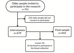
MACHINE LEARNING-BASED PREDICTION MODELS FOR COGNITIVE DECLINE PROGRESSION: A COMPARATIVE STUDY IN MULTILINGUAL SETTINGS USING SPEECH ANALYSIS


B. Ceyhan1, S. Bek2, T. Önal-Süzek1


1. Department of Bioinformatics, Graduate School of Natural and Applied Sciences, Mugla Sitki Kocman University, Mugla 48000, Türkiye; 2. Department of Neurology, Faculty of Medicine, Mugla Sitki Kocman University, Mugla 48000, Türkiye

Corresponding Author: Tuğba Önal-Süzek, 1 Department of Bioinformatics, Graduate School of Natural and Applied Sciences, Mugla Sitki Kocman University, Mugla 48000, Türkiye; tugbasuzek@mu.edu.tr

J Aging Res & Lifestyle 2024;13:43-50
Published online May 14, 2024, http://dx.doi.org/10.14283/jarlife.2024.6


Abstract

BACKGROUND: Mild cognitive impairment (MCI) is a condition commonly associated with dementia. Therefore, early prediction of progression from MCI to dementia is essential for preventing or alleviating cognitive decline. Given that dementia affects cognitive functions like language and speech, detecting disease progression through speech analysis can provide a cost-effective solution for patients and caregivers.
DESIGN-PARTICIPANTS: In our study, we examined spontaneous speech (SS) and written Mini Mental Status Examination (MMSE) scores from a 60-patient dataset obtained from the Mugla University Dementia Outpatient Clinic (MUDC) and a 153-patient dataset from the Alzheimer’s Dementia Recognition through Spontaneous Speech (ADRess) challenge. Our study, for the first time, analyzed the impact of audio features extracted from SS in distinguishing between different degrees of cognitive impairment using both an Indo-European language and a Turkic language, which exhibit distinct word order, agglutination, noun cases, and grammatical markers.
RESULTS: When each machine learning model was tested on its respective trained language, we attained a 95% accuracy using the random forest classifier on the ADRess dataset and a 94% accuracy on the MUDC dataset employing the multilayer perceptron (MLP) neural network algorithm. In our second experiment, we evaluated the effectiveness of each language-specific machine learning model on the dataset of the other language. We achieved accuracies of 72% for English and 76% for Turkish, respectively.
CONCLUSION: These findings underscore the cross-language potential of audio features for automated tracking of cognitive impairment progression in MCI patients, offering a convenient and cost-effective option for clinicians or patients.

Key words: Spontaneous speech, machine learning, dementia, mild cognitive impairment, mental status and dementia tests.


Introduction

Early detection of cognitive impairment on a population scale would benefit both individuals and society, including improved quality of life for affected individuals, decreased healthcare costs linked to late-stage treatment, and the chance for targeted resource allocation in healthcare systems. Nevertheless, existing detection techniques in clinics tend to be intrusive or lengthy, making them impractical for the ongoing observation of asymptomatic individuals. For instance, gathering biological indicators of neuropathology linked to cognitive decline usually requires cerebral spinal fluid samples, while cognitive performance is assessed through in-person evaluations by specialists, and brain metrics are obtained using costly, immobile equipment. Presently, the global population of individuals afflicted with dementia exceeds 55 million, with 60-70% of these cases attributed to Alzheimer’s disease, rendering it the predominant form of dementia (1). It stands as a primary contributor to dependency among older individuals, presenting caregivers with formidable challenges due to decreased physical engagement and mood alterations. Hence, it holds great importance to vigilantly monitor the progress of individuals aged 65 years and older, particularly those exhibiting no discernible symptoms or presenting with mild cognitive impairment (MCI), with the aim of forestalling or mitigating cognitive deterioration (2).
Several clinical tools and imaging techniques help estimate the course of dementia. For example, the Cardiovascular Risk Factors, Aging, and Incidence of Dementia (CAIDE) Risk Score was designed to predict the risk of developing dementia within 20 years for middle-aged people. The Brief Dementia Screening Indicator (BDSI) aims to identify older patients to target for cognitive screening by determining their risk of developing dementia within 6 years (2). The MMSE is a common screening tool for dementia, and it is primarily used by clinicians to assess cognitive decline. However, these tests are generally performed in clinical environments, and patients do not take them unless there are symptoms or avoid repeating them due to an unwillingness to visit these institutions.
Considering the limited accessibility, older patients’ reluctance to undergo standard laboratory cognitive tests, and the urgent need to prevent Mild Cognitive Impairment (MCI) from progressing to advanced stages, nonclinical and non-drug-based tools garner increased attention for thorough investigation. Advances in smartphone technology facilitate effortless passive monitoring of speech, fine motor skills, and gait patterns. Despite several challenges, such as cross-cultural adaptation (4) and standardization associated with these home-based prediction systems, they have the potential to assist in predicting cognitive decline at an earlier stage. Patients with MCI and dementia are known to have language difficulties such as word finding, sentence comprehension in producing speech, acoustic parameters such as shimmer, and number of voice breaks that significantly differentiate them from healthy adults (5). A study that conducted machine learning (ML) modeling by extracting linguistic features at the syntactic, semantic, and pragmatic levels from patient speech data achieved 79% accuracy in distinguishing Alzheimer’s disease patients from healthy adults using support vector machines (SVMs), neural networks (NNs) and decision tree classifiers (6). For acoustic feature research, another study used the Dementia Bank dataset, and 94.71% accuracy was achieved using the Bayes Net classification on 263 features of the audio files (7). For a non-English speaking study, a Spanish study used machine learning to extract linguistic features from spontaneous speech to detect cognitive impairments and achieved accuracies between 65% and 80% (8).
In this study we used the Alzheimer’s Dementia Recognition through Spontaneous Speech (ADReSS) dataset obtained from the ADRess challenge (11) and a Turkish patient dataset collected from the Mugla University Dementia Clinic (MUDC) when creating two ML models for evaluating the accuracy of speech-based acoustic features for predicting the MMSE score of patients. The ADRess dataset has 153 audio recordings of English-speaking older adults, while the MUDC dataset has 60 Turkish audio recordings of dementia clinic patients collected via the application and a website created for this purpose. Both datasets are composed of cognitively normal (CN) and AD patients balanced in terms of age and sex along with their written MMSE scores. This study preferred acoustic feature-based modeling because MMSE prediction is applied to Turkish-speaking patients. Linguistic modeling is affected by language and culture, and the automation of linguistic extraction features has not been successful in several studies (10).


Methods

Dataset

Two audio datasets, one obtained from the ADReSS dataset and one from 60 participants in the MUDC, were used for MMSE prediction via speech ML modeling. The breakdown of patients in the ADReSS dataset is given in Table 1; it is a balanced dataset composed of cognitively normal (CN) patients and those suffering from Alzheimer’s disease (AD) who were requested to talk about Cookie theft pictures used in the Boston Diagnostic Aphasia Exam (BDAE) (11). In addition to the SS recordings, the Pitt corpus of the ADReSS contained the written MMSE scores, age, and gender of all participants. The Pitt corpus dataset was obtained from the ADReSS challenge website by becoming a member of the Dementia Data Bank (12).

Table 1. Breakdown of ADReSS Training Dataset participants by Gender and AD status

To compare the performance of both datasets and assess whether the underlying language’s linguistic features were critical factors in this assessment, we repeated the same Cookie Theft picture exam in Turkish on 60 patients with mild cognitive impairment in the MUDC. The MUDC performed the written MMSE tests on all these patients within one month before the SS recordings were retrieved. Table 2 summarizes the participants’ metadata.

Table 2. Breakdown of MUDC Dataset participants by Gender and MMSE Scores


Audio Data Collection and Preprocessing

A total of 153 audio recordings in the ADReSS dataset and 60 in the MUDC dataset were separately preprocessed and analyzed because both recordings were recorded in different environments with different languages. While the ADReSS dataset was obtained from the Dementia Bank website (13), Figure 2 shows the website developed for this study to capture recordings and check MMSE predictions for the MUDC dataset. A clinician conducted the assessments in Turkish within a calm and controlled environment. Immediately after their formal MMSE cognitive test at the clinic, each participant was presented with the Cookie Theft Picture and was instructed to provide a comprehensive description of the image within 1 minute (Figure 1). The participants’ voices were recorded during the test administration, and the collected data were utilized for further analyses.

Figure 1. Patient recording and MMSE prediction page

Figure 2. (A) Audio file augmentation process steps. (B) Perturbation step from one file to 15 files

The preprocessing of the audio data involved framing, trimming, and augmenting the audio files programmatically to enable repeating this process in both datasets. One of the main purposes of preprocessing was to increase the sample size for each dataset to avoid overfitting and reduce bias. The first step in augmentation was to divide the audio files into smaller but more stationary ones. The average duration of the audio files in the ADReSS dataset is 80 seconds, so we performed both manual and programmatic analyses to determine the optimal segment duration. For the latter, we developed code to go over audio files to find the average lowest total harmonic distortion, a measure that represents the distortion rate in a segment divided by the total distortion in the file (14), which was 15 seconds. After the recordings, the files were programmatically framed into 20-second segments using Python Librosa libraries to have enough words in each segment for a sentence (Figure 2A). After we trimmed the interviewer audio from the beginning and end using a 5-second buffer, we programmatically perturbed each section at speeds of 0.7, 0.8, 1.2, and 1.3 to create 1520 samples from 153 samples for the training dataset and 800 samples from 48 samples for the test dataset (Figure 2B). The same process was applied to the MUDC dataset, and 60 samples were augmented to 300.


Feature Extraction and ML Model Creation from Audio Files

Figure 3 displays the steps we followed after the audio files were preprocessed and augmented. The resulting model is deployed to the application server as a web service called by the website in Figure 2.

Figure 3. Audio file feature extraction and ML model creation steps

The spectrograms in MEL format and waveforms in Figure 4 clearly show differences in the number of peaks, energy levels and pauses between the AD and CN audio files. To measure the differences between these two samples, Table 3 lists extracted audio features such as the root mean square (RMS), zero crossing rate (ZCR), spectrum features, number of silence of segment, skewness and mean, and standard deviation of 30 mel-frequency cepstral coefficients (MFCC), which have been used in other studies that analyzed the ADRess dataset (7, 13). MFCCs are one of the most popular feature extraction techniques used in speech recognition based on frequency domain using the Mel scale which is based on the human ear scale (16). Due to the high variation in MFCC signals, we included the mean and standard variation of this measure. Feature selection using the KBest algorithm did not eliminate any of these features, as the average accuracy rate of the model using 10-fold cross-validation was lower with fewer selected features in each case.

Figure 4. Waveform and Spectrogram of (A) AD recording (B) CN recording

Table 3. Audio features extracted from datasets

After preparing and preprocessing the audio data and extracting the audio features, we performed GridSearchCV hyperparameter optimization using 10-fold cross-validation for logistic regression (LR), random forest (RF) and neural network (NN)-multilayer perceptron (MLP) algorithms for both datasets. Table 4 shows the hyperparameter tuning results, best hyperparameters, means and standard deviations between each set of fold results with and without normalization and feature selection.

Results

Acoustic Analysis

For the ADReSS dataset, the best classification algorithm was the random forest (RF) algorithm, which achieved 95.79% accuracy with normalization and feature reduction. For feature normalization, the Python StandardScaler function was used. Table 5 displays the average accuracy, F1, and AUC scores when using a 10-fold cross-validation score for the RF algorithm, which resulted in the best accuracy of 95%. The difference in the mean cross-validation score between the augmented and unaugmented data clearly demonstrates the value of increasing the sample size for model creation.
To perform independent dataset validation with a 70-30 train-test split, we implemented validation with the Adress Test Dataset, which had 60 recordings with a balanced population of gender and MMSE scores. We achieved 73% accuracy for the labels (dementia or not) and a root mean square error (RMSE) of 5.6 for the MMSE predictions (Table 4, 5).

Table 4. ADReSS dataset hyperparameter optimization results

Table 5. ADReSS dataset Classification Accuracy Scores


For the MUDC dataset, the best classification algorithm was the neural network MLP classifier algorithm, which achieved 94% accuracy with normalized data and 30 reduced features (Table 6, 7).

Table 6. MUDC dataset hyperparameter optimization results

Table 7. MUDC dataset Classification Accuracy Scores

Cross-Language Evaluation

We evaluated the accuracy of the two models trained on the ADRess and MUDC datasets separately by validating them in other languages for independent validation. Using the complete ADRess dataset as the training dataset and the MUDC dataset as the validation dataset, the random forest model achieved the highest accuracy of 72% for the labels and a root mean square error (RMSE) of 6.02 for the MMSE predictions. The Neural Network MLP model trained on the complete MUDC dataset achieved 76% label accuracy when tested on the ADRess dataset for validation. These results indicate the potential power of acoustic features independent of the underlying linguistic properties of the language, such as word order, agglutination, noun cases, and grammatical markers.

Linguistic Analysis

Linguistic analysis was performed on the ADRess dataset to compare with acoustic features. The ADRess dataset provided transcriptions in CHAT file format for both the test and training datasets, which needed parsing of patient words from the file. For preprocessing, interviewer and redundant words were programmatically removed from these transcriptions. We used Python BERT libraries to extract 20 linguistic features, such as the number of words, number of unique words, speech rate, number of sentences, sentence complexity, and clarity score. Our initial experiments using linguistic features for classification achieved a very low accuracy of 45% with the RF classifier algorithm with normalized data and 30 reduced features (Table 8).

Table 8. Linguistic Analysis Accuracy Rates


Discussion

This study used a publicly available English dataset and an in-house dataset that was collected in a Turkish-speaking dementia clinic. This study is the first machine learning study in the literature presenting a benchmark dataset of audio features from Turkish patients diagnosed with mild cognitive impairment, this study diverges from the predominant literature focusing on English language speakers by conducting research in Turkish. Turkish, classified within the Altaic language group alongside Finnish, Korean, and other Turkic languages, exhibits distinctive phonological therefore sound based properties such as vowel harmony, where vowels within a word tend to coalesce based on shared features such as frontness or rounding. In contrast, Indo-European languages commonly share phonological (sound-based) features, including distinct sounds like the Indo-European laryngeals.
As of 2022, the native speakers of Turkish number approximately 400 million, constituting approximately 5% of the global population (15). Expanding our research of sound-based dementia diagnosis to cover other non-Indo-European languages has the potential to enhance the accuracy of early dementia detection for patients across the linguistically diverse non-English-speaking world.
Furthermore, our study offers an additional benefit: non-English-speaking dementia patients within the Indo-European language group may also derive utility from our findings due to the language-independence of our underlying machine learning model.
This approach enabled us to assess whether audio features alone can be utilized to estimate the course of dementia in different populations independent of the linguistic structure of the language. By conducting the audio recordings firsthand at the clinic immediately after the written MMSE test, some patients testified that audio recordings were more convenient than the written format test, while others found it even harder to recall the word ‘Cookie Jar’, which became frustrated and needed to repeat the recording several times.
The random forest and MLP neural network classification methods yielded high accuracy rates of approximately 94%, showing that acoustic features can be used independent of the linguistic features of the underlying language to create a prediction model. The accuracy rate was 52% before augmentation and feature extraction on the ADRess dataset and 65% on the MUDC dataset. Therefore, feature extraction and augmentation contributed significantly to the accuracy of the models. For comparison, we performed a linguistic analysis on the ADRess dataset for which the transcriptions were available. However, the random forest algorithm’s highest accuracy rate was 45%.

Limitatons

Even though we increased Turkish dataset from a sample size to 300 by augmentation, it is small compared to the Indo-European dataset. It is limited to a population from a small city in western region with regional language characteristics which might not be representing general Turkish linguistic characteristics. Moreover, Turkish training dataset is limited to mild cognitive delay although Adress dataset contains a wider spectrum of the disease.

Conclusion

Achieving a high accuracy rate with two different machine learning classifiers in two distinct languages demonstrates the potential of utilizing spontaneous speech (SS) recordings to predict MMSE scores and track the cognitive impairment progress of dementia patients collected at-home by users themselves or their caregivers. Our study highlights the critical importance of the audio features in machine learning models, which can outperform the linguistic features regardless of the language. Our results suggest that adopting a multilingual approach with larger datasets could result in more precise machine learning models. This, in turn, could assist other researchers in software development aimed at monitoring dementia progression more conveniently.


Contributions: Barış Ceyhan: Conceptualization, Data curation, Resources, Software, Writing- Original draft preparation: Semai Bek: Data acquisition, Tuğba Önal-Süzek: Conceptualization, Supervision, Writing- Reviewing and Editing. All authors have read and agreed to the published version of the manuscript.

Acknowledgment: We thank İrem Nur Dede for her assistance in the MUDC SS recording collection and Dr. Barış Süzek for his invaluable contributions to the discussions about the method. The ADRess Pitt dataset was funded by grants NIA AG03705 and AG05133.

Ethics declaration: All the MUDC SS recordings of the patients were collected with the approval of the Noninterventional Ethics for Sports and Health Sciences – 2 Committee of Mugla Sitki Kocman University (approval protocol number: 230086, October 18th, 2023) and were conducted in compliance with the principles of the Declaration of Helsinki. All the spontaneous speech (SS) recordings of the patients from the ADRess dataset consisted of the diagnosis task subset provided to the members of the Dementia Talkbank consortium for academic research purposes. The ADRess dataset has been approved by the CMU IRB number STUDY2022_00000172. This manuscript is a part of Barış Ceyhan’s Ph.D. thesis.

Conflicts of interest: S.B. declares no conflicts of interest. TÖS was partially supported by the company Kedi Mobil Uygulama Anonim Şirketi, Muğla, Turkey, and BC was supported by Infor Global Solutions, Canada, during the study. These companies had no role in the design of the study; in the collection, analyses, or interpretation of data; in the writing of the manuscript; or in the decision to publish the results.

Open Access: This article is distributed under the terms of the Creative Commons Attribution 4.0 International License (http://creativecommons.org/licenses/by/4.0/), which permits use, duplication, adaptation, distribution and reproduction in any medium or format, as long as you give appropriate credit to the original author(s) and the source, provide a link to the Creative Commons license and indicate if changes were made.


References

1. S. Grueso and R. Viejo-Sobera, “Machine learning methods for predicting progression from mild cognitive impairment to Alzheimer’s disease dementia: a systematic review,” Alzheimer’s Research & Therapy 2021, vol. 13, no. 1, p. 162, 12. doi: 10.1186/s13195-021-00900-w
2. C. James, J. M. Ranson, R. Everson and D. J. Llewellyn, “Performance of Machine Learning Algorithms for Predicting Progression to Dementia in Memory Clinic Patients,” JAMA Network Open 2021, vol. 4, no. 12, p. e2136553, 12. doi: 10.1001/jamanetworkopen.2021.36553
3. J. J. G. Meilán, F. Martínez-Sánchez, J. Carro, D. E. López, L. Millian-Morell and J. M. Arana, “Speech in Alzheimer’s Disease: Can Temporal and Acoustic Parameters Discriminate Dementia?,” Dementia and Geriatric Cognitive Disorders 2014, vol. 37, no. 5-6, pp. 327-334. doi: 10.1159/000356726
4. R. Ben Ammar and Y. Ben Ayed, “Speech Processing for Early Alzheimer Disease Diagnosis: Machine Learning Based Approach,” 2018. doi: 10.1109/AICCSA.2018.8612831
5. S. Al-Hameed, M. Benaissa and H. Christensen, “Simple and robust audio-based detection of biomarkers for Alzheimer’s disease,” ISCA, 2016. doi: 10.21437/SLPAT.2016-6
6. F. García-Gutiérrez, M. Alegret, M. Marquié, N. Muñoz, G. Ortega, A. Cano, I. De Rojas, P. García-González, C. Olivé, R. Puerta, A. García-Sanchez, M. Capdevila-Bayo, L. Montrreal, V. Pytel, M. Rosende-Roca, C. Zaldua, P. Gabirondo, L. Tárraga, A. Ruiz, M. Boada and S. Valero, “Unveiling the sound of the cognitive status: Machine Learning-based speech analysis in the Alzheimer’s disease spectrum,” Alzheimer’s Research & Therapy 2024, vol. 16, no. 1, p. 26. doi: 10.1186/s13195-024-01394-y
7. S. Luz, F. Haider, S. de la Fuente, D. Fromm and B. MacWhinney, “Detecting cognitive decline using speech only: The ADReSSo Challenge,” 3 2021. doi: 10.48550/arXiv.2104.09356
8. S. de la Fuente Garcia, C. W. Ritchie and S. Luz, “Artificial Intelligence, Speech, and Language Processing Approaches to Monitoring Alzheimer’s Disease: A Systematic Review,” Journal of Alzheimer’s Disease 2020, vol. 78, no. 4, pp. 1547-1574, 12. doi: 10.3233/JAD-200888
9. Y. Turgeon and J. D. Macoir, “Classical and Contemporary Assessment of Aphasia and Acquired Disorders of Language,” Handbook of the Neuroscience of Language 2008, pp. 3-11. doi: 10.1016/B978-0-08-045352-1.00001-X
10. J. T. Becker, “The Natural History of Alzheimer’s Disease,” Archives of Neurology 1994, vol. 51, no. 6, p. 585, 6. doi: 10.1016/B978-0-08-045352-1.00001-X
11. INTERSPEECH 2020, “ Alzheimer’s Dementia Recognition through Spontaneous Speech The ADReSS Challenge,” 2 2024. [Online]. Available: https://luzs.gitlab.io/adress/.
12. Hongwei Wang, “Measurement of Total Harmonic Distortion (THD) and Its Related Parameters using Multi-Instrument,” 2020.
13. M. S. S. Syed, Z. S. Syed, M. Lech and E. Pirogova, “Automated Screening for Alzheimer’s Dementia Through Spontaneous Speech,” ISCA, 2020. doi: 10.21437/Interspeech.2020-3158
14. World Health Organization, “Dementia – Key Facts,” 2 2024. [Online]. Available: https://www.who.int/news-room/fact-sheets/detail/dementia.
15. English Archived 2023-03-09 at the Wayback Machine, Ethnologue, Dallas, Texas: SIL International., 2022.
16. W. Han, C. Chan, C. Choy, and K.P. Pun. “An efficient MFCC extraction method in speech recognition,” p. 4. IEEE, 2022. doi: 10.1109/ISCAS.2006.1692543

© The Authors 2024
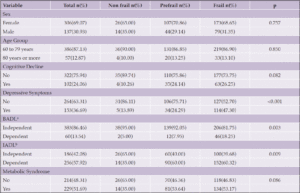
 

DESIGN AND RATIONALE OF A TWO-ARMED RANDOMIZED CONTROLLED TRIAL ON YOGA/BRISK WALKING-BASED LIFESTYLE MODIFICATION ON DEMENTIA RISK REDUCTION, AND INFLUENCE OF APOE GENOTYPES ON THE INTERVENTION


M. Singh1, V. Majumdar1


1. Swami Vivekananda Yoga Anusandhana Samsthana, Bangalore, Karnataka, India-560105.

Corresponding Author: Dr. Vijaya Majumdar, Associate Professor, Division of Life Science, Swami Vivekananda Yoga Anusandhana Samsthana, Bangalore, Karnataka, India-560105, Email ID: vijaya.majumdar@svyasa.edu.in

J Aging Res & Lifestyle 2024;13:33-42
Published online May 14, 2024, http://dx.doi.org/10.14283/jarlife.2024.5


Abstract

BACKGROUND/INTRODUCTION: Though considered a late-onset disease, the 2020 report of the Lancet Commission emphasizes the necessity of conducting primary prevention trials with an approach of never too early in the life course for dementia prevention. Driven by the same notion, we hereby aim to compare the dementia risk reduction potential of two potential interventions, 48 weeks (12 months) of yoga and brisk walking, in middle-aged high-risk subjects.
DESIGN: A randomized controlled trial.
SETTING: Community in India.
PARTICIPANTS: In total, 323 at-risk dementia subjects will be recruited from community settings through health awareness camps and door-to-door surveys across Delhi, India. Participants will be randomized into yoga or brisk-walking groups (1:1). The yoga intervention group will receive 60 contact yoga sessions per 60-min/day at the community parks, followed by continued tele-supervised home practice, further followed by at-home self-practice, and will be tested at 3-time points (baseline, 24-week and 48-week, post-randomization) to test the efficacy of the intervention. The control group will be asked to do brisk walking daily for 45 minutes at their convenience, followed by weekly telephone follow-ups. Applying the intention-to-treat principle, the primary endpoint will be the change from baseline at the 12th month in the Cardiovascular Risk Factors, Aging, and Dementia (CAIDE) Scores. Secondary outcomes will include the composite scores derived from a comprehensive neuropsychology battery, comprising the Trail Making Test, Digit Span Test, N Back, Color Trail, Animal Fluency Test, COWA (Controlled Oral Word Association Test), and Digit Symbol Substitution. The primary outcome will be analyzed using mixed-effect models for repeated measures, adjusted for covariates as fixed effects. The study has been prospectively registered (CTRI/2023/02/049746) on February 15, 2023. The protocol was conceptualized in 2021 and approved by the Institutional Ethics Committee of SVYASA. Recruitment began in February 2023 and is underway with patient enrollment.
CONCLUSION: To our knowledge, this is the first controlled trial to investigate the longitudinal effects of a yoga-based intervention on dementia risk reduction using the CAIDE risk score. The findings of this trial will also provide insight into a better understanding of genotype-dependent responses to yoga intervention and open up avenues for understanding the implications of gene-intervention interactions for precision prevention using yoga.

Key words: Dementia, lifestyle modification, randomized control trial, ApoE and middle-age.


Introduction

The global trends of population aging have tremendously impacted life expectancy in the Southern Asian region (1). India, the most populous country in the world, is at an alarming stage of population aging, with an estimated share of 20% of individuals aged 60 years or older by 2050 (2). Increased dementia is one of the primary consequences of population ageing. The latest estimates by the Longitudinal Aging Study in India (LASI) indicate a 7.4% prevalence of dementia, with 8.8 million individuals being afflicted (3). Dementia refers to a diverse range of conditions, with Alzheimer’s disease and vascular dementia being the most prevalent types (4). Unfortunately, there are no effective therapies available yet to treat dementia. Positively, the pathological model of dementia provides an optimal window for its prevention given the lengthy course of its duration, which takes several years to emerge (5). Hence, early and accurate identification of people at high risk of dementia is critical for the effective implementation of preventive measures. The importance of modifiable risk factors highlights the risk of dementia (5). Detecting changes in these risk factors before the disease manifests clinically allows for timely and careful management of vascular risk factors, thereby delaying the onset of the disease (5). The Lancet Commission on Dementia Prevention, Intervention, and Care Report states that up to one-third of dementia cases are preventable, considering the key potentially reversible risk factors.
Non-pharmacological physical activity-based therapies have shown potential for reducing the risk of dementia (6, 7). The prevention model for dementia is based on the estimation of risk reduction using the Cardiovascular Risk Factors, Ageing, and Dementia (CAIDE) score. This score was developed to address the increasing need for early detection in the treatment of neurodegenerative diseases. The CAIDE score is a validated tool for estimating dementia risk derived from age, sex, education, systolic blood pressure, body mass index, serum total cholesterol, and physical activity in middle-aged community subjects and has been validated to estimate the risk of dementia 20 years later (10). However, the reported associations between physical activity and dementia risk reduction have not been established using the intervention. Mechanistically, exercise and physical activity-based interventions have been proposed to possibly work via attenuating the vascular risk and associated vascular cognitive impairments, thereby halting the advancement of neurodegenerative diseases and dementias. Yoga has a mechanistic basis that can effectively manage cardiovascular risk factors (8). We deemed that testing the same over a composite score like the Cardiovascular Risk Factors, Ageing, and Dementia (CAIDE) score would aid in the translation of the existing evidence to support the preventive potential of yoga for dementia. We also hypothesize a concomitant improvement in cognitive functions.
The causes of dementia are varied, but genetic variations in the apolipoprotein E (ApoE) gene have been identified as major factors contributing to the disease. This conclusion comes from extensive genome-wide association studies (GWASs) (11), which have been confirmed by studies in different populations worldwide, including Asian Indians (12, 13). ApoE plays a key role in both cerebral and peripheral cholesterol metabolism and underlies many functions involved in brain amyloid metabolism, blood-brain barrier integrity, and transport of various lipid species to neuronal cells, as well as in hepatic uptake of triglyceride-rich lipoproteins (13). Interestingly, the genotype of ApoE has also been reported to play an important role in response to interventions (14). ApoE is a significant genetic component that has been validated across many cohorts around the globe, including the Indian population (15). Exercise has been reported to be more beneficial in ApoE4 carriers than non-carriers towards amyloid deposition (16). To our knowledge, no study has explored the risk reduction associated with adherence to yoga and its plausible modification by ApoE ε4 allele status.

Study Design

The study DERRY (Dementia Risk Reduction using yoga will be a prospective, parallel-group, single-center, and 2-arm randomized controlled trial wherein the subjects will be randomly allocated to two groups: yoga or active walking control. The protocol has been drafted following the CONSORT guidelines (Consolidated Standards of Reporting Trials) (17) (Figure 1). The study will be conducted between February 2023 and October 2024. A total of 323 adults (aged 30-65 years) with dementia risk (CAIDE score≥6) will be recruited from the community set up through door-to-door surveys and dementia awareness camps across Delhi, India. Eligible participants will be randomized into 1:1 yoga or active control groups. Primary and secondary outcomes will be assessed at baseline (before intervention) and at 24 and 48-week visits (with a 60-day window of flexibility) (see Figure 1). All participants will follow their usual medical regime during the study period. This protocol (RES/IEC-SVYASA/243/2022) has been approved by the Ethics Committee of Swami Vivekananda Yoga Anusandhana Samsthana. Interested and eligible individuals will be asked to attend an orientation session, and informed consent will be obtained from them before participation.

Figure 1. Participant flow chart for parallel design, based on the Consolidated Standards of Reporting Trials guidelines for transparent reporting of trials. CAIDE: cardiovascular risk factor, aging, and incidence of dementia

MMSE: mini mental status examination


Recruitment and screening

Participants will be enrolled using convenience sampling between February 2023 and October 2024. Middle-aged individuals, aged 39 to 65 years, will be eligible if they have not regularly practiced any form of yoga in the previous 6 months and do not have a history of dementia. Patients will undergo a Mini-Mental State Exam (MMSE) with a score of at least 26 to rule out gross dementia. Additionally, they will be assessed using a CAIDE Risk Score (Cardiovascular Risk Factors, Aging, and Dementia) with a score of at least 6 points (18). Further, they will be excluded based on i) the presence of any neurological disorder; ii) significant arthritis; iii) recent acute infection or other inflammation; iv) persistent cognitive impairment due to psychoactive substance use; and v) individuals who have functional limitations that prevent them from walking or doing yoga and who have been advised against exercising by their physician or have undergone recent surgical interventions. Upon enrollment in the study, participants will be followed for 48 weeks (12 months) or until they withdraw from the study.

Study outcomes and assessments

Primary outcome

The primary objective of the study is to assess the efficacy of a yoga intervention vs. brisk walking at 48 weeks on the risk reduction of dementia. The primary endpoint is a difference in the CAIDE risk scores between the yoga and brisk walking groups after 48 weeks (12 months). As shown in CONSORT, assessments will be conducted at baseline, 24 and 48 weeks over 365 days with a 60-day flexibility window (Table 1). However, the primary time endpoint represents a longer-term outcome.

Table 1. Schedule of enrolment, interventions, and assessments, according to SPIRIT 2013 guidelines

CAIDE- Cardiovascular risk factor, aging, and incidence of dementia; TMT- Trail making Test; DSTT- Digit symbol substitution test, COWA- Controlled Oral Word Association Test, ApoE- Apolipoprotein E


The research staff will evaluate the CAIDE risk score during screening. The CAIDE risk score ranges from 0 to 18, with higher scores indicating an increased risk of developing dementia (18), with scores of 8–9 indicating a 4.2% risk of developing dementia in the next 20 years (18, 20, 21). CAIDE is a comprehensive tool for predicting dementia risk in middle-aged individuals. The CAIDE Risk Score incorporates various non-modifiable and modifiable factors, including age, education, blood pressure, cholesterol levels, body mass index (BMI), physical activity, and ApoE status (18).

Secondary Outcomes

Secondary outcomes will include individual scores of CAIDE components and assessment scores of cognitive tests for memory, attention, language, verbal fluency, and executive ability, measured by neuropsychological tests or other objective measurements. The secondary clinical outcome of this study focuses on the 48-week change in the composite score of a comprehensive neuropsychology battery.

Assessments

At the baseline and 48-week visits, the CAIDE score will be calculated using data on age, gender, self-reported years of formal education, systolic blood pressure, BMI, total cholesterol, physical activity, and ApoE status. Each component of the CAIDE score will undergo assessment, and a predetermined set of points will be allocated to each category of risk factors (17). The CAIDE score for each participant will be computed by summing up the points assigned to each risk factor category (18). Participants self-reported demographic details and physical activity levels, while healthcare professionals measured objective factors like systolic blood pressure, BMI, and blood cholesterol levels. The physical activity index was calculated based on activity duration and intensity. Genetic testing for ApoE ε4 carrier status was conducted during a specific exam. The CAIDE score, ranging from 0 to 18, will be used in a male-only cohort, with observed scores from 1 to 15 (18, 19).
At the outset, supplementary information encompassing demographic data and contemporary risk factors such as hypertension, diabetes, head injury, smoking, alcohol consumption, dyslipidemia, etc. will be documented for the participants.
This battery includes well-established tests to assess the global cognitive ability index as well as various domains of cognition such as memory, attention, language, verbal fluency, and executive ability, which will be measured by neuropsychological tests such as the Trail Making Test, Digit Span Test, N Back, Color Trail, Animal Fluency Test, COWA, and Digit Symbol Substitution.
In addition to the neuropsychology battery assessment, participants will also be asked to complete questionnaires to assess their quality of sleep using the Pittsburgh Sleep Quality Index (PSQI) and their overall quality of life using the World Health Organization Quality of Life (WHOQOL-BREF) questionnaire. These validated questionnaires provide valuable insights into participants’ subjective experiences of sleep quality and their overall well-being across physical, psychological, social, and environmental domains.
Furthermore, genotyping will be performed using DNA amplification through real-time PCR. This genetic analysis aims to explore potential associations between specific genetic variations and cognitive outcomes, providing insights into the role of genetics in cognitive function and dementia risk (22).

Executive Function

Trail-making test

Trail-making test: A neuropsychological test of visual attention and task switching is called the Trail Making Test (23). There are two parts to it; part A seems to primarily depend on the effectiveness of visual scanning and psychomotor speed. In which the subject is asked to accurately connect a sequence of 25 dots as quickly as they can. Part B has circles with both numbers (1–13) and letters (A–L); the subject is instructed to connect the circles in an ascending pattern by alternating between the numbers and letters (i.e., 1-A-2-B-3-C, etc.) (23). Specifically, mental flexibility and a higher demand for working memory are required for executive control in TMT B. It is also capable of accurately identifying many cognitive disorders, including dementia and Alzheimer’s disease (23).

Working Memory: Digit Span test, Verbal N Back

Digit Span: The original digit span task was similar to the Wechsler Memory Scale (24). The participant will be instructed to be attentive to a series of random numbers that will be played once per second. The subject was asked to recall the numbers in reverse order for the digit span backwards (DSB), while the subject was asked to repeat the numbers in a forward series for the digit span forwards (DSF) (25). Each correctly repeated series began with a two-number series and ended with a one-digit series. If the subject failed the first time, they were given another chance with a new set of random numbers for each sequence. If the test subject fails again after the second try, the test will be stopped, as will the longest series they attempt (25).
Verbal N back: Several Indian languages share thirty consonants, which are pronounced one per second. Thirty consonants total, nine of which are repeated (25). The repeated consonants are selected at random. In the 1-back test, the subject responds by tapping the table whenever a consonant is repeated consecutively. In the 2-back test, the subject responds by tapping the table whenever a consonant is repeated after an intervening consonant (26). Scores were based on the number of successes and failures on each test. A negative score was assigned based on the number of errors. After this, the overall score was determined (26).

Attention: Color Trail Test

The Color Trails Test (CTT): The Color Trail Test is a language-free version of the Trail Making Test (TMT) that was developed to allow for broader cross-cultural application to measure sustained attention in adults (27). Numbered circles are printed with vivid pink or yellow backgrounds that are perceptible to color-blind individuals. For Part 1, the respondent uses a pencil to connect circles rapidly numbered 1–25 in sequence. For Part 2, the respondent rapidly connects numbered circles in sequence but alternates between pink and yellow (28).

Language: Animal fluency test, COWA

Animal fluency test: The subject will be asked to come up with as many animal names as they can in a minute. The subject is instructed to omit any mention of fish, snakes, or birds. The score was formed by the number of names produced (26).
COWA: Frontal lobe dysfunction can hinder a person’s ability to quickly form words. A typical neuropsychological test for verbal fluency is the Controlled Oral Word Association Test (COWAT), also called the “FAS.” Three-word criteria make up the COWAT. The subject’s objective is to come up with as many words as possible that begin with the specified letter (F, A, or S) in one minute (29). Additionally, subjects are told to avoid using proper nouns, numbers, and the same word with a different suffix (29). Frontal lobe impairment has been successfully detected using the COWAT and other verbal fluency tests. Jerry Janowsky, Arthur Shimamura, and Larry Squire discovered in 1989 that individuals with circumscribed left or bilateral frontal lobe lesions produced noticeably fewer words than control subjects (30).

Processing Speed: Digit Symbol Substitution

Digit Symbol Substitution: We will administer the DSST to capture processing speed. In this task, participants see a table with a mapping between nine symbols and the digits 1–9. The participants are given 9 seconds to fill in the respective numbers that correspond to the symbols in a large list of symbols. The dependent variable is how many symbols are successfully associated with the respective number (31, 32).

Quality of Sleep

Pittsburgh Sleep Quality Index (PSQI); Improvement in the quality of sleep using PSQI Score after 6 weeks of intervention (33). The PSQI is a self-reported instrument that measures the quality of sleep as well as sleep disturbances over one month. The scale assesses seven domains: sleep quality, sleep duration, sleep latency, habitual sleep efficiency, sleep disturbances, use of sleep medication, and daytime dysfunction (33).

Quality of life

World Health Organization Quality of Life (WHOQOL-BREF); improvement in the quality of sleep using the WHOQOL-BREF Score after 6 weeks of intervention. WHOQOL-BREF is a comprehensive tool for assessing quality of life. It was standardized with 26 items and developed by WHO (34). The scale provides a measure of an individual’s perception of quality of life for the four domains: (1) physical health (seven items), (2) psychological health (six items), (3) social relationships (three items), and (4) environmental health (eight items). In addition, it also includes two questions for the ‘overall quality of life’ and ‘general health’ facets. The domain scores are scaled in a positive direction (i.e., higher scores denote a higher quality of life). The range of scores is 4–20 for each domain (34).

Mini-mental state evaluation score (MMSE)

The MMSE is a measure used for cognitive screening in both clinical and research areas. Scores ≤ 26 on the MMSE indicate the absence of severe dementia (35, 36).

Genotyping of ApoE ε4

Genomic DNA will be extracted from 2 ml of human whole blood using the NucleoSpin® Blood L kit (Macherey-Nagel) according to the manufacturer’s instructions. Following this, genotyping of ApoE variants will be performed. To perform genotyping, we will use the primers reported by Calero (22). The forward primers are from positions 2886 to 2903, whereas the reverse primers are from positions 3041 to 3058. The polymorphic site contains the 3-end nucleotides, and specific primers will be designed to match one of the two variants at the ApoE positions 2903 and 3041. The primers will be combined in three reaction mixtures that are anticipated to produce an amplification product of 173 bp. These three reaction mixtures are «Reaction ApoE 2» (primers ApoE 112C and ApoE 158C), «Reaction ApoE3» (primers ApoE 112C and ApoE 158R), and «Reaction ApoE4» (primers ApoE 112R and ApoE 158R). Each PCR reaction mixture contained the following: 1× Power SYBR® Green PCR Master Mix (Applied Biosystems), 0.3 M of each primer, and 50 ng of genomic DNA. Negative controls will be performed by using the same reaction mixtures without DNA. All the reactions will be run in duplicate. The PCR amplification protocol would be as reported: initial AmpliTaq Gold DNA Polymerase activation at 95 ◦C for 10 min, followed by 40 cycles with denaturation at 95 ◦C for 15 s, and annealing + extension at 62 ◦C for 1 min. Amplification will be performed either on a 7500 Real-Time PCR System (96-well format) (Applied Biosystems) using the comparative Ct (Ct) method (22).


Randomization and blinding

A computer-based program will be utilized by a statistician to generate a randomized list. Participants will be randomized by the system into blocks of four, six, eight, or ten, stratified by. Only research staff will be able to retrieve the generated list, which will be kept confidential. The participant will receive the allocation of intervention only after completion of their baseline assessment. Due to the nature of yoga or walking interventions, participants could not be blinded to the intervention allocation. However, outcome assessments will be carried out by blinded.


Adherence

Adherence will be calculated based on participation in sessions. The trainer will maintain the logbook in Phase 1, while individual participants will receive notepads to keep records in Phase 2. All these will be collected during follow-up assessments. Attendance rates will be calculated by dividing the number of sessions attended by the total number of sessions. Overall attendance will then be calculated by averaging all participants’ attendance rates. Attrition rates will be calculated by dividing the number of individuals who dropped out of the study by the total number of enrolled participants. The reasons cited for study dropout will also be summarized.

Intervention

Participants in the yoga group will be given intervention for 6 months, 5 days a week. Intervention would be delivered by qualified yoga professionals (Table 2). The participants will be trained to perform yoga practices by qualified yoga instructors. In the initial orientation at the community center, participants will be given 1 hour for each group-based sessions, which will consist of a 2-minute introduction followed by 10 minutes of gentle loosening movements followed by 1 minute of relaxation, 15 minutes of postures followed by 2 minutes of relaxation, followed by breathing practices of 15 minutes, and end with guided meditation (5 minutes) followed by 3 minutes of relaxation and a 2-minute closing prayer followed by 5 minutes of query and discussion. Overall, the adjunct yoga intervention will be divided into 2 phases: Phase 1, including supervised sessions at the community center, including the orientation; and Phase 2 unsynchronized teleyoga at-home practice (120 sessions) (Table 1). There would be 24 weeks of yoga training followed by asynchronous teleyoga home practice for 24 weekss, 5 days a week.

Table 2. Schedule of Yoga Sessions


The standardized and validated yoga-based intervention module, including çithilikaraa vyäyäma (loosening practices), äsana (postures), pranäyama (breathing practices), and dhyäna (meditation), aimed at bringing harmony to mind and body, will be provided to the participants. While delivering the intervention, the therapist will observe the condition of each subject and make sure to make them understand each practice by adding more explanations to each approach. Modifications will be made individually, according to each participant’s specific limitations. We will be using the previously published module of yoga intervention already reported to reduce cardiovascular risk factors such as high blood pressure and dyslipidemia. The authors reported that using the specific module titled Integrated Yoga Therapy there would be significant improvements in baroreflex sensitivity, systolic blood pressure, and total peripheral vascular resistance in hypertensive patients (37). In a similar vein, Sharma et al. (2020) reported on the management of lipid profiles in patients with coronary artery disease (CAD) using IAYT (38).
We also aim to involve a few strategies to reduce attrition and minimize loss to follow-up based on our prior experiences with yoga-based interventions: (a) being responsive to participants and/or spouse/care partner questions; and (c) the yoga coordinators will meet with participants throughout the study, sometimes traveling to the participant’s fitness facility or home, which should enhance adherence to the program and allow the development of a strong researcher-participant relationship. We have included periodic tele-synchronized sessions as well to decrease the travel burden on participants considering vacations, work commitments, or other reasons influencing adherence to the trial. We have included participants to choose where they exercise and provide the means to do so to promote retention and long-term adherence to exercise. A research coordinator will contact the participants to inquire about their current health status at least once per month.

Statistical analysis

Sample size: A sample size of 269 (n = 135; 135) was derived based on a moderate effect size assumption, using G power software’s formula for F-test, ANCOVA with fixed effect, main effect and main effect interaction model for 2 group comparisons. The calculated sample size also aligns with the estimate presented by Leon et al. 2009 (39). Further, assuming an attrition rate of 20%, the final sample size is n = 323 subjects randomized in a 1:1 ratio to yoga or brisk walking.Baseline characteristics will be presented using appropriate descriptive statistics. Before analysis, variable distributions will be examined to ensure that assumptions of normality are met using statistical software (SPSS, Statistical Package for the Social Sciences, Version 20.0) with the Shapiro-Wilk test. The equivalence of variance will be found using the F distribution test. Depending on the distribution of data, parametric or nonparametric tests will be performed within and between group comparisons for baseline data. The baseline characteristics of the study completers will also be compared with drop-outs. If the data will be skewed, non-parametric analysis through the Kruskal-Wallis test
Study outcomes will be compared between groups based on the intention-to-treat (ITT) principle. The change in CAIDE risk score from baseline vs. control will be analyzed using a mixed-effects model for repeated measures, adjusted for covariates as fixed effects. The covariates will be the baseline values of the covariates: age (years), education (years), sex (male vs. female), smoking status, and baseline CAIDE risk score. For each continuous endpoint, the baseline of the endpoint variable was included in the model. For genotype x intervention interaction effect, an interaction model will be created, and the influence will be evaluated through a generalized estimated equation model, wherein effect modification by the presence of ApoE 4 allele status will be analyzed by adding the group (yoga vs. walking) x time x variable interaction to the model, together with the main variable effect and variable x time and variable x group interactions. The model will also include interaction terms for treatment by month and a baseline CAIDE risk score by month. IBM SPSS 24.0 software will be used for all statistical analysis. Two-tailed tests will be used, and statistical significance will be set at a p-value < 0.05. Further, assuming an attrition rate of 20% and missing data, we also aim to conduct sensitivity analyses using the multiple imputation method. Subgroup analyses for age, gender, years of education, marital status, socio-economic class, BMI, SBP, Smoking and Alcohol status, etc.
The task of entering data will be carried out by clerical staff who have undergone training in research data entry. All participants will be assigned a participant number, and all data will be stored on an onsite server accessible only to the research team members. A range check will be performed for data values.


Discussion

This paper presents the protocol for the DERREY Dementia Risk Reduction using yoga, a 48-week prospective longitudinal intervention study to evaluate the potential of a yoga-based program for dementia risk reduction. The available evidence on the risk-reducing potential of exercise or physical activity has been primarily derived from prospective cohort studies and case-control studies with a baseline measure of physical activity and a follow-up measure from all-cause dementia observational studies (40). Results from the FINGER trial with a multidomain intervention are supportive of the effect of the multidomain intervention on dementia risk reduction in older adults (41, 42). However, the findings were derived from post-hoc analysis. To our knowledge, the present trial would be one of the pioneer trials shedding insights into the potential of a lifestyle modification-based intervention for the prospective risk reduction of dementia. The study has also been designed with an adequate sample size to effectively capture the primary outcome, i.e., change in CAIDE risk scores, the main effect of the intervention. Since the study outcome CAIDE is an estimated risk score, and similar to the major recent trials, the FINGER study, the present trial targets at-risk individuals without substantial cognitive impairment, incident dementia has not been deemed as a feasible come after one year. However, targeting at-risk participants from the general population would aid in the direct translation of the findings for potential risk reduction in a public health context. If found effective, the study findings will aid in designing further long-term trials to actuate and extend the findings to the true reduction potential of yoga for the incidence of dementia.
The secondary outcome of the study will shed light on the aspect of genetic risk-based identification of yoga-based personalized intervention. To our knowledge, very few trial information can personalize lifestyle modification approaches to mitigate dementia risk. Some studies have reported that adhering to a healthy lifestyle may modify the risk reduction associated with ApoE ε4 allele status (41). However, we could find rare evidence of intervention-based studies that shed light on this aspect.
Another important attribute of the proposed trial is the inclusion of middle-aged individuals for dementia risk reduction, given the fact that the underlying modifiable vascular risk factors, such as national high blood pressure, smoking and obesity, are more predictive of cognitive decline at midlife compared to older age (43). Additionally, the study will also provide insight into the influence of yoga intervention on the quality of life and associated sleep problems in high-risk individuals that have been scarcely investigated in a longitudinal design. This study will have minimal side effects and a low cost of yoga intervention compared with other current treatments. The validated results will confirm the effectiveness of the protocol to be tested and tailored. One limitation of the trial is that it focuses on preventing dementia by using CAIDE risk scores to estimate risk reduction, which limits the direct clinical implications of the findings. However, recent reports support the association of CAIDE scores with the progression of both white mater hypertrophy and systemic inflammation in mid-life adults (44). Findings highlight the CAIDE score’s potential as both a prognostic and predictive marker in the context of cerebrovascular disease, identifying at-risk individuals who might benefit most from managing modifiable risk The CAIDE risk score can be used as a tool to communicate dementia risk and to select people who may benefit from lifestyle interventions. Based on the current results, it can perhaps also be used to track risk factor changes.
The major limitation of the study is the use of estimation-based risk reduction, using the CAIDE score, rather than the incidence of dementia. Another limitation is the limited follow-up time. However, findings from this study would serve as a proof-of-concept and pave a foundation for a larger study with long-term follow-up.


Trial registration: CTRI/2023/02/049746.

Conflict of Interest: On behalf of all authors, the corresponding author states that there is no conflict of interest.

Open Access: This article is distributed under the terms of the Creative Commons Attribution 4.0 International License (http://creativecommons.org/licenses/by/4.0/), which permits use, duplication, adaptation, distribution and reproduction in any medium or format, as long as you give appropriate credit to the original author(s) and the source, provide a link to the Creative Commons license and indicate if changes were made.


References

1. United Nations. Ageing in Asia and the Pacific : overview [Internet]. 2017. Available from: https://repository.unescap.org/handle/20.500.12870/841
2. World population prospects 2022: summary of results. New York: United Nations; 2022.
3. Lee J, Meijer E, Langa KM, et al. Prevalence of dementia in India: National and state estimates from a nationwide study. Alzheimers Dement 2023;19:2898–2912. https://doi.org/10.1002/alz.12928
4. WHO. Dementia [Internet]. [cited 2023 Dec 18]. Available from: https://www.who.int/news-room/fact-sheets/detail/dementia.
5. Livingston G, Huntley J, Sommerlad A, et al. Dementia prevention, intervention, and care: 2020 report of the Lancet Commission. Lancet 2020;396:413–446. https://doi.org/10.1016/S0140-6736(20)30367-6
6. Buchman, A. S., et al. “Total Daily Physical Activity and the Risk of AD and Cognitive Decline in Older Adults.” Neurology, 2012;vol. 78, no. 17, Apr., pp. 1323–29. DOI.org (Crossref), https://doi.org/10.1212/WNL.0b013e3182535d35.
7. Ihira, Hikaru, et al. “Association Between Physical Activity and Risk of Disabling Dementia in Japan.” JAMA Network Open,2022; vol. 5, no. 3, Mar., p. e224590. DOI.org (Crossref), https://doi.org/10.1001/jamanetworkopen.2022.4590.
8. Isath A, Kanwal A, Virk HUH, et al. The Effect of Yoga on Cardiovascular Disease Risk Factors: A Meta-Analysis. Current Problems in Cardiology 2023;48:101593. https://doi.org/10.1016/j.cpcardiol.2023.101593
9. Sindi S, Mangialasche F, Kivipelto M. Advances in the prevention of Alzheimer’s Disease. F1000Prime Rep2015; 7:. https://doi.org/10.12703/P7-50
10. Ravindranath V, Sundarakumar JS. Changing demography and the challenge of dementia in India. Nat Rev Neurol 2021;17:747–758. https://doi.org/10.1038/s41582-021-00565-x
11. European Alzheimer’s Disease Initiative (EADI), Genetic and Environmental Risk in Alzheimer’s Disease (GERAD), Alzheimer’s Disease Genetic Consortium (ADGC), et al. Meta-analysis of 74,046 individuals identifies 11 new susceptibility loci for Alzheimer’s disease. Nat Genet 2013;45:1452–1458. https://doi.org/10.1038/ng.2802
12. Rasmussen KL, Tybjærg-Hansen A, Nordestgaard BG, Frikke-Schmidt R. APOE and dementia – resequencing and genotyping in 105,597 individuals. Alzheimer’s & Dementia 2020;16:1624–1637. https://doi.org/10.1002/alz.12165
13. Agarwal R, Tripathi CB. Association of Apolipoprotein E Genetic Variation in Alzheimer’s Disease in Indian Population: A Meta-Analysis. Am J Alzheimers Dis Other Demen 2014;29:575–582. https://doi.org/10.1177/153331751453144
14. Bugg JM. Exercise Engagement as a Moderator of the Effects of APOE Genotype on Amyloid Deposition. Arch Neurol 2012;69:636. https://doi.org/10.1001/archneurol.2011.845
15. Loy CT, Schofield PR, Turner AM, Kwok JB. Genetics of dementia. The Lancet 2014;383:828–840. https://doi.org/10.1016/S0140-6736(13)60630-3
16. Tokgöz S, Claassen JAHR. Exercise as Potential Therapeutic Target to Modulate Alzheimer’s Disease Pathology in APOE ε4 Carriers: A Systematic Review. Cardiol Ther 2021;10:67–88. https://doi.org/10.1007/s40119-020-00209-z
17. Hopewell S, Boutron I, Chan A-W, et al. An update to SPIRIT and CONSORT reporting guidelines to enhance transparency in randomized trials. Nat Med 2022;28:1740–1743. https://doi.org/10.1038/s41591-022-01989-8
18. Stephen R, Ngandu T, Liu Y, et al. Change in CAIDE Dementia Risk Score and Neuroimaging Biomarkers During a 2-Year Multidomain Lifestyle Randomized Controlled Trial: Results of a Post-Hoc Subgroup Analysis. The Journals of Gerontology: Series A 2021;76:1407–1414. https://doi.org/10.1093/gerona/glab130
19. Chosy EJ, Edland SD, Gross N, et al. The CAIDE Dementia Risk Score and the Honolulu-Asia Aging Study. Dement Geriatr Cogn Disord 2019;48:164–171. https://doi.org/10.1159/000504801
20. Brenes GA, Sohl S, Wells RE, et al (2019) The effects of yoga on patients with mild cognitive impairment and dementia: A scoping review. Am J Geriatr Psychiatry 2019;27:188–197. https://doi.org/10.1016/j.jagp.2018.10.013
21. Kivipelto M, Ngandu T, Laatikainen T, et al. Risk score for the prediction of dementia risk in 20 years among middle-aged people: a longitudinal, population-based study. The Lancet Neurology 2006;5:735–741. https://doi.org/10.1016/S1474-4422(06)70537-3
22. Calero O, Hortigüela R, Bullido MJ, Calero M. Apolipoprotein E genotyping method by Real Time PCR, a fast and cost-effective alternative to the TaqMan® and FRET assays. Journal of Neuroscience Methods 2009;183:238–240. https://doi.org/10.1016/j.jneumeth.2009.06.033
23. Trail Making Test | Psychology Wiki | Fandom. https://psychology.fandom.com/wiki/Trail_Making_Test. Accessed 19 Dec 2023
24. Wechsler, D. 1997. WMS-III: Wechsler memory scale administration and scoring manual (3rd ed). Psychological Corp. https://www.worldcat.org/title/wms-iii-wechsler-memory-scale-administration-and-scoring-manual/oclc/38729493
25. Leung JLM, Lee GTH, Lam YH, et al. The use of the Digit Span Test in screening for cognitive impairment in acute medical inpatients. Int Psychogeriatr 2011;23:1569–1574. https://doi.org/10.1017/S1041610211000792
26. Varsha S, Shashikala K. Effect of Swimming on Cognition in Elderly. Int Jour of Physiol 2017;5:94. https://doi.org/10.5958/2320-608X.2017.00063.4
27. Tyburski E, Karabanowicz E, Mak M, et al. Color Trails Test: A New Set of Data on Cognitive Flexibility and Processing Speed in Schizophrenia. Front Psychiatry 2020;11:521. https://doi.org/10.3389/fpsyt.2020.00521
28. Color Trails Test (CTT) Professional Manual | PAR [Internet]. [cited 2023 Dec 19]. Available from: https://www.parinc.com/Products/Pkey/77
29. Ross TP. The reliability of cluster and switch scores for the Controlled Oral Word Association Test. Archives of Clinical Neuropsychology 2003;18:153–164. https://doi.org/10.1016/S0887-6177(01)00192-5
30. Bush EC, Allman JM. Frontal Cortex – an overview | ScienceDirect Topics. Encyclopedia of Neuroscience 2008;363–366. https://doi.org/10.1016/B978-008045046-9.00960-8
31. Thorndike EL. A standardized group examination of intelligence independent of language. Journal of Applied Psychology 1919;3:13–32. https://doi.org/10.1037/h0070037
32. Kühn S, Berna F, Lüdtke T, et al. Fighting Depression: Action Video Game Play May Reduce Rumination and Increase Subjective and Objective Cognition in Depressed Patients. Front Psychol 2018;9:129. https://doi.org/10.3389/fpsyg.2018.00129
33. Daniel J. Buysse, Charles F. Reynolds III, Timothy H. Monk, Carolyn C. Hoch, Amy L. Yeager and David J. Kupfer. Quantification of Subjective Sleep Quality in Healthy Elderly Men and Women Using the Pittsburgh Sleep Quality Index (PSQI). Sleep 1991;14:331–338. https://doi.org/10.1093/sleep/14.4.331
34. WHOQOL-BREF| The World Health Organization [Internet]. [cited 2023 Dec 19]. Available from: https://www.who.int/tools/whoqol/whoqol-bref
35. Risk reduction for Alzheimer’s disease – Full text view – ClinicalTrials.gov [Internet]. [cited 2023 Dec 19]. Available from: https://classic.clinicaltrials.gov/ct2/show/NCT02913664
36. Creavin ST, Wisniewski S, Noel-Storr AH, et al. Mini-Mental State Examination (MMSE) for the detection of dementia in clinically unevaluated people aged 65 and over in community and primary care populations. Cochrane Database Syst Rev 2016;CD011145. https://doi.org/10.1002/14651858.CD011145.pub2
37. Metri K, Pradhan B, Singh A, Nagendra H. Effect of 1-week yoga-based residential program on cardiovascular variables of hypertensive patients: A Comparative Study. Int J Yoga 2018;11:170. https://doi.org/10.4103/ijoy.IJOY_77_16
38. Sharma KNS, Pailoor S, Choudhary NR, et al. Integrated Yoga Practice in Cardiac Rehabilitation Program: A Randomized Control Trial. The Journal of Alternative and Complementary Medicine 2020;26:918–927. https://doi.org/10.1089/acm.2019.0250
39. Leon AC, Heo M. Sample sizes required to detect interactions between two binary fixed-effects in a mixed-effects linear regression model. Computational Statistics & Data Analysis 2009;53:603–608. https://doi.org/10.1016/j.csda.2008.06.010
40. Iso-Markku P, Kujala UM, Knittle K, et al. Physical activity as a protective factor for dementia and Alzheimer’s disease: systematic review, meta-analysis and quality assessment of cohort and case–control studies. Br J Sports Med 2022;56:701–709. https://doi.org/10.1136/bjsports-2021-104981
41. Rosenberg A, Mangialasche F, Ngandu T, et al. Multıdomaın ınterventıons to prevent cognıtıve ımpaırment, alzheımer’s dısease, and dementıa: from fınger to world-wıde fıngers. J Prev Alz Dis 2019;1–8. https://doi.org/10.14283/jpad.2019.41
42. Ngandu T, Lehtisalo J, Solomon A, et al. A 2 year multidomain intervention of diet, exercise, cognitive training, and vascular risk monitoring versus control to prevent cognitive decline in at-risk elderly people (FINGER): a randomised controlled trial. The Lancet 2015;385:2255–2263. https://doi.org/10.1016/S0140-6736(15)60461-5
43. Debette S, Seshadri S, Beiser A, et al. Midlife vascular risk factor exposure accelerates structural brain aging and cognitive decline. Neurology 2011;77:461–468. https://doi.org/10.1212/WNL.0b013e318227b227
44. Low A, Prats-Sedano MA, Stefaniak JD, et al. CAIDE dementia risk score relates to severity and progression of cerebral small vessel disease in healthy midlife adults: the PREVENT-Dementia study. J Neurol Neurosurg Psychiatry 2022;93:481–490. https://doi.org/10.1136/jnnp-2021-327462

© The Authors 2024